Review
. 2010 Aug;45(7-8):489-502.
doi: 10.1016/j.exger.2010.01.019. Epub 2010 Feb 1.

Animal models of mitochondrial DNA transactions in disease and ageing

Affiliations
Review

Animal models of mitochondrial DNA transactions in disease and ageing

Marcos T Oliveira et al. Exp Gerontol. 2010 Aug.

Abstract

Mitochondrial DNA (mtDNA) transactions, processes that include mtDNA replication, repair, recombination and transcription constitute the initial stages of mitochondrial biogenesis, and are at the core of understanding mitochondrial biology and medicine. All of the protein players are encoded in nuclear genes: some are proteins with well-known functions in the nucleus, others are well-known mitochondrial proteins now ascribed new functions, and still others are newly discovered factors. In this article we review recent advances in the field of mtDNA transactions with a special focus on physiological studies. In particular, we consider the expression of variant proteins, or altered expression of factors involved in these processes in powerful model organisms, such as Drosophila melanogaster and the mouse, which have promoted recognition of the broad relevance of oxidative phosphorylation defects resulting from improper maintenance of mtDNA. Furthermore, the animal models recapitulate many phenotypes related to human ageing and a variety of different diseases, a feature that has enhanced our understanding of, and inspired theories about, the molecular mechanisms of such biological processes.


Figures

Figure 1
Figure 1
Schematic representation of Drosophila and human mtDNA. These genomes represent a typical gene organization found in insect and mammalian mtDNAs, respectively. The major non-coding regions of mtDNA, denoted as “A+T” for Drosophila and “D-loop” for human, are the regions where most sequence variation is found among animal species. The arrows below each gene indicate the direction of transcription. tRNA genes are indicated by one-letter symbols, and the 12S and 16S rRNA genes appear as 12S and 16S, respectively. In the fruitfly genome: Ori, origin of replication (initiation of leading strand synthesis); LSI, initiation of lagging strand synthesis, according to Goddard and Wolstenholme (1980). In the human genome: OH, origin of heavy (leading) strand synthesis; OL, origin of light (lagging) strand synthesis, according to the strand-displacement model of mtDNA replication; LSP, light strand promoter; HSP1 and 2, heavy strand promoters 1 and 2. Only the canonical binding site for MTERF1 is shown; for more details on other binding sites, see the text and Hyvärinen et al. (2007).
Figure 2
Figure 2
Factors involved in mtDNA replication in animals. Solid lines represent DNA, and the dashed line represents RNA. The diagram is not to scale, nor is it meant to depict protein or DNA structure, or specific protein-protein interactions. See Table I and the text for descriptions of the factors.
Figure 3
Figure 3
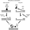
Possible pathways for post-replicative mtDNA repair. Both single-nucleotide (left) and long-patch (right) BER are believed to end with the concerted action of pol γ and DNA ligase III. The diagram is not to scale, nor is it meant to depict protein or DNA structure, or specific protein-protein interactions. See Table I and the text for descriptions of the factors.
Figure 4
Figure 4
Overview of transcription in animal mitochondria and the proteins involved in the process. Solid lines represent DNA, and the dashed line represents RNA. The diagram is not to scale, nor is it meant to depict protein or DNA structure, or specific protein-protein interactions. See Table I and the text for descriptions of the factors.

References

    1. Achanta G, Sasaki R, Feng L, Carew JS, Lu W, Pelicano H, Keating MJ, Huang P. Novel role of p53 in maintaining mitochondrial genetic stability through interaction with DNA pol gamma. EMBO J. 2005;24:3482–3492. - PMC - PubMed
    1. Adán C, Matsushima Y, Hernández-Sierra R, Marco-Ferreres R, Fernández-Moreno MA, González-Vioque E, Calleja M, Aragón JJ, Kaguni LS, Garesse R. Mitochondrial transcription factor B2 is essential for metabolic function in Drosophila melanogaster development. J Biol Chem. 2008;283:12333–12342. - PMC - PubMed
    1. Alam TI, Kanki T, Muta T, Ukaji K, Abe Y, Nakayama H, Takio K, Hamasaki N, Kang D. Human mitochondrial DNA is packaged with TFAM. Nucleid Acids Res. 2003;31:1640–1645. - PMC - PubMed
    1. Andres-Mateos E, Perier C, Zhang L, Blanchard-Fillion B, Greco TM, Thomas B, Ko HS, Sasaki M, Ischiropoulos H, Przedborski S, Dawson TM, Dawson VL. DJ-1 gene deletion reveals that DJ-1 is an atypical peroxiredoxin-like peroxidase. Proc Natl Acad Sci USA. 2007;104:14807–14812. - PMC - PubMed
    1. Ashley N, Poulton J. Anticancer DNA intercalators cause p53-dependent mitochondrial DNA nucleoid re-modelling. Oncogene. 2009;28:3880–3891. - PMC - PubMed

Publication types

MeSH terms

Substances