Review
. 2010 Aug;51(4):444-51.
doi: 10.1016/j.ymeth.2010.01.033. Epub 2010 Feb 1.

The QPCR assay for analysis of mitochondrial DNA damage, repair, and relative copy number

Affiliations
Review

The QPCR assay for analysis of mitochondrial DNA damage, repair, and relative copy number

Senyene E Hunter et al. Methods. 2010 Aug.

Abstract

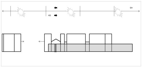
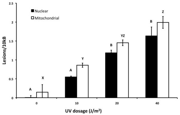
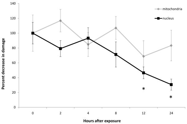
The quantitative polymerase chain reaction (QPCR) assay allows measurement of DNA damage in the mitochondrial and nuclear genomes without isolation of mitochondria. It also permits measurement of relative mitochondrial genome copy number. Finally, it can be used for measurement of DNA repair in vivo when employed appropriately. In this manuscript we briefly review the methodology of the QPCR assay, discuss its strengths and limitations, address considerations for measurement of mitochondrial DNA repair, and describe methodological changes implemented in recent years. We present QPCR assay primers and reaction conditions for five species not previously described in a methods article: Caenorhabditis elegans, Fundulus heteroclitus, Danio rerio, Drosophila melanogaster, and adenovirus. Finally, we illustrate the use of the assay by measuring repair of ultraviolet C radiation-induced DNA damage in the nuclear but not mitochondrial genomes of a zebrafish cell culture.


Figures

Figure 1
Figure 1. Schematic of the rationale of the QPCR assay for detecting damage in the nuclear genome
The continuous line ( ↔ ) represents a segment of the nuclear genome and perpendicular hashmarks along the DNA segment mark 10 kb intervals. Open boxes beneath the DNA segment depict genetic coding regions (exons) that are interrupted by interspersed introns (thick black lines). The long amplicon (10-15 kb) is represented as a transparent grey bar that amplifies the majority of a selected gene as well as a portion of the upstream DNA sequence. The bar representing the small amplicon (~200 bases) is shaded black. The filled arrows below and above the DNA segment indicate the position of the corresponding forward and reverse primers for the long (grey) and short (black) amplicon. Lesions are represented as stars that would inhibit or block the progression of the DNA polymerase used in the QPCR assay, thus reducing the amplification of the long product under quantitative conditions. For the smaller amplicon, the relatively close placement of the primers creates a small target size and reduces the probability that such lesions will be detected. Amplification of the short product is not inhibited except by very high levels of damage.
Figure 2
Figure 2. DNA damage in UVC-treated zebrafish cells
Mitochondrial and nuclear DNA damage was assessed in UVC-treated zebrafish cells. Petri dishes (10 mm diameter) containing roughly 106 cells/ml were drained free of culture medium and treated with 10, 20, and 40 J/m2 of UVC. Total DNA was isolated from cells and analyzed using QPCR. Letters indicate significant differences (p < 0.05) according to Fisher’s least significant differences analysis, carried out after ANOVA.
Figure 3
Figure 3. DNA damage repair in the zebrafish cell line ZF-4
A petri dish (10 mm diameter) containing roughly 106 cells/ml were drained free of culture medium and treated with 10 J/m2 of UVC. The dishes were immediately filled with new culture media and wrapped in aluminum foil to prevent any exposure to light. Then the plates were kept in normal incubation condition for 0, 2, 4, 8, 12, and 24 hours. After each designated time, DNA was isolated from three random plates per time point, and mtDNA and nucDNA damage was measured using QPCR. * indicates significant differences from 0 hrs (p < 0.01).

Similar articles

Cited by

References

    1. Ponti M, Forrow SM, Souhami RL, D’Incalci M, Hartley JA. Nucleic Acids Res. 1991;19:2929–33. - PMC - PubMed
    1. Ayala-Torres S, Chen Y, Svoboda T, Rosenblatt J, Van Houten B. Methods. 2000;22:135–47. - PubMed
    1. Meyer JN, Boyd WA, Azzam GA, Haugen AC, Freedman JH, Van Houten B. Genome Biol. 2007;8:R70. - PMC - PubMed
    1. Friedberg EC, W G, Siede W, Wood RD, Schultz RA, Ellenberger T. ASM Press; Washington, D.C.: 2006.
    1. Hanawalt PC, Spivak G. Nat Rev Mol Cell Biol. 2008;9:958–70. - PubMed

Publication types

LinkOut - more resources