

Review
doi: 10.1016/j.bbagrm.2009.11.011.
Developmental function of HMGN proteins
Affiliations
- PMID: 20123069
- PMCID: PMC2818498
- DOI: 10.1016/j.bbagrm.2009.11.011
Item in Clipboard
Review
Developmental function of HMGN proteins
Biochim Biophys Acta.
2010 Jan-Feb.
Abstract
High mobility group N (HMGN) proteins are the only nuclear proteins known to specifically recognize the generic structure of the 147-bp nucleosome core particle. Both in vitro and in vivo experiments demonstrate that HMGN proteins are involved in epigenetic regulation by modulating chromatin structure and levels of posttranslational modifications of nucleosomal histones. Expression of HMGN proteins is developmentally regulated, and the loss or overexpression of these proteins can lead to developmental abnormalities. This review will focus on the role and on the possible molecular mechanism whereby HMGN proteins affect cellular differentiation and development.
Published by Elsevier B.V.
Figures

(A) Representative HMGN protein is composed of three functional domains: a bipartite nuclear localization signal (NLS), a nucleosome binding domain and C-terminal chromatin regulatory domain. (B) Schematic diagram of mouse HMGN protein variant. Number of amino acids is indicated on right. While HMGN1, HMGN2, HMGN3a and HMGN4 show similar structure, HMGN3b lack C-terminal domain and HMGN5 possesses a long acidic tail which is composed of negatively charged amino acid repeats. (C) Chromosome locations of human HMGN genes. Each green bars represents chromosome and its number is written on the bottom. Yellow boxes indicate approximate HMGN gene location and exact locus of each HMGN variant is written on the right. Short arm and long arm is represented by separated bars.
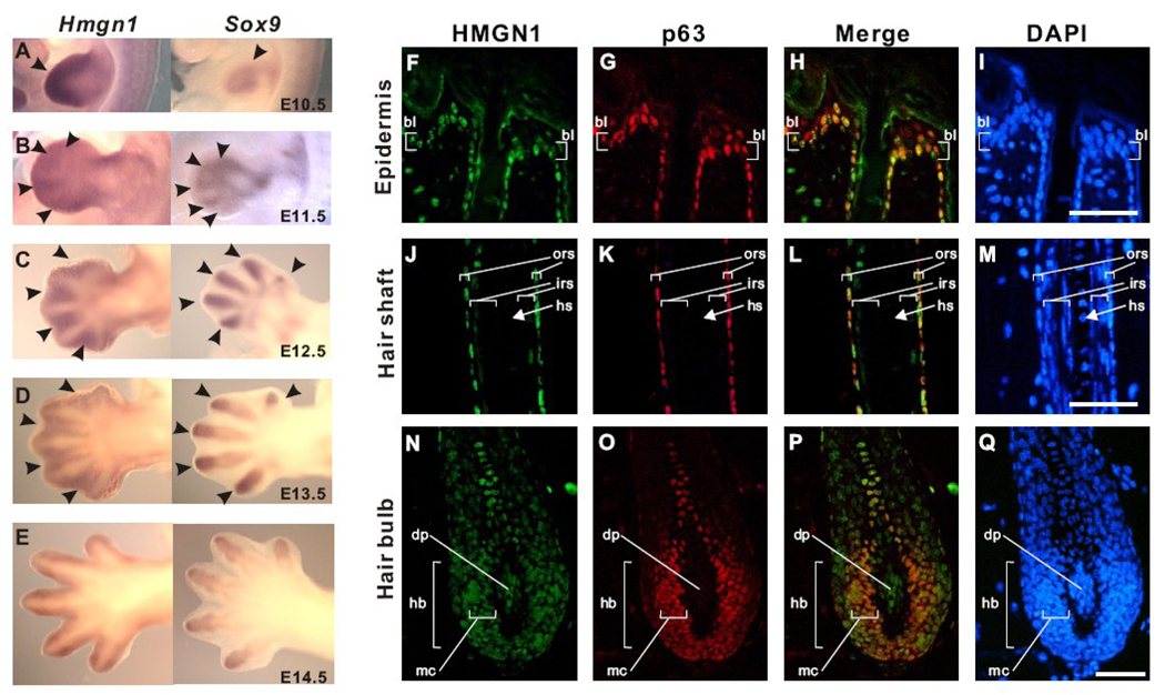
(A to E) The comparison of expression patterns of Hmgn1 (left) and Sox9 (right) in the developing limb bud. Hmgn1 expression is detected in the distal mesenchyme (filled arrowhead in A; left) and interdigit mesenchyme (filled arrowheads in B–E; left) while Sox9 is detected in the compensating mesenchyme (open arrow head in A; right) and digit mesenchyme (open arrowheads B–E; right). (F to Q) Expression of HMGN1 in the adult mouse hair follicle. HMGN1 (green) and p63 (red) protein are detected by immunofluorescence. Nuclear DNA is stained with DAPI (blue). Note that HMGN1 signal is detected in undifferentiated region and reduced in differentiated region. HMGN1 and p63 colocalize in most of the hair follicle except the dermal papilla. Scale bar: 50 µm. bl, basal layer; ors, outer root sheath; irs, inner root sheath; hs, hair shaft; hb, hair bulb; dp, dermal papilla; mc, matrix cells. (See details in [10])
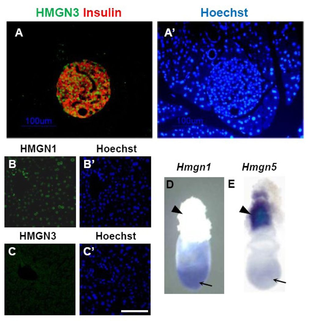
(A) Expression of HMGN3 proteins in the adult pancreas detected by immunofluorescence. HMGN3 protein is detected by green signal and pancreatic β-cells are detected by red signal with anti-Insulin. (A’) Nuclear DNA is stained with Hoechst. Note that HMGN3 expression is islet specific. (Refer details in [28]) (B and C) Expression of HMGN1 and HMGN3 in the adult liver. While HMGN1 is detected all over the liver, significant HMGN3 signal is not detected. (B’ and C’) Nuclear DNA is stained with Hoechst. (D and E) Expression of Hmgn1 and Hmgn5 in the mouse embryo at 7.5 days post coitus. Arrows indicate embryonic region and arrowheads indicate ectoplacental cone. Hmgn1 and Hmgn5 mRNA are detected by whole mount in situ hybridization. (Refer details in [31])
References
-
- Bustin M. Chromatin unfolding and activation by HMGN(*) chromosomal proteins. TrendsBiochem Sci. 2001;26:431–437. - PubMed
-
- Rochman M, Postnikov Y, Correll S, Malicet C, Wincovitch S, Karpova TS, McNally JG, Wu X, Bubunenko NA, Grigoryev S, Bustin M. The interaction of NSBP1/HMGN5 with nucleosomes in euchromatin counteracts linker histone-mediated chromatin compaction and modulates transcription. Mol Cell. 2009;35:642–656. - PMC - PubMed
-
- Shirakawa H, Landsman D, Postnikov YV, Bustin M. NBP-45, a novel nucleosomal binding protein with a tissue-specific and developmentally regulated expression. J Biol Chem. 2000;275:6368–6374. - PubMed
Publication types
MeSH terms
Substances
Grants and funding
LinkOut - more resources
Full Text Sources
