. 2010 Apr 8;129(2):100-7.
doi: 10.1016/j.imlet.2010.01.006. Epub 2010 Feb 1.

Cytokine induced expression of programmed death ligands in human neutrophils

Affiliations

Cytokine induced expression of programmed death ligands in human neutrophils

Paul E Bankey et al. Immunol Lett. .

Abstract

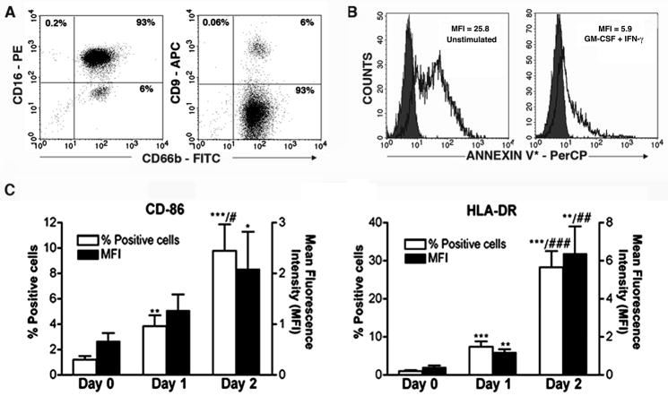
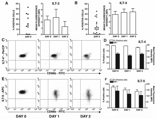
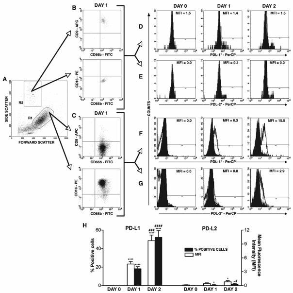
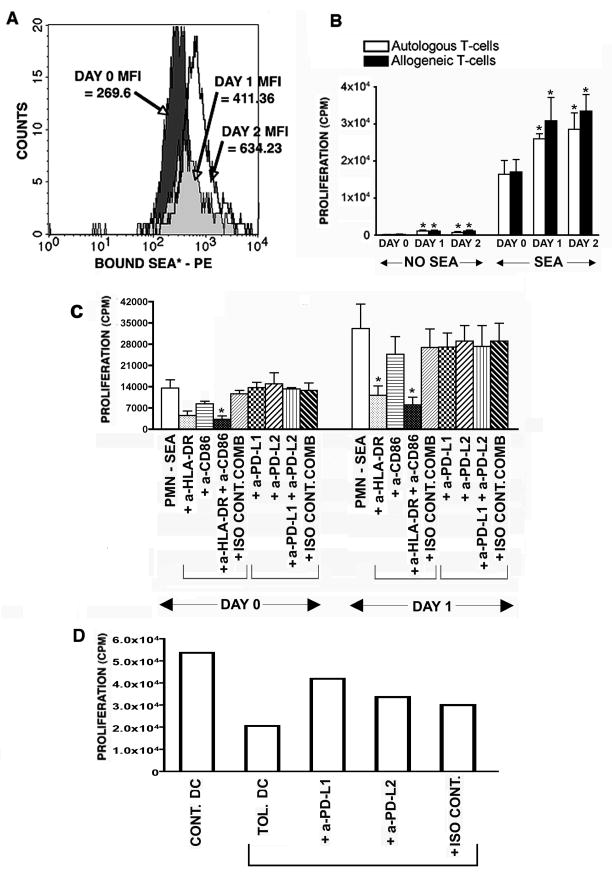
Recent evidence indicates that human neutrophils can serve as non-professional antigen presenting cells (APC). Although expression of MHC class II and co-stimulatory molecules on human neutrophils is limited, these molecules can be significantly induced following in vitro exposure to the cytokines IFNgamma and GM-CSF. Since professional APCs such as dendritic cells express both co-stimulatory and co-inhibitory molecules for activation and regulation of adaptive immunity, we determined whether cytokines induce increased expression of specific co-signaling molecules on human neutrophils. We report here that circulating human neutrophils express co-inhibitory molecules such as immunoglobulin-like transcript (ILT) 4 and 5, and also comparatively low and highly variable levels of ILT2 and ILT3, but the expression of these ILTs was not significantly changed by cytokine treatment. In contrast, we demonstrate for the first time that human peripheral blood neutrophils, although do not express the co-inhibitory molecule, programmed death ligand (PD-L) 1 on their surface, can express this molecule at moderate levels following cytokine exposure. Although moderate PD-L1 levels on healthy volunteers' neutrophils were not inhibitory to T cells, our findings do not exclude a possible robust increase in neutrophil PD-L1 expression in pathological conditions with immunosuppressive functions. These results suggest a possible immunoregulatory role for human neutrophils in adaptive immunity.


Figures

Fig. 1
Fig. 1
Cytokine induced expression of HLA-DR and CD86 in human neutrophils with delayed apoptosis. A) Neutrophil purity in the isolated granulocyte population was assessed by flow cytometry using CD66b-FITC, CD16-PE, CD9-APC antibody. B) Neutrophils cultured for 18–20hrs in medium alone (unstimulated) and in presence of IFNγ (100U/ml) + GM-CSF (20 ng/ml) (I+G) were stained for biotinylated Annexin V in binding buffer followed by labeling with streptavidin – PerCP and assessed for Annexin V binding (indicator of apoptosis) by flow cytometry. Representative of 4 experiments. C & D ) Untreated neutrophils isolated from peripheral blood (Day 0), treated with I+G for 18–20 hrs (Day 1) and for 40–44 hrs (Day 2), were assessed for CD86 and HLA-DR respectively by flow cytometry. Data are expressed as % positive and MFI. N=13; *p<0.05, **p<0.01, ***p<0.001 as compared to Day 0 data and #p<0.05, ##p<0.01, ###p<0.001 as compared to Day 1 data.
Fig. 2
Fig. 2
Human neutrophils express co-inhibitory molecules ILT2, 3, 4 and 5, and their expressions are not significantly altered by I+G exposure. Untreated neutrophils isolated from peripheral blood (Day 0), treated with I+G for 18–20 hrs (Day 1) and for 40–44 hrs (Day 2) were assessed by flow cytometry for expression of ILT2 (A; n=11), ILT3 (B; n=19), ILT4 (C – representative data, D – all donors’ data; n=20) and ILT5 (C – representative data, D – all donors’ data; n=11). Data are expressed as MFI and/or % positive cells. Gates were set up according to isotype control data. No statistical significance was observed after cytokine exposure.
Fig. 3
Fig. 3
Cytokine induces PD-L1 expression in neutrophils but not in contaminating eosinophils. A) Forward/side scatter of neutrophil and contaminating eosinophil population. Day 1 data is used as a representative of the time points (Day 0, 1 &2). B&C) Cell types in the indicated gated population were characterized by flow cytometry. R1 gate consists of neutrophils (CD66b+ CD16high CD9low) whereas R2 gate represents eosinophils (CD66b+CD16low CD9high). Day 0, 1 & 2 cell populations were assessed for expression of PD-L1 [(D&F - representative data and H - data of all donors (n=16)] and PD-L2 [(E&G - representative data and H - data of all donors (n=11)] by flow cytometry. Gates were set up according to isotype control data. Grey area - isotype control and open area - specific antibody. *p<0.05, ****p<0.0001 as compared to Day 0 data and #p<0.05, ###p<0.001 and ####p<0.0001 as compared to Day 1 data.
Fig. 4
Fig. 4
Cytokine exposure to neutrophils increases binding of SEA and its presentation to autologous and allogeneic T cells. A) Untreated neutrophils isolated from peripheral blood (Day 0), treated with I+G for 18–20hrs (Day 1) and for 40–44hrs (Day 2) were assessed for SEA binding by incubation of the cells in absence or presence of 100ng of SEA for 40 minutes at 37°C. Cells were washed, blocked and stained with rabbit anti-SEA antibody or in FACS buffer alone (to be used for background binding subtraction). Cells were washed, stained with PE labeled anti-rabbit antibody, fixed and analyzed by flow cytometry. SEA binding is expressed as MFI. SEA binding data of SEA treated Day 0, 1 and 2 neutrophils from the same donor (as indicated by arrows) are overlayed. Representative of 3 experiments. B) Day 0, 1 and 2 neutrophils (1×10⁵) were kept as controls or pulsed with 20 ng of SEA for 3hrs followed by co-culture with autologous (n=4) and allogeneic T cells (1×10⁵) (n=6) for 3 days. Cells were then pulsed with [3H]-thymidine for 18–20 hrs and harvested. T cell proliferation is expressed as counts per minute [CPM] of triplicate cultures. *p<0.05, **p<0.01 as compared to Day 0 data. C) Day 0 and 1 neutrophils (1×10⁵) were first pulsed with 20 ng of SEA for 3hrs followed by incubation for 1hr with different neutralizing antibodies or istype controls [anti-HLA-DR or anti-CD86 (each 5μg/ml) or their combination; anti-PD-L1 or anti-PD-L2 (each 10μg/ml) or their combination]. Neutrophils were then co-cultured with autologous T cells (1×10⁵) (n=3) for 3 days. T cell proliferation was measured as above. *p<0.05 as compared to APC activity of SEA – pulsed neutrophil alone. D) MOs were cultured in presence of IL-4 (50 ng/ml) + GM-CSF (50 ng/ml) for 5 days for their differentiation into immature DC and then cultured for 2 more days in presence [tolerogenic (Tol.) DC] or absence [control (Cont.) DC] of IL-10 (50 ng/ml). Tolerogenic DCs were then incubated for 1hr with neutralizing antibodies against PD-L1, PD-L2 or isotype control (each 10μg/ml) and the allogeneic T cells were added to DC at 20:1 ratios and the MLR activities of DCs were assessed by [3H]-thymidine ([1μCi/well] incorporation during the last 18 hrs of 6 day co-culture [27].

Similar articles

Cited by

References

    1. Nauseef WM. How human neutrophils kill and degrade microbes: an integrated view. Immunol Rev. 2007;219:88–102. - PubMed
    1. Herrmann JM, Bernardo J, Long HJ, Seetoo K, McMenamin ME, Batista EL, Jr, et al. Sequential chemotactic and phagocytic activation of human polymorphonuclear neutrophils. Infect Immun. 2007;75:3989–98. - PMC - PubMed
    1. Steinman RM. Dendritic cells: understanding immunogenicity. Eur J Immunol. 2007;37 (Suppl 1):S53–60. - PubMed
    1. Adler HS, Steinbrink K. Tolerogenic dendritic cells in health and disease: friend and foe! Eur J Dermatol. 2007;17:476–91. - PubMed
    1. van Gisbergen KP, Sanchez-Hernandez M, Geijtenbeek TB, van Kooyk Y. Neutrophils mediate immune modulation of dendritic cells through glycosylation-dependent interactions between Mac-1 and DC-SIGN. J Exp Med. 2005;201:1281–92. - PMC - PubMed

Publication types