

Aetiology of corneal ulcers assume FHV-1 unless proven otherwise
- PMID: 20123484
- PMCID: PMC10845471
- DOI: 10.1016/j.jfms.2009.12.004
Aetiology of corneal ulcers assume FHV-1 unless proven otherwise
Abstract
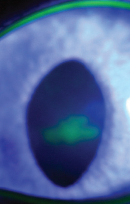
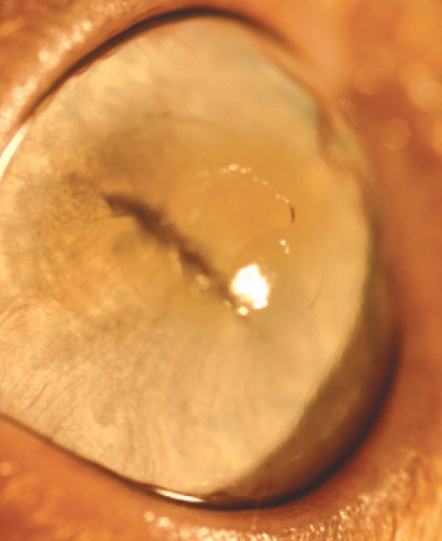
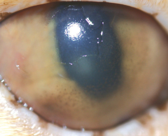
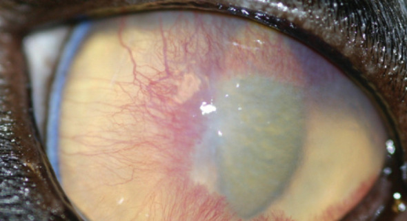
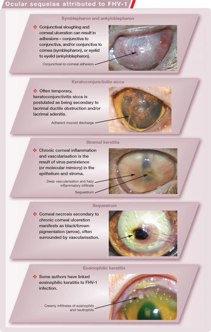
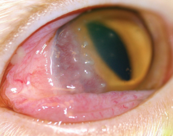
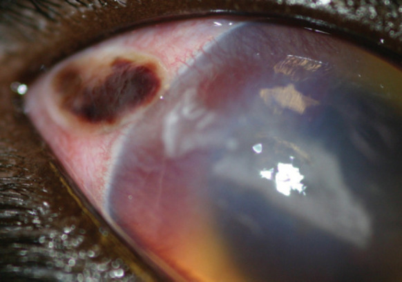
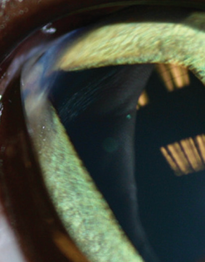
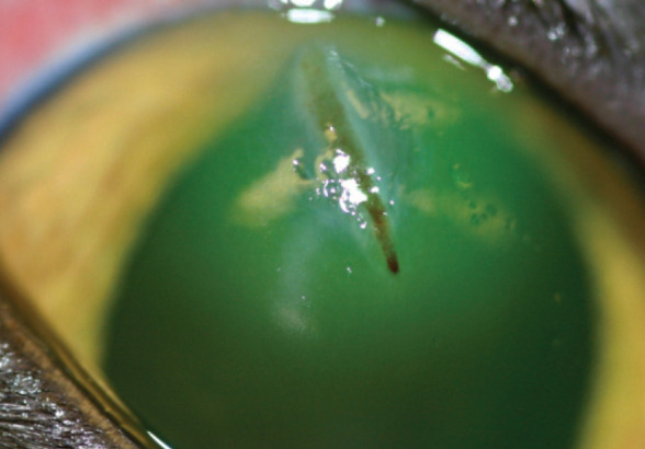
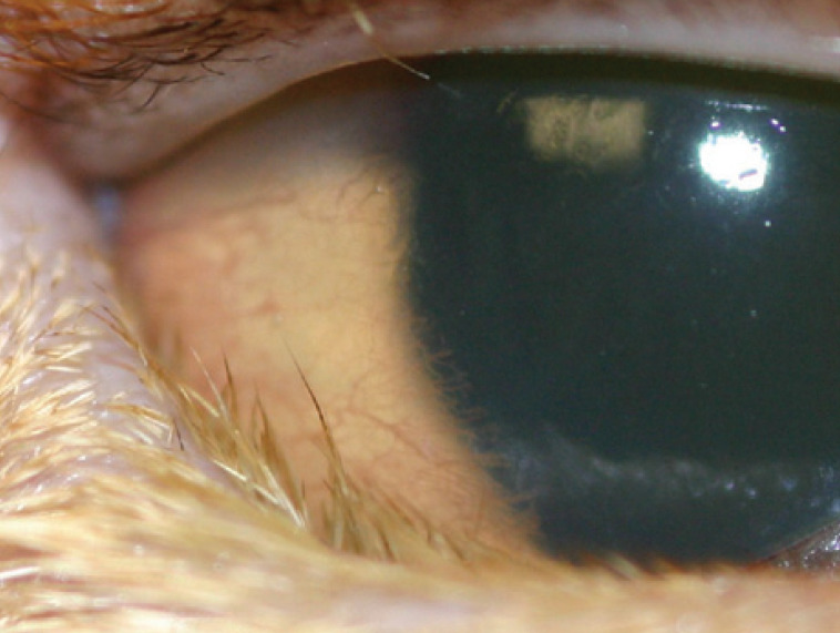
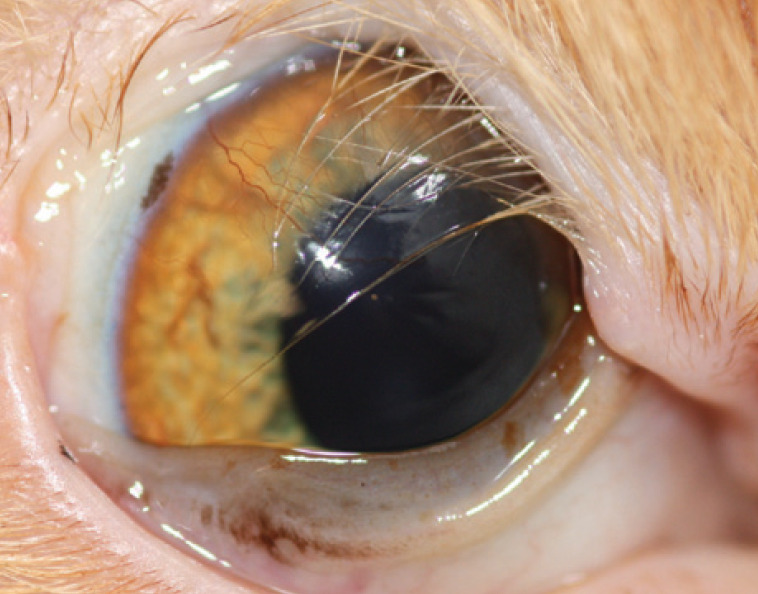
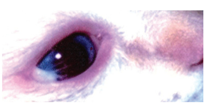
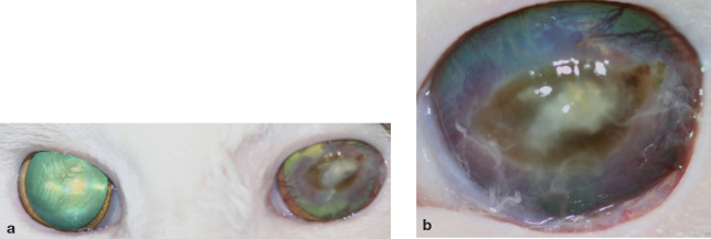
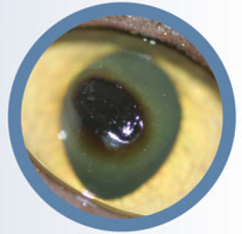
Overview: Feline ulcerative keratitis is a common presenting complaint and is frequently a sequela of feline herpesvirus 1 (FHV-1) infection; so much so, in fact, that it is fair to assume an FHV-1 aetiology until proven otherwise. Other potential causes of ulceration are trauma or underlying eyelid abnormalities (entropion, ectropion, agenesis, dermoids, neoplasia), lash abnormalities (ectopic cilia, trichiasis), tear film abnormalities or neurological deficiencies (trigeminal nerve paralysis, facial nerve paralysis).
Clinical challenges: The management of corneal ulceration in cats is frequently challenging, and treatment needs to be tailored carefully to the individual cat, its temperament, and the disease process present.
Evidence base: The scientific literature on feline ulcerative keratitis is extensive, particularly that related to FHV-1 infection. The aim of this article is to review the aetiology and diagnosis of corneal ulceration in cats with particular reference to the evidence base available.
Patient group: All age groups and breeds can suffer with ulcerative keratitis. Breed predispositions are present for some forms of corneal ulceration, and these are discussed.
Copyright 2009 ESFM and AAFP. Published by Elsevier Ltd. All rights reserved.
Figures


Similar articles
-
Treatment of corneal ulcers: what are the medical options?J Feline Med Surg. 2010 May;12(5):384-97. doi: 10.1016/j.jfms.2010.03.012. J Feline Med Surg. 2010. PMID: 20417899 Free PMC article. Review.
-
Treatment of corneal ulcers: when is surgery indicated?J Feline Med Surg. 2010 May;12(5):398-405. doi: 10.1016/j.jfms.2010.03.013. J Feline Med Surg. 2010. PMID: 20417900 Free PMC article. Review.
-
Treatment of feline herpesvirus-1 associated disease in cats with famciclovir and related drugs.J Feline Med Surg. 2009 Jan;11(1):40-8. doi: 10.1016/j.jfms.2008.11.012. J Feline Med Surg. 2009. PMID: 19154974 Free PMC article.
-
Ocular manifestations of feline herpesvirus infection.J Am Vet Med Assoc. 1971 Nov;159(10):1223-37. J Am Vet Med Assoc. 1971. PMID: 4330456 No abstract available.
-
Isolation of the feline herpesvirus-1 modified live vaccine strain F2 from one of four cats with dendritic ulcers.J Feline Med Surg. 2025 Jan;27(1):1098612X241306954. doi: 10.1177/1098612X241306954. J Feline Med Surg. 2025. PMID: 39751391 Free PMC article.
Cited by
-
Selkirk Rex: morphological and genetic characterization of a new cat breed.J Hered. 2012 Sep-Oct;103(5):727-33. doi: 10.1093/jhered/ess039. Epub 2012 Jul 26. J Hered. 2012. PMID: 22837475 Free PMC article.
-
Phylogenetic and recombination analysis of the herpesvirus genus varicellovirus.BMC Genomics. 2017 Nov 21;18(1):887. doi: 10.1186/s12864-017-4283-4. BMC Genomics. 2017. PMID: 29157201 Free PMC article.
-
Diagnostic Ophthalmology.Can Vet J. 2021 Jul;62(7):762-764. Can Vet J. 2021. PMID: 34219788 Free PMC article. No abstract available.
-
In vitro efficacy of ganciclovir against feline herpesvirus type 1 and assessment of ocular tolerability in healthy cats.J Feline Med Surg. 2021 Apr;23(4):400-404. doi: 10.1177/1098612X20944363. Epub 2020 Aug 4. J Feline Med Surg. 2021. PMID: 32749191 Free PMC article.
-
The Latest Prevalence, Isolation, and Molecular Characteristics of Feline Herpesvirus Type 1 in Yanji City, China.Vet Sci. 2024 Sep 7;11(9):417. doi: 10.3390/vetsci11090417. Vet Sci. 2024. PMID: 39330796 Free PMC article.
References
-
- Kafarnik C, Fritsche J, Reese R. In vivo confocal microscopy in the normal corneas of cats, dogs and birds. Vet Ophthalmol 2007; 10: 222–30. - PubMed
-
- Cullen CL, Lim C, Sykes J. Tear film breakup times in young healthy cats before and after anaesthesia. Vet Ophthalmol 2005; 8: 159–65. - PubMed
-
- Low HC, Powell CC, Veir JK, Hawley JR, Lappin MR. Prevalence of feline herpesvirus 1, Chlamydophila felis, and Mycoplasma spp DNA in conjunctival cells collected from cats with and without conjunctivitis. Am J Vet Res 2007; 68: 643–48. - PubMed
-
- Di Martino B, Di Francesco CE, Meridiani I, Marsilio F. Aetiological investigation of multiple respiratory infections in cats. New Microbiol 2007; 30: 455–61. - PubMed
Publication types
MeSH terms
LinkOut - more resources
Full Text Sources
Miscellaneous
