

doi: 10.1128/IAI.00731-09.
Epub 2010 Feb 1.
Acute immune response to Mycobacterium massiliense in C57BL/6 and BALB/c mice
Affiliations
- PMID: 20123718
- PMCID: PMC2849394
- DOI: 10.1128/IAI.00731-09
Item in Clipboard
Acute immune response to Mycobacterium massiliense in C57BL/6 and BALB/c mice
Infect Immun.
2010 Apr.
Abstract
Mycobacterium massiliense is an environmental opportunistic pathogen that has been associated with soft tissue infection after minor surgery. We studied the acute immune response of C57BL/6 and BALB/c mice infected intravenously with 10(6) CFU of an M. massiliense strain isolated from a nosocomial infection in Brazil. The results presented here show that M. massiliense is virulent and pathogenic to both C57BL/6 and BALB/c mice, inducing a granulomatous inflammatory reaction that involves the activation of macrophages, dendritic cells, and natural killer cells induced by gamma interferon and interleukin-17 (IL-17) in C57BL/6 mice and by IL-12 in BALB/c mice.
Figures
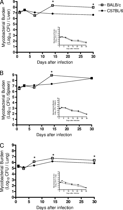
The M. massiliense bacterial load is maintained during infection. C57BL/6 and BALB/c mice were injected intravenously with 106 CFU of M. massiliense, and the bacterial load of their livers, spleens, and lungs (A, B, and C, respectively) were determined at 1, 3, 7, 14, and 30 days postinfection. The data show the means ± the SD bacterial load of five mice per group per time point. The results represent one of three independent experiments. The inset graphs represent infection with 104 CFU of M. massiliense for the same organs. *, P < 0.05.
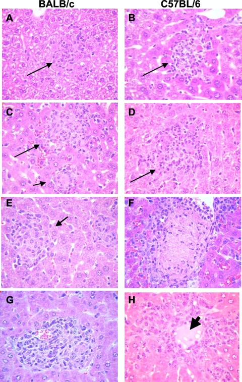
Development of early granulomatous lesions in the livers of M. massiliense-infected mice. Histological findings in the livers of C57BL/6 and BALB/c mice infected with M. massiliense. Liver sections were stained with H&E and examined at 200× magnification. Inflammatory cell infiltrates (arrows) are shown for days 1 (A and B), 3 (C and D), 7 (E and F), and 14 (G and H) postinfection. The heavy arrow in panel H indicates necrosis observed in the liver of C57BL/6 mice 14 days postinfection.
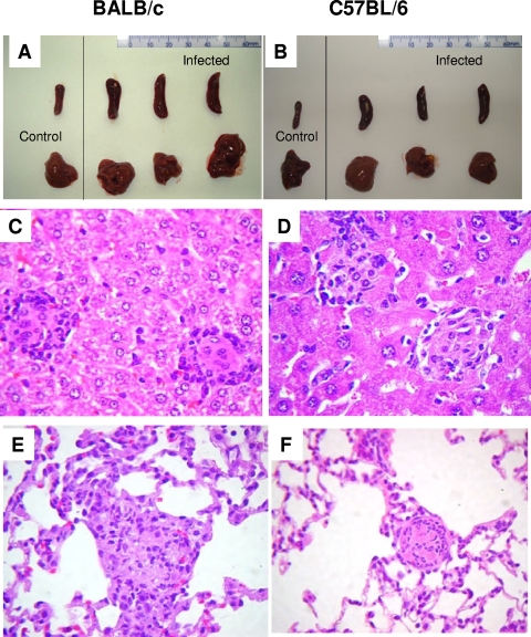
M. massiliense infection compromises the lungs, spleens, and livers of infected animals. Macroscopic and histological findings observed in C57BL/6 and BALB/c mice infected with M. massiliense. (A) Comparison of the spleen and liver of a control uninfected mouse (left) and an infected BALB/c mouse. (B) Comparison of the spleen and liver of a control uninfected mouse (left) and an infected C57BL/6 mouse. Lung and liver sections were stained with H&E and examined at 400× magnification (C to F). (C and D) Liver granulomatous reactions at 30 days postinfection in C57BL/6 and BALB/c mice, respectively. (E and F) Granulomatous lesions in the lungs of C57BL/6 and BALB/c mice, respectively.
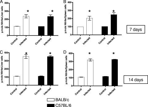
Evidence that NO is induced by the immune response against M. massiliense in C57BL/6 and BALB/c mice. NO production by liver (A and C) and spleen (B and D) cells at 7 and 14 days postinfection intravenously with 106 CFU of M. massiliense. The results are presented as total micromoles produced per 106 cells during 48 h of culture. The data shown are from one of three independent experiments. *, P < 0.05.
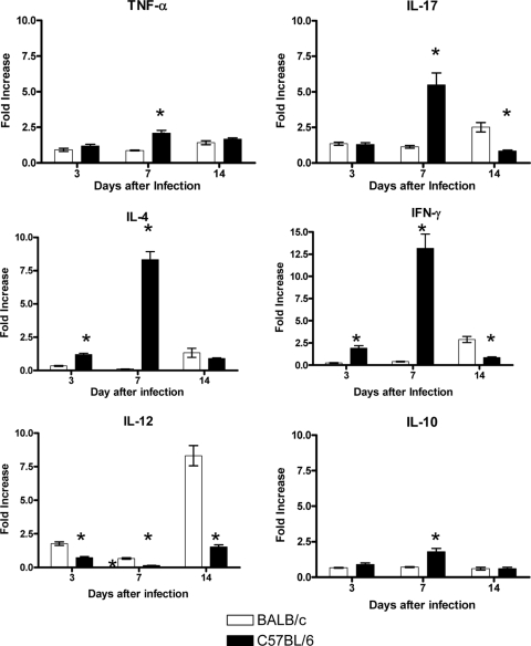
Modulation of host cytokine gene expression in the spleens of C57BL/6 and BALB/c mice in response to M. massiliense infection. mRNA was extracted from the spleens of infected mice on days 3, 7, and 14 postinfection. The levels of mRNA were quantified by real-time PCR using gene specific primers for inflammatory cytokine genes. *, P < 0.05 for C57BL/6 versus BALB/c cytokine transcript levels.
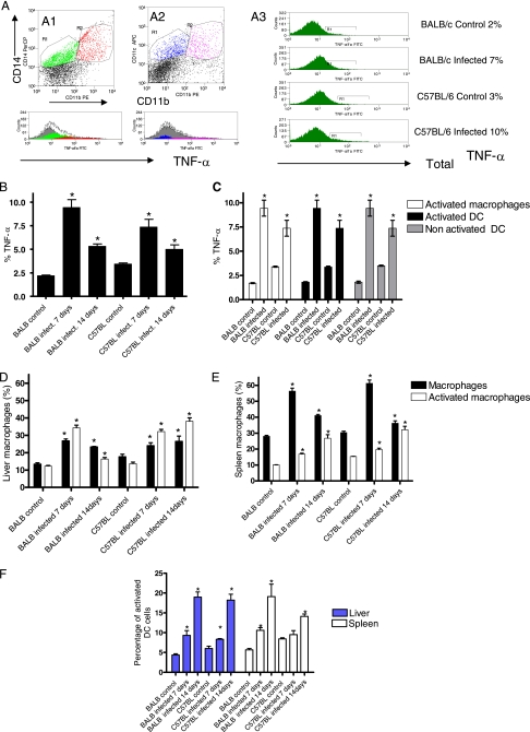
Macrophages and DCs are activated in response to M. massiliense infection. Macrophages, characterized by expression of CD14 (A1), and DCs, characterized by expression of CD11c (A2), were evaluated for their activation status (CD11b) and expression of TNF-α (A3 and B). The percentage of TNF-α+ macrophages and DCs in the spleens of infected mice was quantified according to the activation status of each cell population (C). The number of macrophages, according to their activation status, in the livers and spleens of infected mice was determined (D and E, respectively). (F) Proportion of DCs in the liver and spleen during infection.

Increase of NK cells in response to M. massiliense infection in C57BL/6 and BALB/c mice. Animals were inoculated intravenously with 106 CFU of M. massiliense, and spleen and liver NK2AG+ and CD62L+ cells were analyzed by flow cytometry on days 7 and 14 postinfection. Experiments were repeated three times. Each dot plot represents one of four mice analyzed. The graph shows the means ± the SD percentage of cells obtained from four mice in one experiment. *, P < 0.05.
Similar articles
-
Mycobacterium genavense infection in normal and immunodeficient mice.Microbes Infect. 2000 May;2(6):575-80. doi: 10.1016/s1286-4579(00)00369-5. Microbes Infect. 2000. PMID: 10884607
-
Importance of T cells, gamma interferon, and tumor necrosis factor in immune control of the rapid grower Mycobacterium abscessus in C57BL/6 mice.Infect Immun. 2007 Dec;75(12):5898-907. doi: 10.1128/IAI.00014-07. Epub 2007 Sep 17. Infect Immun. 2007. PMID: 17875636 Free PMC article.
-
Inflammatory dendritic cells migrate in and out of transplanted chronic mycobacterial granulomas in mice.J Clin Invest. 2011 Oct;121(10):3902-13. doi: 10.1172/JCI45113. Epub 2011 Sep 12. J Clin Invest. 2011. PMID: 21911937 Free PMC article.
-
The many niches and strategies used by pathogenic mycobacteria for survival within host macrophages.Immunobiology. 2009;214(7):526-42. doi: 10.1016/j.imbio.2008.12.005. Epub 2009 Mar 3. Immunobiology. 2009. PMID: 19261352 Review.
-
Monocyte-derived inflammatory dendritic cells in the granuloma during mycobacterial infection.Adv Exp Med Biol. 2012;946:277-93. doi: 10.1007/978-1-4614-0106-3_16. Adv Exp Med Biol. 2012. PMID: 21948374 Review.
Cited by
-
The mycma_1113 Gene from Mycobacterium abscessus subsp. massiliense is Related to Siderophore Synthesis.Indian J Microbiol. 2019 Jun;59(2):180-187. doi: 10.1007/s12088-019-00788-z. Epub 2019 Feb 27. Indian J Microbiol. 2019. PMID: 31031432 Free PMC article.
-
Mycobacterium abscessus subsp. massiliense: Biofilm Formation, Host Immune Response, and Therapeutic Strategies.Microorganisms. 2025 Feb 18;13(2):447. doi: 10.3390/microorganisms13020447. Microorganisms. 2025. PMID: 40005812 Free PMC article. Review.
-
Increased virulence of an epidemic strain of Mycobacterium massiliense in mice.PLoS One. 2011;6(9):e24726. doi: 10.1371/journal.pone.0024726. Epub 2011 Sep 12. PLoS One. 2011. PMID: 21931831 Free PMC article.
-
Non-disulfide-Bridge Peptide 5.5 from the Scorpion Hadrurus gertschi Inhibits the Growth of Mycobacterium abscessus subsp. massiliense.Front Microbiol. 2017 Feb 22;8:273. doi: 10.3389/fmicb.2017.00273. eCollection 2017. Front Microbiol. 2017. PMID: 28275372 Free PMC article.
-
Antimycobacterial Activity of a New Peptide Polydim-I Isolated from Neotropical Social Wasp Polybia dimorpha.PLoS One. 2016 Mar 1;11(3):e0149729. doi: 10.1371/journal.pone.0149729. eCollection 2016. PLoS One. 2016. PMID: 26930596 Free PMC article.
References
-
- Cardona, P. J., A. Cooper, M. Luqui, A. A. Filipos, I. M. Orme, and V. Ausuna. 1999. The intravenous model of murine tuberculosis is less pathogenic than the aerogenic model owing to a more rapid induction of systemic immunity. Scand. J. Immunol. 49:362-366. - PubMed
-
- Cardoso, A. M., E. M. de Sousa, C. Viana-Niero, F. B. de Bortoli, Z. C. P. das Neves, S. C. Leão, A. P. Junqueira-Kipnis, and A. Kipnis. 2008. Emergence of nosocomial Mycobacterium massiliense infection in Goiás, Brazil. Microbes Infect. 10:1552-1557. - PubMed
Publication types
MeSH terms
Substances
LinkOut - more resources
Full Text Sources
Medical
