

Local host response to chlamydial urethral infection in male guinea pigs
- PMID: 20123720
- PMCID: PMC2849414
- DOI: 10.1128/IAI.01339-09
Local host response to chlamydial urethral infection in male guinea pigs
Abstract
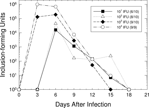
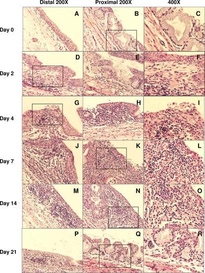
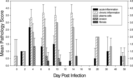
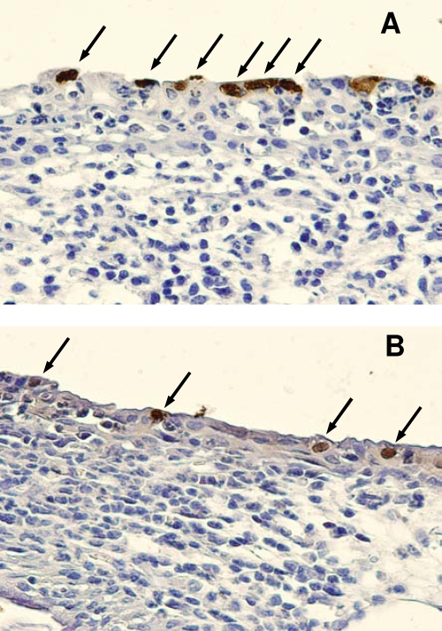
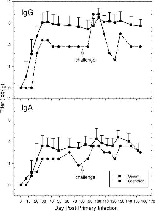
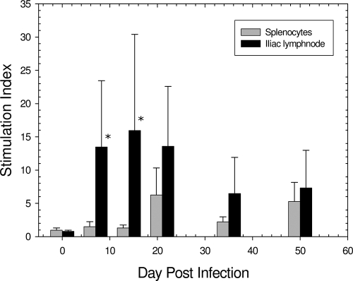
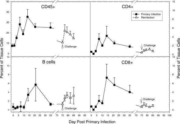
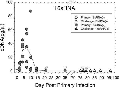
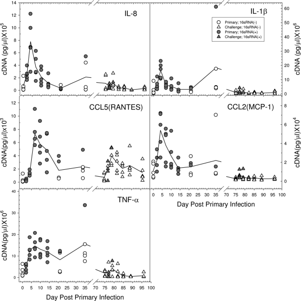
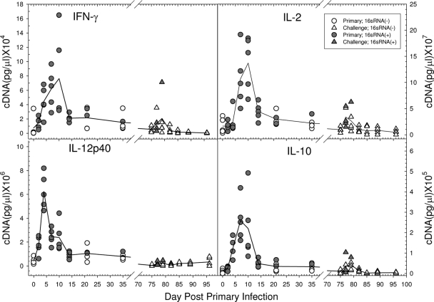
Very little is known about the host response to chlamydial genital infection in the male, particularly about the nature of the local response in the urethra. In this study, the pathological and immunologic responses to urethral infection of the male guinea pig with Chlamydia caviae (Chlamydophila caviae) were characterized both during a primary infection and following a challenge infection. A dose-response experiment found that the 50% infectious dose for male urethral infection was 78 inclusion-forming units. The histopathologic response was similar to that of the female, with an initial acute inflammatory response followed by a chronic inflammatory response and plasma cell infiltration. Production of IgG and IgA antibodies in local urethral secretions developed following infection, and levels of both increased in a typical anamnestic response following a challenge infection. CD4 and CD8 T cells, as well as B cells, were observed in the local site by flow cytometry, with a slightly increased number of CD8 cells. Following challenge infection, the dominant anamnestic response was solely in the B-cell compartment, with only a minimal number of T cells. The T-cell response was clearly a Th1 response, as judged by increased levels of gamma interferon (IFN-gamma), interleukin-12 p40 (IL-12p40), and IL-2. The proinflammatory cytokines and chemokines IL-8, IL-1beta, tumor necrosis factor alpha (TNF-alpha), CCL2 (monocyte chemoattractant protein 1 [MCP-1]), and CCL5 (RANTES) were elicited in the urethra following primary infection, but only CCL5 showed increased levels upon challenge. This study represents the first comprehensive analysis of the local immune response in the male urethra to a chlamydial genital infection.
Figures
Similar articles
-
Chlamydial serum IgG, IgA and local IgA antibodies in patients with genital-tract infections measured by solid-phase radioimmunoassay.J Med Microbiol. 1981 Feb;14(1):77-87. doi: 10.1099/00222615-14-1-77. J Med Microbiol. 1981. PMID: 7463469
-
Humoral and cellular immunity in secondary infection due to murine Chlamydia trachomatis.Infect Immun. 1997 Jul;65(7):2876-82. doi: 10.1128/iai.65.7.2876-2882.1997. Infect Immun. 1997. PMID: 9199462 Free PMC article.
-
Genital tract infection with Chlamydia trachomatis fails to induce protective immunity in gamma interferon receptor-deficient mice despite a strong local immunoglobulin A response.Infect Immun. 1997 Mar;65(3):1032-44. doi: 10.1128/IAI.65.3.1032-1044.1997. Infect Immun. 1997. PMID: 9038313 Free PMC article.
-
[The role of cytokines in Chlamydia-induced inflammation].Xi Bao Yu Fen Zi Mian Yi Xue Za Zhi. 2025 Jun;41(6):564-570. Xi Bao Yu Fen Zi Mian Yi Xue Za Zhi. 2025. PMID: 40525346 Review. Chinese.
-
Role of CD8(+)T cells in the host response to Chlamydia.Microbes Infect. 2008 Nov-Dec;10(14-15):1420-30. doi: 10.1016/j.micinf.2008.08.006. Epub 2008 Aug 26. Microbes Infect. 2008. PMID: 18790073 Free PMC article. Review.
Cited by
-
Cross-Reactive Effects of Vaccines: Heterologous Immunity between Tetanus and Chlamydia.Vaccines (Basel). 2020 Dec 1;8(4):719. doi: 10.3390/vaccines8040719. Vaccines (Basel). 2020. PMID: 33271962 Free PMC article.
-
Antibiotic treatment of Chlamydia-induced cystitis in the koala is linked to expression of key inflammatory genes in reactive oxygen pathways.PLoS One. 2019 Aug 15;14(8):e0221109. doi: 10.1371/journal.pone.0221109. eCollection 2019. PLoS One. 2019. PMID: 31415633 Free PMC article.
-
Effect of inflammatory response on in vivo competition between two chlamydial variants in the guinea pig model of inclusion conjunctivitis.Infect Immun. 2012 Feb;80(2):612-9. doi: 10.1128/IAI.06054-11. Epub 2011 Dec 5. Infect Immun. 2012. PMID: 22144478 Free PMC article.
-
Use of a Guinea pig-specific transcriptome array for evaluation of protective immunity against genital chlamydial infection following intranasal vaccination in Guinea pigs.PLoS One. 2014 Dec 11;9(12):e114261. doi: 10.1371/journal.pone.0114261. eCollection 2014. PLoS One. 2014. PMID: 25502875 Free PMC article.
-
HIV infection and immune defense of the penis.Am J Reprod Immunol. 2011 Mar;65(3):220-9. doi: 10.1111/j.1600-0897.2010.00941.x. Epub 2011 Jan 9. Am J Reprod Immunol. 2011. PMID: 21214659 Free PMC article. Review.
References
-
- Al-Mously, N., and A. Eley. 2007. Interaction of Chlamydia trachomatis serovar E with male genital tract epithelium results in secretion of proinflammatory cytokines. J. Med. Microbiol. 56:1025-1032. - PubMed
-
- Cunningham, K. A., A. J. Carey, J. M. Finnie, S. Bao, C. Coon, R. Jones, O. Wijburg, R. A. Strugnell, P. Timms, and K. W. Beagley. 2008. Poly-immunoglobulin receptor-mediated transport of IgA into the male genital tract is important for clearance of Chlamydia muridarum infection. Am. J. Reprod. Immunol. 60:405-414. - PubMed
-
- Jacobs, N. F., Jr., E. S. Arum, and S. J. Kraus. 1978. Experimental infection of the chimpanzee urethra and pharynx with Chlamydia trachomatis. Sex. Transm. Dis. 5:132-136. - PubMed
Publication types
MeSH terms
Substances
Grants and funding
LinkOut - more resources
Full Text Sources
Medical
Research Materials
Miscellaneous
