. 2010 Mar;133(Pt 3):808-21.
doi: 10.1093/brain/awp333. Epub 2010 Jan 31.

Reactive microgliosis: extracellular micro-calpain and microglia-mediated dopaminergic neurotoxicity

Affiliations

Reactive microgliosis: extracellular micro-calpain and microglia-mediated dopaminergic neurotoxicity

Shannon Levesque et al. Brain. 2010 Mar.

Abstract

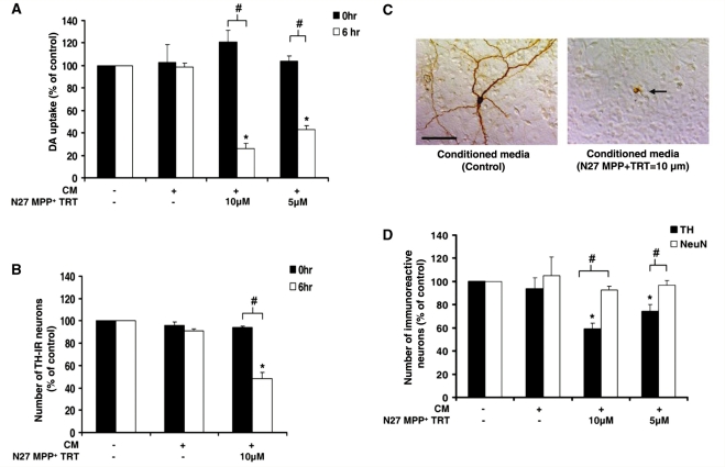
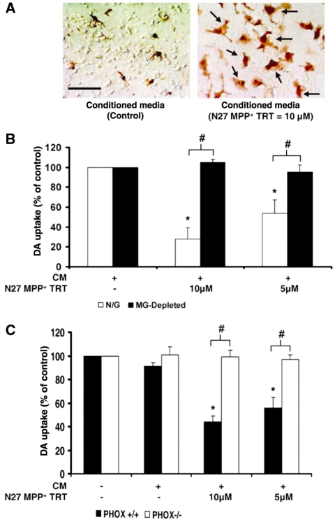
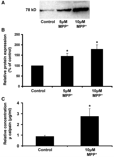
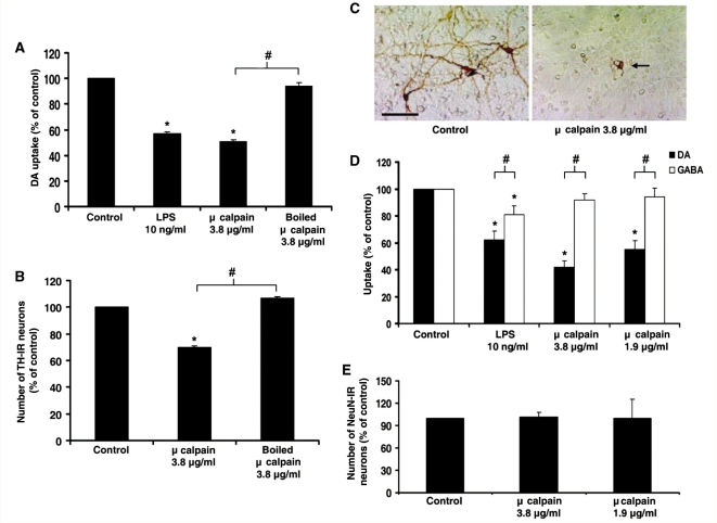
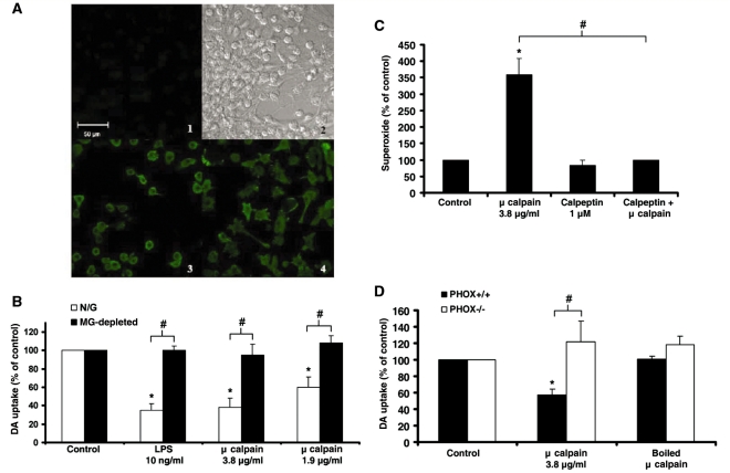
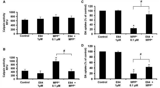
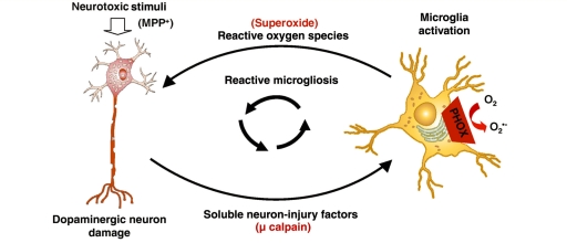
Microglia, the innate immune cells in the brain, can become chronically activated in response to dopaminergic neuron death, fuelling a self-renewing cycle of microglial activation followed by further neuron damage (reactive microgliosis), which is implicated in the progressive nature of Parkinson's disease. Here, we use an in vitro approach to separate neuron injury factors from the cellular actors of reactive microgliosis and discover molecular signals responsible for chronic and toxic microglial activation. Upon injury with the dopaminergic neurotoxin 1-methyl-4-phenylpyridinium, N27 cells (dopaminergic neuron cell line) released soluble neuron injury factors that activated microglia and were selectively toxic to dopaminergic neurons in mixed mesencephalic neuron-glia cultures through nicotinamide adenine dinucleotide phosphate oxidase. mu-Calpain was identified as a key signal released from damaged neurons, causing selective dopaminergic neuron death through activation of microglial nicotinamide adenine dinucleotide phosphate oxidase and superoxide production. These findings suggest that dopaminergic neurons may be inherently susceptible to the pro-inflammatory effects of neuron damage, i.e. reactive microgliosis, providing much needed insight into the chronic nature of Parkinson's disease.


Figures

Figure 1
Figure 1
Dopaminergic neurons are selectively vulnerable to soluble neuron injury signals. Mesencephalic neuron-glia cultures were treated with medium alone (unconditioned medium, −/−) or conditioned medium (CM) from N27 dopaminergic (DA) cells exposed to MPP+ to determine if signals released from damaged neurons propagate further dopaminergic neuronal damage. N27 cells were treated with either medium alone (+/−) or MPP+ (5 or 10 μM) for 24 h (N27 MPP+ TRT). After washing MPP+ from N27 dopaminergic neuron cells three times with 1 ml of medium, the conditioned medium was collected from N27 cells at either 0 or 6 h following the final wash. The fresh medium alone (−/−) or the conditioned medium was added to mixed neuron-glia cultures. (A) Soluble neuron-injury signals take time to accumulate in the conditioned medium. Loss of dopaminergic neuron function was measured 9 days later with the [3H] dopamine uptake assay. (B) Loss of dopaminergic neurons was determined 9 days later by counting the number of tyrosine hydroxylase (TH)-immunoreactive (IR) neurons. (C) Tyrosine hydroxylase staining demonstrates the ability of 6 h conditioned medium (from N27 cells treated with 10 μM MPP+) to cause morphological damage to dopaminergic neurons. The arrow denotes the damaged dopaminergic neuron and the scale bar indicates 50 μm. Representative images are from three separate experiments. (D) Loss of dopaminergic neurons (tyrosine hydroxylase-immunoreactive neurons) and total neurons (NeuN-immunoreactive neurons) was determined 9 days later by cell count. Graphs show the results expressed as percentage of the control cultures (unconditioned medium, −/−) and are the mean ± standard error of mean from three independent experiments in triplicate. *P < 0.05, control compared to treatment, #P < 0.05 indicates significant differences due to either time (0 versus 6 h) or selective neurotoxicity (tyrosine hydroxylase versus NeuN).
Figure 2
Figure 2
Soluble neuron injury signals are toxic to dopaminergic neurons through microglial activation and NADPH oxidase. Mesencephalic neuron-glia cultures were treated with medium alone (−/−) or conditioned medium (CM) from N27 dopaminergic (DA) cells exposed to MPP+ to determine if signals released from damaged neurons propagate further dopaminergic neuron damage. N27 cells were treated with either medium alone (+/−) or MPP+ (5 or 10 μM) for 24 h (N27 MPP+ TRT). After washing MPP+ from N27 dopaminergic neuron cells three times with 1 ml of medium, the conditioned medium was collected from N27 cells at 6 h following the final wash. Fresh unconditioned medium (−/−) or the conditioned medium was added to mixed neuron-glia cultures. (A) At 12 h post-treatment, neuron-glia cultures were stained for ionized calcium-binding adaptor molecule-1. The arrows denote examples of activated microglia and the scale bar indicates 50 μm. Microglial activation in response to the conditioned medium from N27 cells damaged with MPP+ is depicted by an increase in the number of stained cells, enlarged size of stained cells and irregular amoeboid morphology. Representative images are from three separate experiments. (B) Dopamine neurotoxicity was measured 9 days later with the [3H] dopamine uptake assay in neuron-glia cultures (N/G: containing microglia, astrocytes and neurons) and microglia-depleted cultures (MG-depleted: containing astrocytes and neurons). The conditioned medium was only toxic in the presence of microglia. (C) Dopamine neurotoxicity was measured 9 days later with the [3H] dopamine uptake assay in neuron-glia cultures from mice missing functional NADPH oxidase (PHOX−/−) and control strains (PHOX+/+). The conditioned medium was only toxic in the presence of NADPH oxidase. Graphs show the results expressed as percentage of the control cultures and are the mean ± SEM from three independent experiments in triplicate. *P < 0.05, control compared to treatment, #P < 0.05 indicates significant differences due to microglia (N/G versus microglia) or mouse strain (PHOX+/+ versus PHOX−/−).
Figure 3
Figure 3
μ-Calpain is a soluble neuron injury factor released by damaged dopaminergic neurons. N27 dopaminergic neuron cells were treated with either medium alone or MPP+ (5 or 10 μM) for 24 h (N27 MPP+ TRT). After washing MPP+ from N27 dopaminergic neuron cells three times with 1 ml of medium, conditioned medium (CM, serum-free medium) from N27 cells at 6 h following the final wash. Samples were concentrated from 15 ml to 100 μl and ran out on a sodium dodecyl sulphate polyacrylamide gel electrophoresis gel. (A) Representative image of western blot analysis. Blots of concentrated conditioned medium probed with an anti-μ-calpain antibody reveals a 78 kDa protein band that increases with the concentration the MPP+ exposure (i.e. increases with enhanced neuron damage). (B) The densitometry graph shows the results expressed as percentage of the control and are the mean ± SEM from five independent experiments. (C) The relative amount of extracellular μ-calpain present in the unconcentrated N27 conditioned medium, as determined by ELISA. The results are from six separate experiments. *P < 0.05, control compared to treatment.
Figure 4
Figure 4
Extracellular μ-calpain selectively kills dopaminergic neurons. Rat mesencephalic neuron-glia cultures were treated with either medium alone (control), lipopolysaccharide (LPS; 10 ng/ml) or μ-Calpain (3.8 μg/ml, 1.9 μg/ml or boiled). (A) Loss of dopaminergic neuron function was measured 9 days later with the [3H] dopamine uptake assay. (B) Loss of dopaminergic neurons was determined 9 days later by counting the number of tyrosine hydroxylase (TH)-immunoreactive (IR) neurons. (C) Tyrosine hydroxylase staining demonstrates μ-calpain-induced morphological damage to dopaminergic neurons. The arrow denotes the damaged dopaminergic neuron and the scale bar indicates 50 μm. Representative images are from three separate experiments. (D) Dopamine neurotoxicity (DA) and GABA neurotoxicity (GABA) was determined 9 days later by [3H] dopamine or GABA uptake assay. (E) General loss of total neurons (NeuN-immunoreactive neurons) was determined 9 days later by the cell count. Graphs show the results expressed as percentage of the control cultures and are the mean ± SEM from three independent experiments in triplicate. *P < 0.05, control compared to treatment; #P < 0.05 indicates significant differences in selective neurotoxicity (tyrosine hydroxylase versus GABA).
Figure 5
Figure 5
Extracellular μ-calpain is neurotoxic due to activation of microglial NADPH oxidase. (A) Confocal images of neuron-glia cultures stained for ionized calcium-binding adaptor molecule-1. Cells were treated with either vehicle (medium alone) or μ-calpain (3.8 μg/ml) for 12 h at 37°C. (1) Background fluorescence (no secondary antibody added); (2), transmitted light (Differential Interference Contrast) image; (3) untreated control cells; (4) μ-calpain-treated cells. The fluorescence micrographs depict representative changes in morphology caused by μ-calpain that indicates microglial activation. Activated microglial cells are amoeboid with multiple extended processes (4). The scale bar indicates 50 μm. (B) Neuron-glia cultures (N/G—containing microglia, astrocytes, and neurons) and microglia-depleted cultures (MG-depleted—containing astrocytes and neurons) were treated with medium alone (Control), lipopolysaccharide (LPS) 10 ng/ml (positive control for microglia-mediated neurotoxicity) or μ-calpain (3.8 or 1.9 μg/ml). Dopamine neurotoxicity was measured 9 days later with the [3H] dopamine uptake assay. μ-Calpain was only toxic in the presence of microglia. (C) Enriched microglia cultures were treated with medium alone (Control), μ-calpain (3.8 μg/ml), calpeptin (1 μM, a specific calpain inhibitor) or calpeptin + μ-calpain. The production of extracellular superoxide was measured by the superoxide dismutase-inhibitable reduction of tetrazolium salt, WST-1 at 30 min post-treatment. Results are mean ± SEM. Data are from four separate experiments. *P < 0.05, compared with control cultures. (D) Mesencephalic midbrain neuron-glia cultures from PHOX+/+ and PHOX−/− mice were treated with medium alone (Control), μ-calpain (3.8 μg/ml) or boiled μ-calpain (3.8 μg/ml). Graphs show the results expressed as percentage of the control cultures and are the mean ± SEM from three independent experiments in triplicate. *P < 0.05, control compared to treatment; #P < 0.05 indicates significant differences due to microglia (N/G versus microglia), superoxide reduction (calpeptin reduction of μ-calpain) or mouse strain (PHOX+/+ versus PHOX−/−).
Figure 6
Figure 6
Inhibiting extracellular μ-calpain protects dopaminergic neurons. (A) E64 fails to inhibit intracellular calpain activity in N27 cells. N27 cells were pre-treated with medium alone or E64 (1 μM, non-permeable, extracellular only calpain inhibitor) for 30 min prior to treatment with either medium alone or 10 μM MPP+. After 24 h, the conditioned medium was removed, cells were lysed and intracellular calpain activity was calculated with a commercially available kit. Data are presented as relative fluorescence units (RFU) and are the mean ± SEM from three independent experiments performed with duplicate samples. (B) E64 inhibits extracellular calpain activity. The conditioned medium from the N27 cells above was tested for extracellular calpain activity using the commercially available kit. Data are presented as relative fluorescence units and are the mean ± SEM from three independent experiments performed with duplicate samples. (C) E64 protects against dopaminergic neurotoxicity caused by soluble neuron-injury factors (conditioned medium). Rat mesencephalic neuron-glia cultures were pre-treated with medium alone or E64 (1 μM, non-permeable, extracellular only calpain inhibitor) for 30 min prior to treatment with either medium alone or the conditioned medium (CM N27 10 μM MPP+). [3H] Dopamine (DA) uptake assay was performed at 9 days following the MPP+ treatment. The data are expressed as the percent of the control cultures and are the mean ± SEM from three independent experiments performed with triplicate samples. (D) E64 protects against dopamine neurotoxicity caused by soluble neuron-injury factors (MPP+). Rat mesencephalic neuron-glia cultures were pre-treated with medium alone or E64 (1 μM, non-permeable, extracellular only calpain inhibitor) for 30 min prior to treatment with either medium alone or 0.1 μM MPP+. [3H] Dopamine uptake assay was performed at 9 days following the MPP+ treatment. The data are expressed as the percent of the control cultures and are the mean ± SEM from three independent experiments performed with triplicate samples. *P < 0.05, compared to control-treated cultures. #P < 0.05, compared to MPP+ treatment.
Figure 7
Figure 7
μ-Calpain is a key factor driving the progressive nature of dopaminergic neuron damage. Dopaminergic (DA) neuron damage is chronic in part because damaged cells release soluble factors that accumulate over time to active resident microglia, driving further toxicity (reactive microgliosis). μ-Calpain is externalized in the process of dopamine neuron damage and is a fundamental soluble neuron injury factor responsible for the toxic aspects of reactive microgliosis. Specifically, extracellular μ-calpain activates microglial NADPH oxidase, producing superoxide to damage neighbouring dopaminergic neurons selectively and propagate neurotoxicity. This feed-forward cycle provides much needed insight into the progressive nature of dopaminergic neuron damage.

Similar articles

Cited by

References

    1. Block ML, Hong JS. Microglia and inflammation-mediated neurodegeneration: multiple triggers with a common mechanism. Prog Neurobiol. 2005;76:77–8. - PubMed
    1. Block ML, Hong JS. Chronic microglial activation and progressive dopaminergic neurotoxicity. Biochem Soc Trans. 2007;35:1127–32. - PubMed
    1. Block ML, Li G, Qin L, Wu X, Pei Z, Wang T, et al. Potent regulation of microglia-derived oxidative stress and dopaminergic neuron survival: substance P vs. dynorphin. Faseb J. 2006;20:251–8. - PubMed
    1. Block ML, Wu X, Pei Z, Li G, Wang T, Qin L, et al. Nanometer size diesel exhaust particles are selectively toxic to dopaminergic neurons: the role of microglia, phagocytosis, and NADPH oxidase. Faseb J. 2004;18:1618–20. - PubMed
    1. Block ML, Zecca L, Hong JS. Microglia-mediated neurotoxicity: uncovering the molecular mechanisms. Nat Rev Neurosci. 2007;8:57–69. - PubMed

Publication types

MeSH terms