. 2010 Jun;38(10):3340-50.
doi: 10.1093/nar/gkq017. Epub 2010 Jan 31.

TRAP150 activates pre-mRNA splicing and promotes nuclear mRNA degradation

Affiliations

TRAP150 activates pre-mRNA splicing and promotes nuclear mRNA degradation

Kuo-Ming Lee et al. Nucleic Acids Res. 2010 Jun.

Abstract

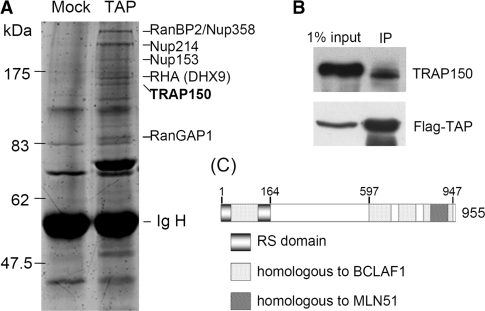
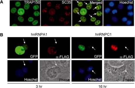
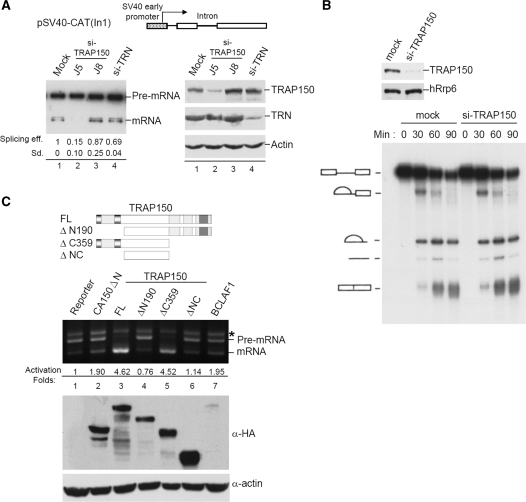
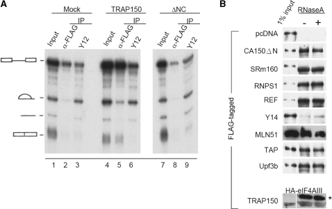
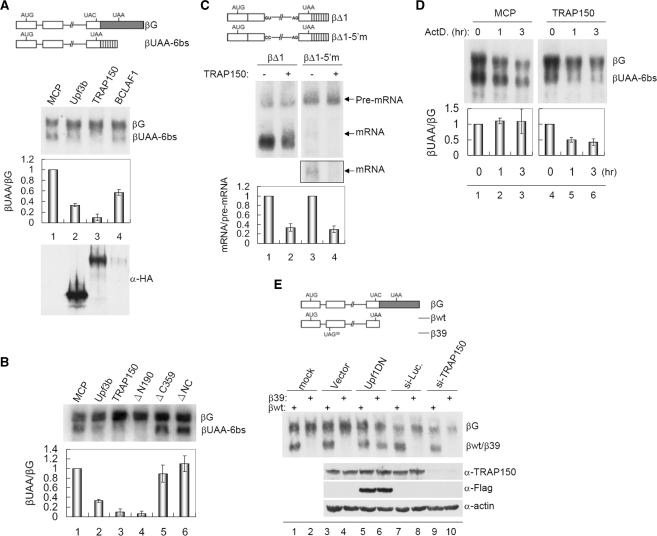
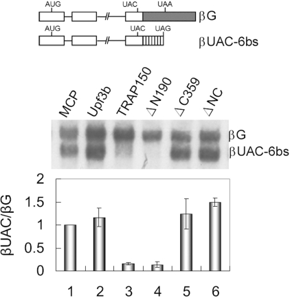
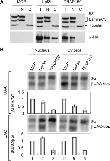
TRAP150 has been identified as a subunit of the transcription regulatory complex TRAP/Mediator, and also a component of the spliceosome. The exact function of TRAP150, however, remains unclear. We recently identified TRAP150 by its association with the mRNA export factor TAP. TRAP150 contains an arginine/serine-rich domain and has sequence similarity with the cell death-promoting transcriptional repressor BCLAF1. We found that TRAP150 co-localizes with splicing factors in nuclear speckles, and is required for pre-mRNA splicing and activates splicing in vivo. TRAP150 remains associated with the spliced mRNA after splicing, and accordingly, it interacts with the integral exon junction complex. Unexpectedly, when tethered to a precursor mRNA, TRAP150 can trigger mRNA degradation in the nucleus. However, unlike nonsense-mediated decay, TRAP150-mediated mRNA decay is irrespective of the presence of upstream stop codons and occurs in the nucleus. Moreover, TRAP150 activates pre-mRNA splicing and induces mRNA degradation by its separable functional domains. Therefore, TRAP150 represents a multi-functional protein involved in nuclear mRNA metabolism.


Figures

Figure 1.
Figure 1.
TRAP150 associates with TAP. HEK 293 cells were transiently transfected with an expression vector encoding FLAG-tagged TAP or mock-transfected. Cell lysates were subjected to immunoprecipitation with anti-FLAG-conjugated resin. (A) Co-precipitated proteins were detected by SDS-PAGE using SYPRO Ruby. FLAG-TAP-associated proteins were identified by mass spectrometry. (B) Immunoblotting of FLAG-TAP-co-precipitates was performed using anti-TRAP150. (C) Schematic representation of the TRAP150 domain structure.
Figure 2.
Figure 2.
Cellular localization of human TRAP150. (A) Double immunofluorescence staining of HeLa cells was performed using anti-TRAP150 and anti-SC35. Co-localized signals are indicated by arrows. Nuclei were stained with Hoechst dye 33258. (B) HeLa cells were co-transfected with the expression vector encoding FLAG-TRAP150 and a vector encoding GFP-hnRNP A1 or GFP-hnRNP C1. Transfected HeLa cells were fused with mouse NIH3T3 cells for 3 or 16 h in the presence of cycloheximide. Immunofluorescence was performed using anti-FLAG; GFP fusion proteins were directly visualized by fluorescence microscopy. Arrows indicate mouse cell nuclei.
Figure 3.
Figure 3.
TRAP150 activates pre-mRNA splicing. (A) The splicing report pSV40-CAT(In1) encodes the chloramphenicol acetyl transferase, in which human β-globin intron 1 was inserted. The pSV40-CAT(In1) reporter was co-transfected with indicated siRNA into HeLa cells. RT-PCR was performed using primers specific for the reporter exons; the PCR products were analyzed by Southern blotting using primers specific for the exons (left). Splicing efficiency in individual transfectants was calculated as the intensity of the spliced RNA over the sum of the spliced RNA and the precursor. Splicing activation fold was obtained from three independent experiments. Immunoblotting using antibodies specific to indicated protein was performed to reveal knockdown efficiency (right). (B) HeLa cells were mock-transfected or transfected with si-TRAP150 (J5). The nuclear extract was prepared from transfected cells and analyzed by immunoblotting (upper). Lower panel shows that in vitro splicing using 32P-labeled PIP85a pre-mRNA in the mock- or TRAP150-depleted nuclear extract. (C) Schematic representation of full-length and truncated TRAP150 proteins. Domains are depicted as in Figure 1. The pSV40-CAT(In1) vector was co-transfected with an expression vector encoding the indicated effector into HeLa cells. PCR was performed as in (A); asterisk denotes the precursor/spliced RNA hybrid. Splicing efficiency was also calculated as in (A) and the splicing activation fold relative to the reporter only was indicated below the gel. Immunoblotting using anti-HA shows the expression level of the effectors; actin was used as the loading control.
Figure 4.
Figure 4.
TRAP150 is associated with the spliced mRNP and the EJC. (A) In vitro splicing using 32P-labeled PIP85a pre-mRNA as substrate was performed in HEK293 cell nuclear extract that contained overexpressed FLAG-TRAP150 or truncated TRAP150 (ΔNC). Immunoprecipitation was performed using anti-FLAG or anti-Sm protein (antibody Y12). The autoradiogram shows the splicing reaction and precipitated RNAs. (B) HEK293 cells were transfected with an expression vector encoding HA-TRAP150 and vector encoding the FLAG-tagged proteins as indicated. The bottom panel shows co-transfection of the vectors encoding FLAG-TRAP150 and HA-eIF4AIII. Immunoprecipitation and immunoblotting were sequentially performed using anti-FLAG and anti-HA, respectively. Asterisk denotes immunoglobulin heavy chain and below is HA-eIF4AIII.
Figure 5.
Figure 5.
TRAP150 promotes mRNA degradation in the tethering NMD assay but is not essential for NMD. (A) Schematic representation of the β-globin reporter βUAA-6bs that contains the UAA stop codon upstream of six copies of the MS2 coat protein (MCP)-binding site; βG without these sites was used as a co-transfection reference. The βUAA-6bs and βG vectors were co-transfected with an expression vector encoding the indicated effector. Northern blotting was performed using a 32P-labeled β-globin probe. In individual transfectants, the intensity of steady-state βUAA-6bs mRNA was normalized to that of βG. Bars show the relative level of the βUAA-6bs mRNA of MCP-effector transfectants to the unfused MCP. (B) The tethering NMD assay was performed by co-transfection of the reporter as in (A) and the vector encoding MCP or MCP-fused proteins as indicated. Bars shown at bottom are as in (A). (C) Schematic representation of the βΔ1 and the 5′ splice site mutant βΔ1-5′m reporters. These two reporters were each co-transfected with the MCP or MCP-TRAP150 expression vector. Northern blotting was performed as in (A); a longer exposure of the spliced product of βΔ1-5′m is shown in the outlined rectangle. The steady-state level of the spliced mRNA was normalized to that of the precursor mRNA in individual transfectants; relative levels of mRNA/pre-mRNA of the MCP-TRAP150 transfectant to the corresponding mock are indicated. (D) The βUAA-6bs and βG vectors were co-transfected with the MCP or MCP-TRAP150 expression vector for 10 h. Cells were collected after addition of actinomycin D for 0, 1 or 3 h. Northern blotting was performed as in (A). Bars show the relative level of βUAA-6bs to βG mRNA of each transfectant was normalized to that of time zero. (E) HeLa cells were transfected with the indicated siRNA for 48 h. Subsequently, the β-globin reporter containing (β39) or not containing (βwt) a translation termination codon UAG in exon 2, as depicted in the scheme, was co-transfected with the reference βG. For lanes 5 and 6, the expression vector of the dominant-negative Upf1 was co-transfected with the reporters. Northern blotting was performed as in (A). Immunoblotting of cell lysates was performed using anti-TRAP150, FLAG and anti-actin (bottom).
Figure 6.
Figure 6.
Premature stop codon is not required for TRAP150-mediated mRNA decay. The scheme shows the βUAC-6bs reporter, which is similar to βUAA-6bs except that the UAA codon is changed to UAC. The TRAP150-tethering assay was performed as in Figure 5B. The relative level of βUAC-6bs to βG mRNA of the transfectants was shown as in Figure 5B.
Figure 7.
Figure 7.
TRAP150-mediated NMD mRNA decay occurs in the nucleus. (A) Tet-Off HeLa cells were transiently transfected with an expression vector encoding MCP, MCP-Upf3b or MCP-TRAP150. Immunoblotting of total (T), nuclear (N) or cytoplasmic (C) extract of transfected HeLa cells was performed using respective antibodies against lamin, α-tubulin and HA epitope that detected MCP-fusions. (B) The βUAA/C-6bs reporter assay was performed essentially as in Figure 5A except that northern blotting was using RNAs prepared from subcellular fractions. Bars show the relative level of βUAA-6bs or βUAC-6bs to βG mRNA.

Similar articles

Cited by

References

    1. Moore MJ, Proudfoot NJ. Pre-mRNA processing reaches back to transcription and ahead to translation. Cell. 2009;136:688–700. - PubMed
    1. Maniatis T, Reed R. An extensive network of coupling among gene expression machines. Nature. 2002;416:499–506. - PubMed
    1. Kornblihtt AR, de la Mata M, Fededa JP, Muňoz MJ, Nogués G. Multiple links between transcription and splicing. RNA. 2004;10:1489–1498. - PMC - PubMed
    1. Wahl MC, Will CL, Lührmann R. The spliceosome: design principles of a dynamic RNP machine. Cell. 2009;136:701–718. - PubMed
    1. Tange TO, Nott A, Moore MJ. The ever-increasing complexities of the exon junction complex. Curr. Opin. Cell. Biol. 2004;16:279–284. - PubMed

Publication types