

Glycine transporter GLYT1 is essential for glycine-mediated protection of human intestinal epithelial cells against oxidative damage
- PMID: 20123783
- PMCID: PMC2849964
- DOI: 10.1113/jphysiol.2009.186262
Glycine transporter GLYT1 is essential for glycine-mediated protection of human intestinal epithelial cells against oxidative damage
Abstract
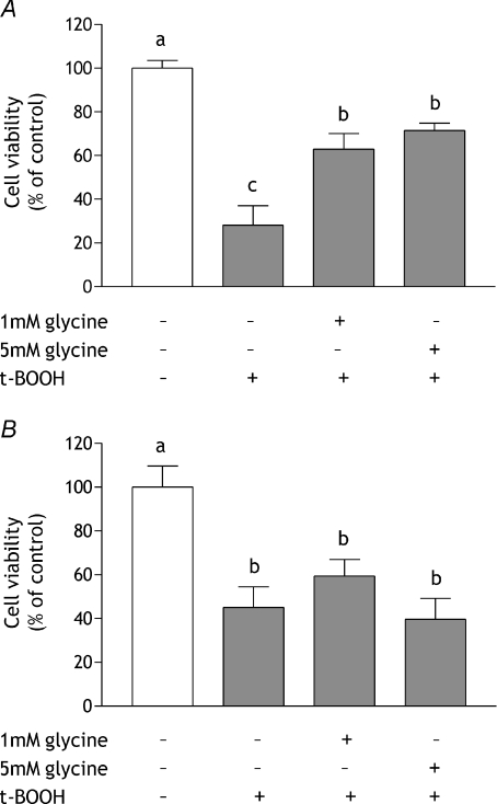
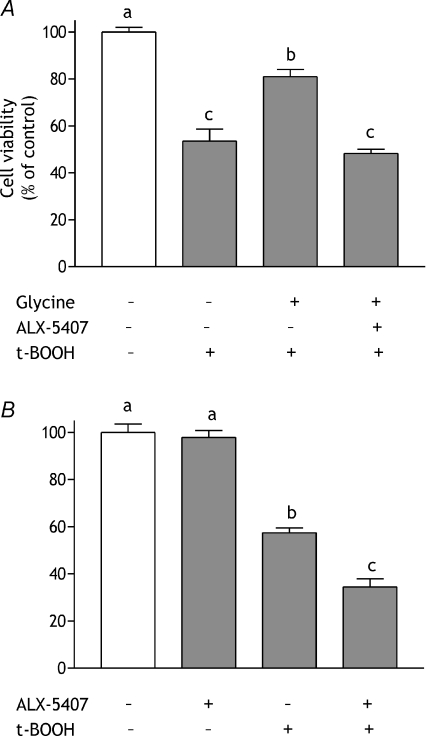
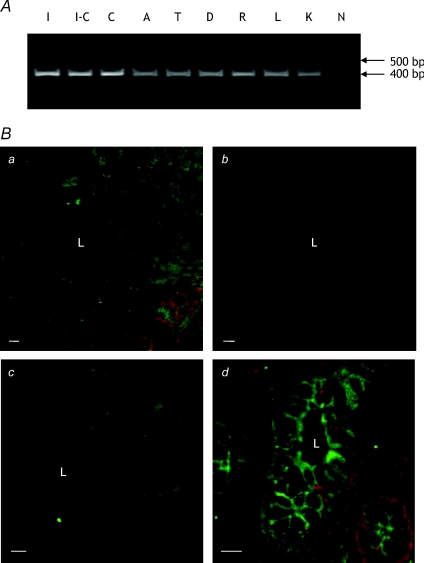
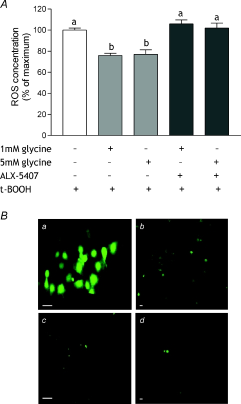
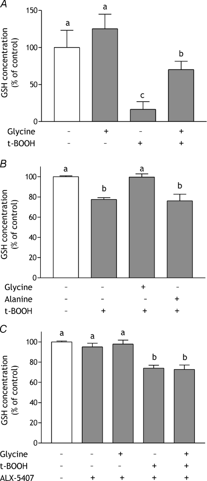
Glycine protects mammalian intestine against oxidative damage caused by ischaemia-reperfusion (IR) injury and prevents or reverses experimentally-induced colitis. However the mechanism of protection remains largely unknown. The objectives of the current study were to demonstrate directly glycine-mediated protection of human intestinal epithelial cells and to determine the requirement for glycine uptake by the specific transporter GLYT1. Exogenous glycine protected human intestinal Caco-2 and HCT-8 cells against the oxidative agent tert-butylhydroperoxide and reduced the intracellular concentration of reactive oxygen species, when applied prior to but not concomitant with the oxidative challenge. Glycine given prior to oxidative challenge preserved intracellular glutathione concentration but had no effect on the rate of glycine uptake. Protection was dependent on GLYT1 activity, being blocked by a specific GLYT1 inhibitor, supporting a requirement for intracellular glycine accumulation. Maintained intracellular glutathione content is indicated as a mechanism through which the protective effect may in part be mediated. However expression of the genes encoding GLYT1 and the glutathione synthesising enzymes glutamate-cysteine ligase, both catalytic and modifier subunits, and glutathione synthetase was not altered by glycine or tert-butylhydroperoxide, suggesting transcriptional regulation is not involved. This work has demonstrated a novel role of GLYT1 in intestine and shown that intestinal epithelial cells respond directly to oxidative challenge without reliance on extra-epithelial tissues or functions such as neurone, blood-flow or immune responses for antioxidant defence. The protective actions of glycine and maintenance of epithelial antioxidant defences suggest it may be beneficial in treatment of inflammatory bowel disease.
Figures


Comment in
-
The epithelial glycine transporter GLYT1: protecting the gut from inflammation.J Physiol. 2010 Apr 1;588(Pt 7):1033-4. doi: 10.1113/jphysiol.2010.188516. J Physiol. 2010. PMID: 20360026 Free PMC article. No abstract available.
Similar articles
-
The glycine transporter GLYT1 in human intestine: expression and function.Biol Pharm Bull. 2011;34(6):784-8. doi: 10.1248/bpb.34.784. Biol Pharm Bull. 2011. PMID: 21628872 Review.
-
Glycine supply to human enterocytes mediated by high-affinity basolateral GLYT1.Gastroenterology. 2001 Feb;120(2):439-48. doi: 10.1053/gast.2001.21207. Gastroenterology. 2001. PMID: 11159884
-
Glycine Transporter-1 and glycine receptor mediate the antioxidant effect of glycine in diabetic rat islets and INS-1 cells.Free Radic Biol Med. 2018 Aug 1;123:53-61. doi: 10.1016/j.freeradbiomed.2018.05.007. Epub 2018 May 9. Free Radic Biol Med. 2018. PMID: 29753073
-
Molecular identification and characterisation of the glycine transporter (GLYT1) and the glutamine/glutamate transporter (ASCT2) in the rat lens.Exp Eye Res. 2006 Aug;83(2):447-55. doi: 10.1016/j.exer.2006.01.028. Epub 2006 Apr 25. Exp Eye Res. 2006. PMID: 16635486
-
Synergistic Control of Transmitter Turnover at Glycinergic Synapses by GlyT1, GlyT2, and ASC-1.Int J Mol Sci. 2022 Feb 25;23(5):2561. doi: 10.3390/ijms23052561. Int J Mol Sci. 2022. PMID: 35269698 Free PMC article. Review.
Cited by
-
Gut barrier and microbiota changes with glycine and branched-chain amino acid supplementation in chronic haemodialysis patients.J Cachexia Sarcopenia Muscle. 2021 Dec;12(6):1527-1539. doi: 10.1002/jcsm.12781. Epub 2021 Sep 18. J Cachexia Sarcopenia Muscle. 2021. PMID: 34535959 Free PMC article. Clinical Trial.
-
Extracellular glycine is necessary for optimal hemoglobinization of erythroid cells.Haematologica. 2017 Aug;102(8):1314-1323. doi: 10.3324/haematol.2016.155671. Epub 2017 May 11. Haematologica. 2017. PMID: 28495915 Free PMC article.
-
Elucidation of gastrointestinal dysfunction in response to irradiation using metabolomics.Biochem Biophys Rep. 2020 Jul 29;23:100789. doi: 10.1016/j.bbrep.2020.100789. eCollection 2020 Sep. Biochem Biophys Rep. 2020. PMID: 32775703 Free PMC article.
-
The epithelial glycine transporter GLYT1: protecting the gut from inflammation.J Physiol. 2010 Apr 1;588(Pt 7):1033-4. doi: 10.1113/jphysiol.2010.188516. J Physiol. 2010. PMID: 20360026 Free PMC article. No abstract available.
-
Reduction of Rapid Proliferating Tumour Cell Lines by Inhibition of the Specific Glycine Transporter GLYT1.Biomedicines. 2021 Nov 25;9(12):1770. doi: 10.3390/biomedicines9121770. Biomedicines. 2021. PMID: 34944586 Free PMC article.
References
-
- Aki T, Egashira N, Yamauchi Y, Hama M, Yano T, Itoh Y, Yamada T, Oishi R. Protective effects of amino acids against gabexate mesilate-induced cell injury in porcine aorta endothelial cells. J Pharmacol Sci. 2008;107:238–245. - PubMed
-
- Allen CN, Harpur ES, Gray TJB, Hirst BH. Toxic effects of non-steroidal anti-inflammatory drugs in a human intestinal epithelial cell line (HCT-8), as assessed by the MTT and neutral red assays. Toxicology in Vitro. 1991;5:183–191. - PubMed
-
- Atkinson BN, Bell SC, De Vivo M, Kowalski LR, Lechner SM, Ognyanov VI, Tham CS, Tsai C, Jia J, Ashton D, Klitenick MA. ALX 5407: a potent, selective inhibitor of the hGlyT1 glycine transporter. Mol Pharmacol. 2001;60:1414–1420. - PubMed
-
- Christie GR, Ford D, Howard A, Clark MA, Hirst BH. Glycine supply to human enterocytes mediated by high-affinity basolateral GLYT1. Gastroenterology. 2001;120:439–448. - PubMed
-
- Deters M, Siegers CP, Strubelt O. Influence of glycine on the damage induced in isolated perfused rat liver by five hepatotoxic agents. Toxicology. 1998;128:63–72. - PubMed
Publication types
MeSH terms
Substances
Grants and funding
LinkOut - more resources
Full Text Sources