

Cancerous breast lesions on dynamic contrast-enhanced MR images: computerized characterization for image-based prognostic markers
- PMID: 20123903
- PMCID: PMC2826695
- DOI: 10.1148/radiol.09090838
Cancerous breast lesions on dynamic contrast-enhanced MR images: computerized characterization for image-based prognostic markers
Abstract
Purpose: To assess the performance of computer-extracted dynamic contrast material-enhanced (DCE) magnetic resonance (MR) imaging kinetic and morphologic features in the differentiation of invasive versus noninvasive breast lesions and metastatic versus nonmetastatic breast lesions.
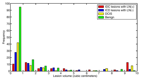
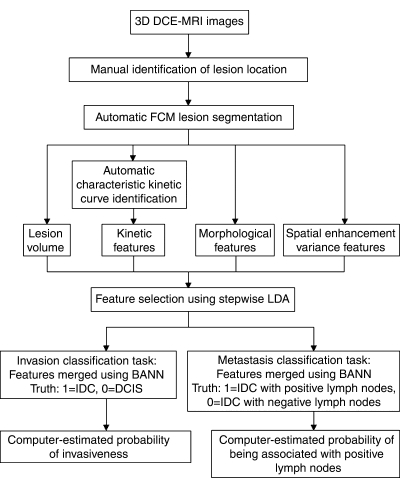
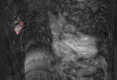
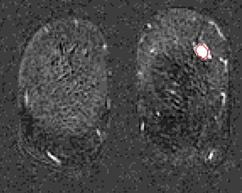
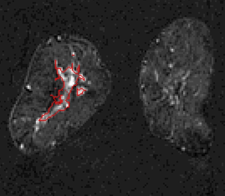
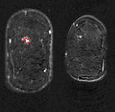
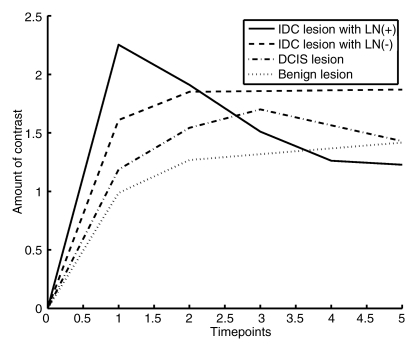
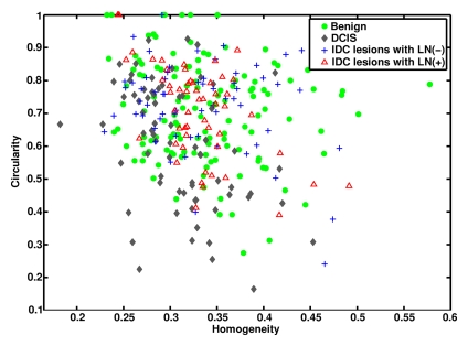
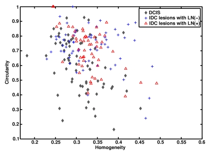
Materials and methods: In this institutional review board-approved HIPAA-compliant study, in which the requirement for informed patient consent was waived, breast MR images were retrospectively collected. The images had been obtained with a 1.5-T MR unit by using a gadodiamide-enhanced T1-weighted spoiled gradient-recalled acquisition in the steady state sequence. The breast MR imaging database contained 132 benign, 71 ductal carcinoma in situ (DCIS), and 150 invasive ductal carcinoma (IDC) lesions. Fifty-four IDC lesions were associated with metastasis-positive lymph nodes (LNs), and 64 IDC lesions were associated with negative LNs. Lesion segmentation and extraction of morphologic and kinetic features were automatically performed by a laboratory-developed computer workstation. Features were first selected by using stepwise linear discriminant analysis and then merged by using Bayesian neural networks. Lesion classification performance was assessed with receiver operating characteristic analysis.
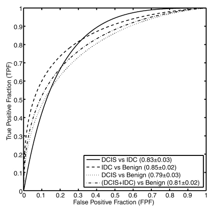
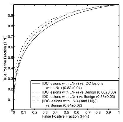
Results: Differentiation of DCIS from IDC lesions yielded an area under the receiver operating characteristic curve (AUC) of 0.83 +/- 0.03 (standard error). AUCs were 0.85 +/- 0.02 for differentiation between IDC and benign lesions and 0.79 +/- 0.03 for differentiation between DCIS and benign lesions. Differentiation between IDC lesions associated with positive LNs and IDC lesions associated with negative LNs yielded an AUC of 0.82 +/- 0.04. AUCs were 0.86 +/- 0.03 for differentiation between IDC lesions associated with positive LNs and benign lesions and 0.83 +/- 0.03 for differentiation between IDC lesions associated with negative LNs and benign lesions.
Conclusion: Computer-aided diagnosis of breast DCE MR imaging-depicted lesions was extended from the task of discriminating between malignant and benign lesions to the prognostic tasks of distinguishing between noninvasive and invasive lesions and discriminating between metastatic and nonmetastatic lesions, yielding MR imaging-based prognostic markers.
Supplemental material: http://radiology.rsna.org/lookup/suppl/doi:10.1148/radiol.09090838/-/DC1.
(c) RSNA, 2010
Figures
References
-
- Schnall MD. Breast MR imaging. Radiol Clin North Am 2003;41:43–50 - PubMed
-
- Morris EA. Breast cancer imaging with MRI. Radiol Clin North Am 2002;40:443–466 - PubMed
-
- Kuhl CK, Schild HH. Dynamic image interpretation of MRI of the breast. J Magn Reson Imaging 2000;12:965–974 - PubMed
-
- Kuhl CK, Mielcareck P, Klaschik S, et al. Dynamic breast MR imaging: are signal intensity time course data useful for differential diagnosis of enhancing lesions?. Radiology 1999;211:101–110 - PubMed
-
- Bartella L, Smith CS, Dershaw DD, Liberman L. Imaging breast cancer. Radiol Clin North Am 2007;45:45–67 - PubMed
Publication types
MeSH terms
Substances
Grants and funding
LinkOut - more resources
Full Text Sources
Other Literature Sources
Medical
