. 2010 Feb 1;24(3):265-76.
doi: 10.1101/gad.544410.

Polycomb complexes act redundantly to repress genomic repeats and genes

Affiliations

Polycomb complexes act redundantly to repress genomic repeats and genes

Martin Leeb et al. Genes Dev. .

Abstract

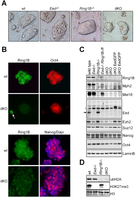
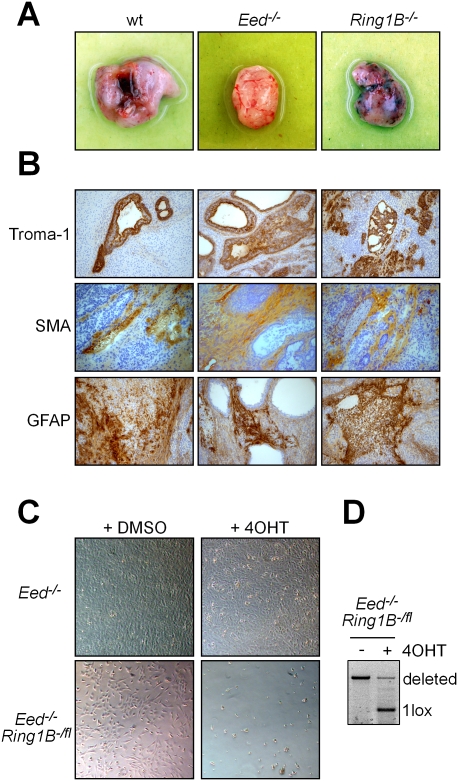
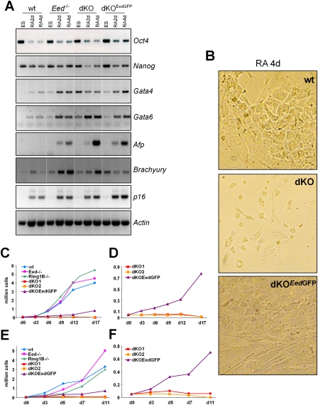
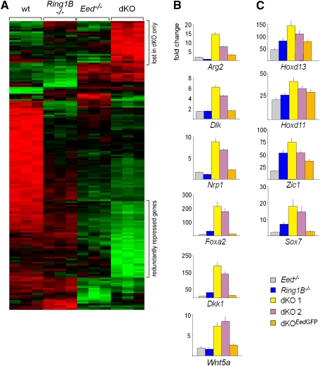
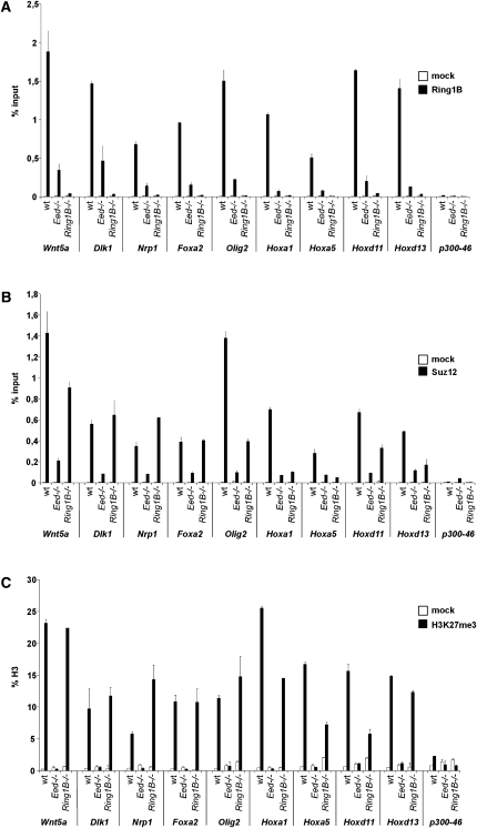
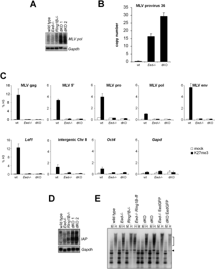
Polycomb complexes establish chromatin modifications for maintaining gene repression and are essential for embryonic development in mice. Here we use pluripotent embryonic stem (ES) cells to demonstrate an unexpected redundancy between Polycomb-repressive complex 1 (PRC1) and PRC2 during the formation of differentiated cells. ES cells lacking the function of either PRC1 or PRC2 can differentiate into cells of the three germ layers, whereas simultaneous loss of PRC1 and PRC2 abrogates differentiation. On the molecular level, the differentiation defect is caused by the derepression of a set of genes that is redundantly repressed by PRC1 and PRC2 in ES cells. Furthermore, we find that genomic repeats are Polycomb targets and show that, in the absence of Polycomb complexes, endogenous murine leukemia virus elements can mobilize. This indicates a contribution of the Polycomb group system to the defense against parasitic DNA, and a potential role of genomic repeats in Polycomb-mediated gene regulation.


Figures

Figure 1.
Figure 1.
Establishment of dKO ES cells lacking PRC1 and PRC2 activity. (A) Morphology of wild-type (wt), Ring1B−/−, Eed−/−, and dKO ES cell colonies. (B) Immunofluorescence staining showing Oct4 and Nanog expression in dKO and wild-type ES cell colonies. (C) Western analysis of PcG proteins and transcription factors in ES cells of indicated genotypes shows that Mph2 and Mel18 are virtually absent in Ring1B−/− and dKO ES cells. Ezh2 and Suz12 are reduced in Eed−/− and dKO ES cells. An arrow indicates Eed (asterisk; nonspecific band). The pluripotency markers Nanog and Oct4 are largely unaffected. Lamin B was used as loading control. (D) Western analysis of ubH2A and H3K27me3 showing absence of PRC1 and PRC2 activity in dKO ES cells.
Figure 2.
Figure 2.
Eed and Ring1B are redundantly required for differentiation. (A) Teratomas formed by wild-type (wt), Eed−/−, and Ring1B−/− ES cells 3 wk after injection. dKO ES cells did not give rise to teratomas. (B) Immunohistochemical analysis of wild-type, Ring1B−/−, and Eed−/− teratomas using markers for endoderm (Troma1), mesoderm (smooth muscle actin [SMA]), and ectoderm (GFAP). (C) Double deficiency for PRC1 and PRC2 is not compatible with NS cell viability. Deletion of Ring1B in Eed-deficient NS cells by induction of CreERT2 with 4OHT caused cell death, whereas 4OHT had no effect on control Eed-deficient NS cells. (D) PCR analysis showing that induction of CreERT2 results in deletion of the conditional Ring1Bfl allele in Eed−/− Ring1B-/fl NS cells.
Figure 3.
Figure 3.
Differentiation of ES cells lacking PcG activity. (A) RT–PCR expression analysis of pluripotency markers (Oct4 and Nanog) and differentiation markers (Gata4, Gata6, Afp, and Brachyury) and p16 in wild-type, Eed-deficient, dKO, and dKOEedGFP ES cells induced to differentiate with retinoic acid. Undifferentiated ES cells and cells after 2 and 4 d of differentiation are shown. Actin is used as loading control. (B) Microscopy image showing differentiated cell morphology on day 4 of retinoic acid-induced differentiation of wild-type, dKO, and dKOEedGFP ES cells. (C–F) Cell numbers in differentiating cultures of wild-type, Eed-deficient, Ring1B-deficient, dKO, and dKOEedGFP ES cells. (C,E) Two experiments are shown. (D,F) Rescaled representation of experiments in C and E showing that expression of an EedGFP transgene in dKOEedGFP ES cells rescues the differentiation defect of dKO ES cells.
Figure 4.
Figure 4.
Analysis of transcriptional changes in ES cells lacking PcG activity. (A) A heat map showing high (green), intermediate (black), and low (red) expression of genes in wild-type, Eed−/−, Ring1B−/−, and dKO ES cells. The top regulated genes in all genotypes are shown. (B,C) Quantitative real-time PCR was used to confirm derepression of genes in PcG-deficient ES cells. Introduction of an EedGFP transgene in dKO ES cells re-establishes repression largely to the levels of Ring1B−/− ES cells. (B) A redundantly repressed set of PcG target genes is specifically derepressed in dKO ES cells. (C) PcG target genes derepressed in all PcG mutant ES cells.
Figure 5.
Figure 5.
PRC1 and PRC2 bind to the promoters of redundantly silenced genes. ChIP analysis of Ring1B (A), Suz12 (B), and H3K27me3 (C) binding to gene promoters in wild-type, Eed-deficient, and Ring1B-deficient ES cells. H3K27me3 signal is normalized to histone H3, and Ring1B and Suz12 are represented as percent input. Error bars represent standard deviation.
Figure 6.
Figure 6.
Redundant repression of endogenous retroviruses by PRC1 and PRC2. (A) Northern analysis showing derepression of endogenous MLV retroelements in dKO ES cells. Gapdh was used as a loading control. (B) Quantitative PCR analysis showing an increase in MLV provirus copy number in Polycomb-deficient ES cells. The copy number was quantitated relative to an intergenic genomic sequence using the standard curve method. (C) Directed ChIP showing enrichment of H3K27me3 over MLV elements in ES cells. (Top row) Different regions of the MLV provirus sequence were investigated by quantitative PCR. (Bottom row) Lef1 was used as a positive control, and Oct4, Gapdh, and an intergenic sequence on chromosome 8 were used as negative controls. H3K27me3 signals are diminished in Eed-deficient ES cells demonstrating specificity. (D) Northern analysis showing the derepression of IAP retroviral sequences in dKO ES cells. Gapdh was used as loading control. (E) Restriction analysis with methylation-sensitive HpaII (H) and methylation-resistant MspI (M) and subsequent Southern analysis show loss of DNA methylation on IAP elements in Eed-deficient and dKO ES cells. Size of demethylated bands in HpaII lanes are indicated by the arrowhead and square bracket.

References

    1. Arrigoni R, Alam SL, Wamstad JA, Bardwell VJ, Sundquist WI, Schreiber-Agus N. The Polycomb-associated protein Rybp is a ubiquitin binding protein. FEBS Lett. 2006;580:6233–6241. - PubMed
    1. Atsuta T, Fujimura S, Moriya H, Vidal M, Akasaka T, Koseki H. Production of monoclonal antibodies against mammalian Ring1B proteins. Hybridoma. 2001;20:43–46. - PubMed
    1. Bernstein E, Duncan EM, Masui O, Gil J, Heard E, Allis CD. Mouse polycomb proteins bind differentially to methylated histone H3 and RNA and are enriched in facultative heterochromatin. Mol Cell Biol. 2006;26:2560–2569. - PMC - PubMed
    1. Boyer LA, Mathur D, Jaenisch R. Molecular control of pluripotency. Curr Opin Genet Dev. 2006a;16:455–462. - PubMed
    1. Boyer LA, Plath K, Zeitlinger J, Brambrink T, Medeiros LA, Lee TI, Levine SS, Wernig M, Tajonar A, Ray MK, et al. Polycomb complexes repress developmental regulators in murine embryonic stem cells. Nature. 2006b;441:349–353. - PubMed

Publication types