. 2010 Feb 15;207(2):429-42.
doi: 10.1084/jem.20090851. Epub 2010 Feb 1.

Type I IFN controls chikungunya virus via its action on nonhematopoietic cells

Affiliations

Type I IFN controls chikungunya virus via its action on nonhematopoietic cells

Clémentine Schilte et al. J Exp Med. .

Abstract

Chikungunya virus (CHIKV) is the causative agent of an outbreak that began in La Réunion in 2005 and remains a major public health concern in India, Southeast Asia, and southern Europe. CHIKV is transmitted to humans by mosquitoes and the associated disease is characterized by fever, myalgia, arthralgia, and rash. As viral load in infected patients declines before the appearance of neutralizing antibodies, we studied the role of type I interferon (IFN) in CHIKV pathogenesis. Based on human studies and mouse experimentation, we show that CHIKV does not directly stimulate type I IFN production in immune cells. Instead, infected nonhematopoietic cells sense viral RNA in a Cardif-dependent manner and participate in the control of infection through their production of type I IFNs. Although the Cardif signaling pathway contributes to the immune response, we also find evidence for a MyD88-dependent sensor that is critical for preventing viral dissemination. Moreover, we demonstrate that IFN-alpha/beta receptor (IFNAR) expression is required in the periphery but not on immune cells, as IFNAR(-/-)-->WT bone marrow chimeras are capable of clearing the infection, whereas WT-->IFNAR(-/-) chimeras succumb. This study defines an essential role for type I IFN, produced via cooperation between multiple host sensors and acting directly on nonhematopoietic cells, in the control of CHIKV.


Figures

Figure 1.
Figure 1.
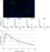
CHIKV induces IFN-α/β in vivo in humans but fails to stimulate production in hematopoietic cells. (A) Patient sera samples were obtained from consenting CHIKV-infected adults (age = 16–86 yr; n = 25) and age-matched controls (age range = 16–89 yr; n = 17). IFN-α was monitored by luminex and plotted as a function of viral load, measured by quantitative PCR. Mean IFN-α serum concentration in the control population was 179 pg/ml, indicated by the dotted line. The solid line represents the linear regression between viral load and IFN-α in patients. (B) Monocyte-derived DCs were generated from healthy donors, and 105 immature DCs (iDCs) or mature DCs (mDCs) were exposed to CHIKV. In parallel, 3 × 10⁴ freshly isolated pDCs or 106 PBMCs were cultured with CHIKV. In the experiment shown, 106 PFU/ml was used and cells were incubated for 40 h. Culture supernatants were analyzed for IFN-α/β production by a reporter assay involving an IFNAR-expressing cell line, HL-116, stably transfected with a plasmid encoding an IFN-inducible luciferase gene (limit of detection, 5 IFN U/ml). 50 HAU influenza A/PR8 or 50 µg/ml poly I:C were used as a positive control. Results were identical for three different donors. Similar results were found in monocyte-derived macrophages (not depicted). (C and D) Human foreskin fibroblasts (black) or MRC-5 cell lines (white) were infected at increasing MOI and cultured for 48 h. Cells were harvested and intracellular staining was performed using an anti-CHIKV mAb and analyzed by FACS (C). In parallel, culture supernatant was analyzed for IFN-β by Elisa (limit of detection, 20 pg/ml; D). Error bars indicate SD. Data is representative of five experiments.
Figure 2.
Figure 2.
CHIKV-infected IFNAR−/− mice produce high levels of type I IFNs. (A) C57BL/6 IFNAR−/− mice were infected ID with 102 PFU or 106 PFU CHIKV. Survival was assessed twice a day and Kaplan-Meier survival curves were drawn (n = 5). (B) STAT1−/−, STAT4−/−, and STAT6−/− (n = 5) mice, along with corresponding WT mice of a similar strain background, were infected ID with 106 PFU CHIKV. Survival was assessed twice a day and Kaplan-Meier survival curves were drawn. (C and D) WT and IFNAR−/− mice were infected with 106 PFU CHIKV and serum concentrations of IFN-β and IFN-α were monitored at day 2 after infection. Each dot represents an individual mouse, horizontal bars represent the mean. Data shown is combined from two independent experiments.
Figure 3.
Figure 3.
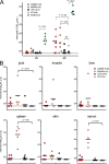
Local production of type I IFNs control viral dissemination in WT animals. (A) WT mice were injected with GFP-expressing CHIKV. Immunofluorescence was performed at day 1 after infection on skin at the site of injection (ear). Nuclei were stained with DAPI (blue) and replicating virus was detected using anti-GFP mAbs (green). Bar, 50 µm. (B) mRNA was isolated from the infection site at 3, 8, 24, and 48 h after infection. RT was performed and IFN-β, IFN-α2, IFN-α4, and Mx-1 mRNA expression was determined by quantitative PCR. Individual mice are represented by dots and horizontal bars represent the mean. The experiment presented is representative of three independent experiments. (C) As in B, total RNA was isolated from infected skin at various times and viral load was assessed. Data from three independent experiments are plotted to indicate viral RNA over time. Individual mice are represented.
Figure 4.
Figure 4.
Skin fibroblasts, but not hematopoietic cells, are infected in WT mice. Immunofluorescence was performed 1 d after ID infection with CHIKV-GFP (A, C, and D) or with WT CHIKV (B) at the site of inoculation into the ear. Cell nuclei were stained with DAPI and replicating virus was detected using anti-GFP mAbs (A, C, and D) or with anti CHIK antibody (B). CD45 (A), Vimentin (B), or CD31 and SMA (C and D) staining was performed to determine the phenotype of infected cells. S, sebaceous gland; C, cartilage; arrow, infected myofibroblast (SMA+int). Bars, 20 µm. Data are representative of two independent experiments.
Figure 5.
Figure 5.
Cell-intrinsic control of CHIKV in fibroblasts is mediated by intracellular sensors. (A) Cardif−/− and the corresponding WT control MEFs were infected with increasing MOI of CHIKV. At 48 h, cells were analyzed by FACS for expression of CHIKV antigens. (B) Culture supernatants were harvested at 48 h and analyzed for IFN-β protein. As a positive control, MEFs were exposed to FluΔNS1. (C) IFN-β mRNA induction was measured by RT-PCR at 24 h after infection. The mean of three independent experiments is shown. Error bars show SD.
Figure 6.
Figure 6.
Redundancy between RIG-I and Mda5 pathway. Mda5−/−, RIG-I−/−, and littermate WT controls were infected. Tissues and serum were collected 72 h after infection and viral titers were determined using TCID50. The limits of detection are indicated by dotted lines. Individual mice are represented and the geometric means are indicated by bars. A Mann-Whitney U test was used and, where significant, p-values are reported. Data shown is combined from two independent experiments.
Figure 7.
Figure 7.
Cross talk between Cardif- and MyD88-dependant pathways for efficient clearance of CHIKV. Cardif−/−, MyD88−/−, TLR3−/−, and the corresponding WT controls were infected with 106 PFU. (A) Viral titer was measured at 24 and 48 h after infection. As a control we show infection of an IFNAR−/− animal. (B) Tissues were collected 72 h after infection and viral titers were determined using TCID50. The limits of detection are indicated by dashed lines. Individual mice are represented and geometric means are indicated by bars. It is of note that IFNAR mice are dead at that time point. Mann-Whitney U test was used and, where significant, p-values are reported. Data shown is combined from two independent experiments.
Figure 8.
Figure 8.
IFNAR−/−→WT bone marrow chimeras maintain their ability to clear CHIKV infection. WT and IFNAR−/− mice were lethally irradiated and reconstituted with bone marrow from IFNAR−/− or WT mice, respectively. After 2–3 mo, chimeric mice were inoculated with 102 or 106 PFU CHIKV. (A) Survival was monitored twice a day for 2 wk and Kaplan-Meier curves are shown (n = 8 mice). Data represents two independent experiments. (B) At day 3 after infection, virus titer was determined using TCID50 in IFNAR→WT and WT controls (n = 3 mice). Dashed lines represent the limit of detection. Horizontal bars represent mean.

References

    1. Alexopoulou L., Holt A.C., Medzhitov R., Flavell R.A. 2001. Recognition of double-stranded RNA and activation of NF-kappaB by Toll-like receptor 3. Nature. 413:732–738 10.1038/35099560 - DOI - PubMed
    1. Aricò E., Robertson K.A., Belardelli F., Ferrantini M., Nash A.A. 2004. Vaccination with inactivated murine gammaherpesvirus 68 strongly limits viral replication and latency and protects type I IFN receptor knockout mice from a lethal infection. Vaccine. 22:1433–1440 10.1016/j.vaccine.2003.10.015 - DOI - PubMed
    1. Barry G., Breakwell L., Fragkoudis R., Attarzadeh-Yazdi G., Rodriguez-Andres J., Kohl A., Fazakerley J.K. 2009. PKR acts early in infection to suppress Semliki Forest virus production and strongly enhances the type I interferon response. J. Gen. Virol. 90:1382–1391 10.1099/vir.0.007336-0 - DOI - PMC - PubMed
    1. Bender A., Sapp M., Schuler G., Steinman R.M., Bhardwaj N. 1996. Improved methods for the generation of dendritic cells from nonproliferating progenitors in human blood. J. Immunol. Methods. 196:121–135 10.1016/0022-1759(96)00079-8 - DOI - PubMed
    1. Bieback K., Lien E., Klagge I.M., Avota E., Schneider-Schaulies J., Duprex W.P., Wagner H., Kirschning C.J., Ter Meulen V., Schneider-Schaulies S. 2002. Hemagglutinin protein of wild-type measles virus activates toll-like receptor 2 signaling. J. Virol. 76:8729–8736 10.1128/JVI.76.17.8729-8736.2002 - DOI - PMC - PubMed

Publication types

MeSH terms