. 2010 Apr;30(7):1769-82.
doi: 10.1128/MCB.01100-09. Epub 2010 Feb 1.

Regulation of Drosophila vasa in vivo through paralogous cullin-RING E3 ligase specificity receptors

Affiliations

Regulation of Drosophila vasa in vivo through paralogous cullin-RING E3 ligase specificity receptors

Jan-Michael Kugler et al. Mol Cell Biol. 2010 Apr.

Abstract

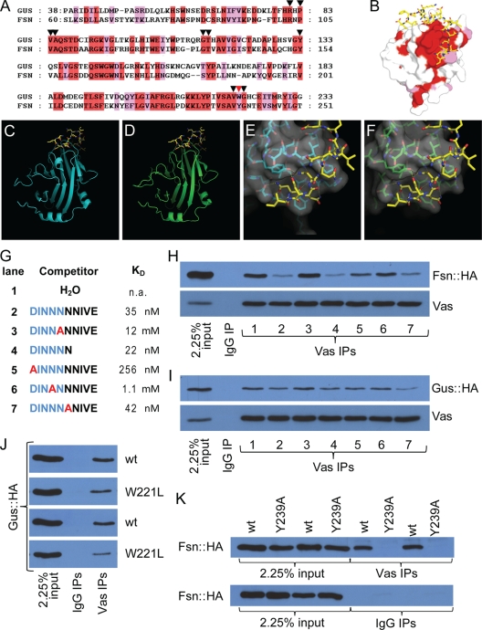
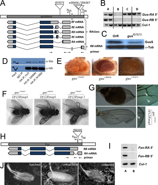
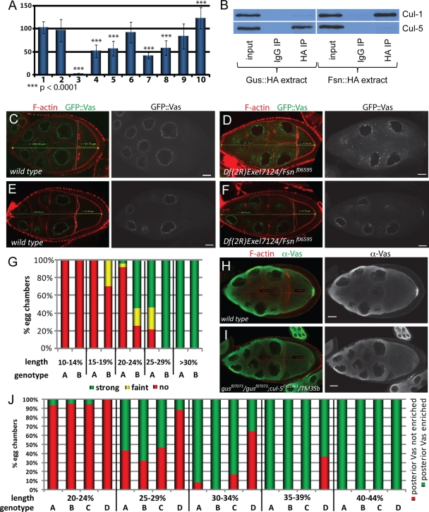
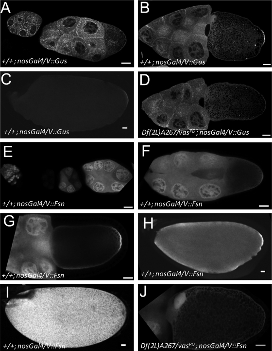
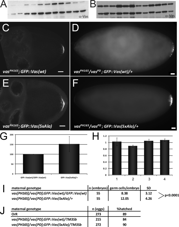
In Drosophila species, molecular asymmetries guiding embryonic development are established maternally. Vasa, a DEAD-box RNA helicase, accumulates in the posterior pole plasm, where it is required for embryonic germ cell specification. Maintenance of Vasa at the posterior pole requires the deubiquitinating enzyme Fat facets, which protects Vasa from degradation. Here, we found that Gustavus (Gus) and Fsn, two ubiquitin Cullin-RING E3 ligase specificity receptors, bind to the same motif on Vasa through their paralogous B30.2/SPRY domains. Both Gus and Fsn accumulate in the pole plasm in a Vasa-dependent manner. Posterior Vasa accumulation is precocious in Fsn mutant oocytes; Fsn overexpression reduces ovarian Vasa levels, and embryos from Fsn-overexpressing females form fewer primordial germ cells (PGCs); thus, Fsn destabilizes Vasa. In contrast, endogenous Gus may promote Vasa activity in the pole plasm, as gus females produce embryos with fewer PGCs, and posterior accumulation of Vas is delayed in gus mutant oocytes that also lack one copy of cullin-5. We propose that Fsn- and Gus-containing E3 ligase complexes contribute to establishing a fine-tuned steady state of Vasa ubiquitination that influences the kinetics of posterior Vasa deployment.


Figures

FIG. 1.
FIG. 1.
Gus and Fsn are Vas-interacting proteins. (A) Sequence alignment of the SPRY domains of Gus and Fsn. Identical and similar residues are highlighted in red and pink, respectively. Inverted triangles highlight the residues that form the Vas binding pocket. The red inverted triangle marks the residue W221 of Gus and Y239 of Fsn. (B) Surface view of a Gus crystal structure (PDB code 2IHS) (44). Surface residues conserved between Gus and Fsn are colored as described for panel A. (C to F) The B30.2/SPRY domains of Gus and Fsn are likely to form highly similar 3D structures. Panels C and E show crystal structures obtained from Gus (cyan) in complex with a Vas fragment (yellow) (PDB code 2IHS) (45). Panels D and F are structural predictions of the B30.2/SPRY domain of Fsn onto the crystals shown in panels C and E, respectively. The surface topology of the predicted Vas-binding site on Fsn (F) is almost identical to that of Gus (E). (G) Peptides used for the competition experiments shown in panels H and I. Kd values (apparent dissociation constants) for the interactions between Gus and peptides (45) are shown. (H) Fsn::HA and Vas coimmunoprecipitate. The interaction is sensitive to competition with peptides containing an intact DINNN motif. (I) GusL::HA and Vas coimmunoprecipitate. While the interaction is sensitive to peptide competition, the effects are much less pronounced than is the case for Fsn. (J) Both the wild type and W221L mutant Gus::HA associate with Vas in coimmunoprecipitation experiments; however, the amino acid substitution reduces the affinity of the interaction. All transgenic lines expressing GusL::HAW221L produced low levels of protein, possibly indicating a destabilizing effect of the substitution. Two transgenic lines are shown for Gus::HAW221L (second and fourth panels from the top), both compared to the same Gus::HA strain that expressed at a similar level to the mutant protein (first and third panels). (K) Unlike wild-type Fsn::HA, transgenic Fsn containing the Y239A mutation does not detectably coimmunoprecipitate with Vas. Two independent transgenic lines for each protein are shown.
FIG. 2.
FIG. 2.
Molecular genetics of gus and Fsn. (A) Schematic representation of the gus locus and its transcripts (not to scale). Insertion sites of the PBac elements used in this study are indicated. (B) RT-PCR amplifications specific for the RA class and gus-RB transcripts indicate that both mRNAs are expressed in wild-type ovaries (lane A). gusf07073 impairs expression of all tested transcripts (lane B), while guse00456 in trans to gusf07073 (lane C) or homozygous (lane D) affects only gus-RB. Positions of the primer pairs used are indicated in panel A; exact sequences are available upon request. A fragment of cul-1 mRNA was amplified as a control. (C) An antibody raised against recombinant Gus recognizes a ∼30-kDa protein that is reduced in gusf07073 ovaries, corresponding most likely to the 281-amino-acid-long isoform of Gus (GusS). The blot was also probed with anti-α-tubulin antibodies (α-Tub) as a loading control. (D) Transgenic GusS::HA efficiently coimmunoprecipitates Vas. A peptide containing an intact DINNN motif moderately interferes with this reaction, while a corresponding peptide bearing the N188A substitution within the DINNN motif does not have this effect. (E) Eyes from flies bearing the gusf07073, guse00456, and gusf04367 insertions. The mini white marker that tags the PBac shows position effect variegation in its expression. (F) In trans to the deficiency Df(2R)nap1, the gusf07073, guse00456, and gusf04367 mutations cause defects in the wing, including blisters, folded wings and ectopic vein material. (G) Hoyer's embedded wings derived from Df(2R)nap1/gusf07073 flies. (H) Schematic representation of the Fsn locus (not to scale). The insertion site of PBac Fsnf06595 is indicated. (I) RT-PCR amplifications specific for the Fsn-RA and Fsn-RB transcripts indicating expression of both isoforms in wild-type ovaries (lane A). In Df(2R)Exel7124/Fsnf06595 ovaries (lane B) these mRNAs are not expressed at detectable levels. (J) Eggs produced by gusf07073/gusf07073 mothers. Many of these eggs collapse.
FIG. 3.
FIG. 3.
Effects of manipulations of gus- and Fsn-dependent pathways on Vas in ovaries. (A) Relative amount of Vas in ovaries normalized to the α-tubulin signal. Bar 1 shows a comparison of an Oregon-R wild-type extract to itself and bar 2 shows a comparison of an Oregon-R extract to three independently prepared Oregon-R wild-type extracts. Bars 3 to 10 show comparisons of extracts from the following genotypes to Oregon-R: vasPD/vasPH165 (bar 3), GusL::HA/nosGal4 (bar 4), GusS::HA/nosGal4 (bar 5), gusf07073/gusf07073 (bar 6), gusf07073/gusf07073; Fsn::HA/nosGal4 (bar 7), Fsn::HA/nosGal4 (bar 8), Fsn::HAY239A/nosGal4 (bar 9), and Fsnf06595/Df(2R)Exel7124 (bar 10). (B) Cul-1 is found in Fsn::HA but not Gus::HA immunoprecipitates. Conversely, Cul-5 associates with Gus::HA but not with Fsn::HA. (C to F) Examples in which posterior GFP-Vas is not apparent in wild-type oocytes (C and E) but is faint (D) or obvious (F) in smaller, earlier-stage Fsn mutant oocytes. (G) Correlation between posterior GFP::Vas accumulation and the ratio of oocyte to germ line cyst length. Posterior GFP::Vas is detectable in smaller Df(2R)Exel7124/Fsnf06595 oocytes (columns B) than in the wild type (columns A) (n = 74 for the wild type; n = 90 for Fsn). (H and I) Stage 10 oocytes of wild-type females and gusf07073/gusf07073; cul-5EY21463/TM3Sb females immunostained for Vas. F-actin is labeled with rhodamine phalloidin. No posterior enrichment of Vas is evident in the mutant oocyte. (J) Correlation between posterior Vas enrichment and the ratio of oocyte to germ line cyst length. Genotypes represented by the columns are Oregon-R (A), gusf07073/gusf07073 (B), cul-5EY21463/TM3Sb (C), and gusf07073/gusf07073; cul-5EY21463/TM3Sb (D). Posterior Vas accumulation is delayed in gusf07073 homozygotes sensitized by introduction of a cul-5 P-element insertion (n = 63 for the wild type; n = 66 for gusf07073/gusf07073; n = 55 for cul-5EY21463/TM3Sb; n = 55 for gusf07073/gusf07073; cul-5EY21463/TM3Sb).
FIG. 4.
FIG. 4.
V::Gus and V::Fsn accumulate in the pole plasm in a vas-dependent manner. (A to C) In a wild-type background, V::Gus driven by nosGal4 is expressed at low levels in germ line nuclei but accumulates to high levels in the cytoplasm surrounding the nurse cell nuclei. In the germ line cytoplasm, V::Gus is ubiquitous, but it is enriched in numerous luminous foci, particularly during the earlier stages of oogenesis. In the oocyte, V::Gus is enriched in the cortex but accumulates in the pole plasm during stage 10. In late oocytes, posterior V::Gus levels decrease. (D) Pole plasm accumulation of V::Gus is lost in vas mutants (compare with panel B). (E to I) V::Fsn driven by nosGal4 in a wild-type background during early oogenesis (notice that transgene expression follows the expression pattern of the driver) (E), stage 9 (F), stage 10 (G), stage 13 (H), and stage 14 (I). V::Fsn accumulates in nuclei and is uniformly distributed throughout the nurse cell cytoplasm. In the oocyte, the protein appears cortically enriched, and from stage 10 on, V::Fsn is enriched in the pole plasm. Posterior enrichment is not evident in mature eggs. (J) In a vas mutant background, pole plasm accumulation of V::Fsn is lost; however, a general cortical stain is still observed. Bars, 20 mm.
FIG. 5.
FIG. 5.
GFP::Vas lacking the DINNN motif accumulates to high levels, localizes normally, and rescues vas phenotypes. (A and B) Western blots of ovarian extracts showing expression levels of individually established lines for GFP::Vas(wt) (A) and GFP::Vas(5xAla) (B) transgenes (upper band) and endogenous Vas (lower band). (C to F) Wild-type GFP::Vas (C) and a mutant form lacking the DINNN motif (E) localize similarly during oogenesis. Both transgenic proteins rescue PGC formation in vas mutant embryos. Bars, 20 mm. (G and H) GFP::Vas(5xAla) accumulates to higher levels than GFP::Vas(wt). (G) Normalized expression levels of the mutant protein in comparison to wild type. (H) RNA expression levels assayed by qRT-PCR. Bars 1 and 2 represent two independently prepared samples of vasPH165/vasPD; GFP::Vas(wt)/GFP::Vas(wt). Bars 3 and 4 represent two independent samples of vasPH165/vasPD; GFP::Vas(5xAla)/+. One copy of the mutant transgene expresses an RNA amount comparable to two copies of the wild-type transgene. (I) One copy of the GFP::Vas(5xAla) transgene permits the formation of more PGCs in vasPH165/vasPD embryos than two copies of GFP::Vas(wt). (J) Both constructs rescue the ability of vas embryos to hatch.

Similar articles

Cited by

References

    1. Besse, F., and A. Ephrussi. 2008. Translational control of localized mRNAs: restricting protein synthesis in space and time. Nat. Rev. Mol. Cell Biol. 9:971-980. - PubMed
    1. Carrera, P., O. Johnstone, A. Nakamura, J. Casanova, H. Jackle, and P. Lasko. 2000. VASA mediates translation through interaction with a Drosophila yIF2 homolog. Mol. Cell 5:181-187. - PubMed
    1. Chen, Y., A. Pane, and T. Schüpbach. 2007. cutoff and aubergine mutations result in retrotransposon upregulation and checkpoint activation in Drosophila. Curr. Biol. 17:637-642. - PMC - PubMed
    1. Doronkin, S., I. Djagaeva, and S. K. Beckendorf. 2002. CSN5/Jab1 mutations affect axis formation in the Drosophila oocyte by activating a meiotic checkpoint. Development 129:5053-5064. - PubMed
    1. Gao, M. X., E. H. Liao, B. Yu, Y. Wang, M. Zhen, and W. B. Derry. 2008. The SCF FSN-1 ubiquitin ligase controls germline apoptosis through CEP-1/p53 in C. elegans. Cell Death Differ. 15:1054-1062. - PubMed

Publication types

MeSH terms