. 2010 Apr;30(7):1828-37.
doi: 10.1128/MCB.01434-09. Epub 2010 Feb 1.

Serum response factor regulates hippocampal lamination and dendrite development and is connected with reelin signaling

Affiliations

Serum response factor regulates hippocampal lamination and dendrite development and is connected with reelin signaling

Christine Stritt et al. Mol Cell Biol. 2010 Apr.

Abstract

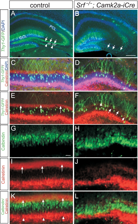
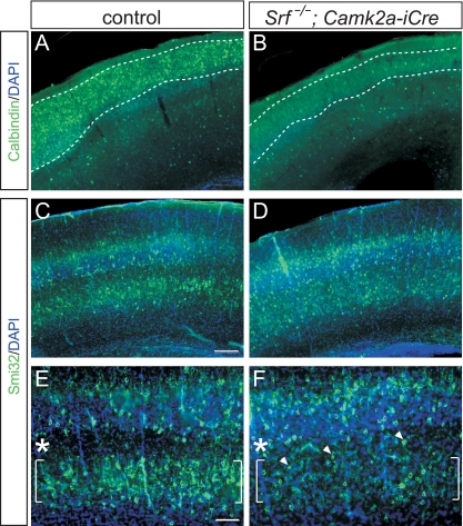
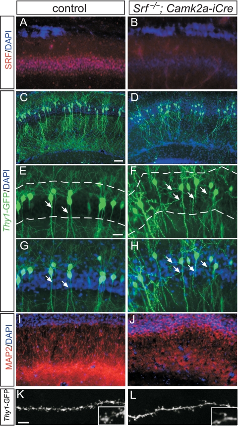
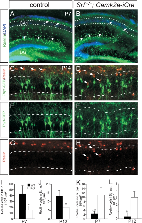
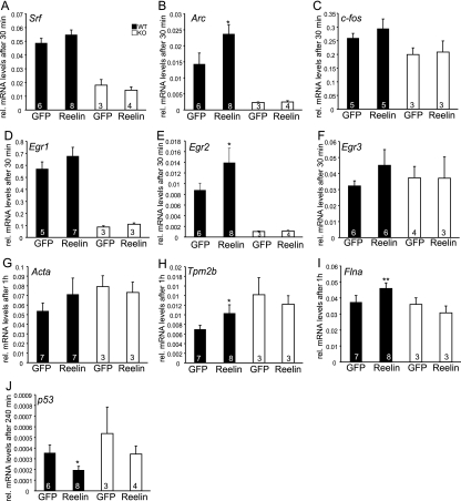
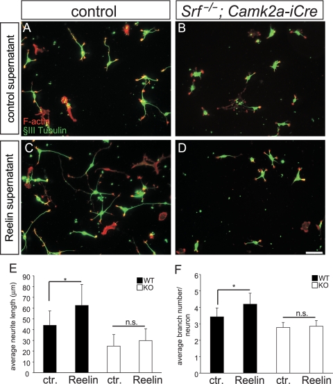
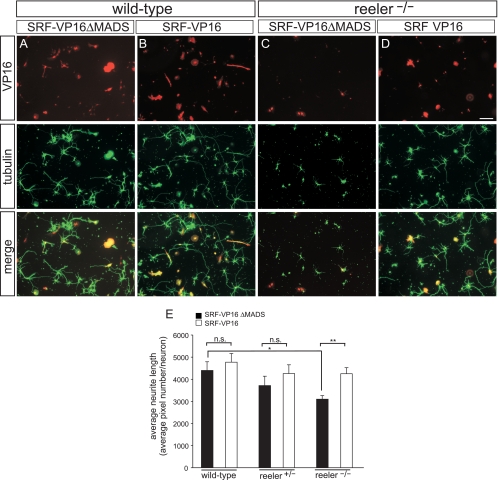
During brain development, neurons and their nerve fibers are often segregated in specific layers. The hippocampus is a well-suited model system to study lamination in health and aberrant cell/fiber lamination associated with neurological disorders. SRF (serum response factor), a transcription factor, regulates synaptic-activity-induced immediate-early gene (IEG) induction and cytoskeleton-based neuronal motility. Using early postnatal conditional SRF ablation, we uncovered distorted hippocampal lamination, including malpositioning of granule cell neurons and disruption of layer-restricted termination of commissural-associational and mossy fiber axons. Besides axons, dendrite branching and spine morphogenesis in Srf mutants were impaired, offering a first morphological basis for SRF's reported role in learning and memory. Srf mutants resemble mice lacking components of the reelin signaling cascade, a fundamental signaling entity in brain lamination. Our data indicate that reelin signaling and SRF-mediated gene transcription might be connected: reelin induces IEG and cytoskeletal genes in an SRF-dependent manner. Further, reelin-induced neurite motility is blocked in Srf mutants and constitutively active SRF rescues impaired neurite extension in reeler mouse mutants in vitro. In sum, data provided in this report show that SRF contributes to hippocampal layer and nerve fiber organization and point at a link between Srf gene transcription and reelin signaling.


Figures

FIG. 1.
FIG. 1.
SRF controls cell body and fiber lamination in the DG. (A and B) In P14 control (A) and Srf mutant (B) mice, mossy fibers (green) were highlighted by GFP expression via the thy1 promoter of transgenic mice. In controls (A), mossy fibers navigated in either the suprapyramidal (arrows) or infrapyramidal (arrowheads) blade outside the stratum pyramidale (str. pyr.). In Srf mutants (B), mossy fibers navigated inside the stratum pyramidale rather than growing outside. (C to F) Using thy1-GFP mice, a subpopulation of DG granule cells was visualized in control (C and E) and Srf mutant (D and F) mice. C/A fibers were stained red by calretinin. In controls, essentially all of the GFP-positive granule cells were localized underneath the C/A fibers (indicated by arrows in panel E). In contrast, GFP-positive granule cells in Srf mutants were intermingled with C/A fibers (arrows in panel F). C/A fibers in Srf mutants (D and F) did not form a tight bundle, as seen for control mice (C and E). GFP-positive granule cells in Srf mutants were aberrantly positioned above the DAPI-positive GCL (D; arrowhead in panel F). (G to L) All granule cell neurons were visualized with calbindin (green), in parallel with calretinin (red; arrows in panel I). In controls, calbindin-positive cells (green) were embedded by the calretinin-positive C/A fiber layer (arrows in panel K) and neurons in the hilus/subgranular zone (arrowhead in panel K). In contrast, in Srf mutants (J and L), granule cell neurons were more dispersed and were found within or above C/A fibers. Scale bars: A and B, 200 μm; C to L, 50 μm.
FIG. 2.
FIG. 2.
Impaired cell layering in the Srf mutant cortex. (A and B) P14 cortices were stained with calbindin, an interneuron and pyramidal cell marker. In the WT (A), calbindin-positive cells localize to the upper cortical layers, whereas fewer cells populate the Srf mutant cortex (B). (C to F) Smi32 labels layer III and layer V pyramidal neurons. In the WT (C and E), Smi32-psoitive neurons segregated into layers III and V (brackets), leaving a Smi32-free zone in between (asterisks). In Srf mutants (D and F), layer V contained fewer Smi32-positive cells and many Smi32-positive neurons gathered between layers III and V (arrowheads). Scale bars: A to D, 200 μm; E and F, 100 μm.
FIG. 3.
FIG. 3.
Aberrant dendrite arborization and spine formation in Srf mutant hippocampi. (A and B) SRF is expressed in WT CA1 neurons (A). Cre-mediated recombination effectively diminished SRF levels in CA1 neurons of Srf mutants (B). (C and D) Dendritic arborizations were visualized in a subpopulation of CA1 pyramidal neurons of P14 thy1-GFP transgenic mice. In the WT (C), CA1 neurons elaborated dendritic arborizations above (basal) and below (apical) the DAPI-positive cell body layer. In Srf mutants (D), the complexity of dendritic arborizations was decreased. Additionally, ectopic dendritic protrusions were observed within the cell body layer (D; see also panels E to H). (E to H) Higher magnifications of results shown in panels C and D. In control mice (E and G), dendrites assembled a basal and an apical tuft. In the WT, dendrites did not protrude within the DAPI-positive cell body layer (arrows in panels E and G; dashed lines label the cell body layer). In Srf mutants, dendrites were ectopically localized within the cell body layer (arrows in panels F and H). (I and J) Dendrites of all CA1 neurons were highlighted with anti-MAP2 antibodies in controls (I) and Srf mutants (J). In Srf mutants (J), dendritic arborization is reduced compared to that in control mice (I). (K and L) Dendritic spine formation was investigated using thy1-GFP mice interbred with WT (K) or Srf mutant mice (L) at P14. In WT mice (K), the dendritic spine number exceeded that of Srf mutants (L). Inserts are higher magnifications. Scale bars: A, B, and E to J, 20 μm; C and D, 50 μm; K and L, 5 μm.
FIG. 4.
FIG. 4.
Reelin-positive cells are misplaced in Srf mutant hippocampi. (A and B) Overview of reelin distribution in the hippocampi of P7 WT (A) and Srf mutant (B) mice. In Cajal-Retzius cells of the DG (arrowheads), no obvious changes in reelin expression were noticed between the genotypes. In Srf mutants, reelin-positive cells entered the CA1 cell body region (arrows in panel B), whereas in WT mice (arrows in panel A), they remained restricted to areas outside CA1 (indicated by the dashed lines in panels A and B). (C to H) In WT P14 mice (C, E, and G), reelin-positive cells were confined to positions above the CA1 stratum pyramidale (indicated by dashed lines). In Srf mutants (D, F, and H), reelin-positive cells enter the CA1 stratum pyramidale (arrows in panels D and H). Thus, in Srf mutants, ectopic dendritic arborizations inside the stratum pyramidale are next to reelin-expressing cells. (I to L) Quantification of reelin localization in the stratum oriens (I and J) and the stratum pyramidale (K and L) at P7 (I and K) and P12 (J and L). Scale bars: A and B, 100 μm; C to H, 20 μm.
FIG. 5.
FIG. 5.
Reelin elevated IEG and cytoskeletal mRNA levels through SRF. Cortical cultures derived from embryonic day 17.5 WT or Srf mutant (KO) mice were stimulated with reelin-containing or control supernatant for the indicated times. RNA was isolated, and cDNA was subjected to qRT-PCR using primers to the genes indicated. Reelin increased the mRNA abundance of the IEGs Srf (A), Arc (B), c-fos (C), Egr1 (D), Egr2 (E), and Egr3 (F) to a variable extent. In addition to IEGs, reelin induces mRNA levels of cytoskeletal genes, including Acta (G), Tpm2b (H), and Flna (I). p53 levels are suppressed by reelin in both WT and Srf mutant neurons (J). In Srf mutants, reelin does not upregulate mRNA levels of IEGs and cytoskeletal genes, indicating that SRF is the transcriptional regulator mediating reelin stimulation. Statistical significance was calculated by comparing control values with those obtained with the respective reelin-containing media. The values in the bars are the numbers of animals analyzed.
FIG. 6.
FIG. 6.
Reelin-stimulated neurite elongation and branching requires SRF. (A) WT hippocampal neurons derived from P1-to-P3 pups were stained for F-actin (red) and βIII-tubulin (green). Neurons were incubated with control (ctr.) supernatant derived from GFP-expressing HEK293 cells. (B) Srf mutant neurons—in the presence of control supernatant—are, compared to control-treated WT neurons (A), shorter and contain fewer branches. (C) Addition of reelin to WT neurons increases the average neurite length and number of branches (see quantification in panels E and F). (D) In SRF-deficient cultures, exogenous reelin fails to elevate the neurite length and branch number, indicating that reelin signaling requires SRF activity to modulate neuron morphology. (E and F) Quantification of the average neurite length (E) and the average number of branches per neuron (F) under the various conditions. Scale bars in panels A to D, 50 μm.
FIG. 7.
FIG. 7.
SRF-VP16 rescues impaired neurite outgrowth in reeler mutants. Hippocampal neurons derived from P1-to-P3 pups were stained for tubulin (green) and expression of the SRF constructs via antibodies directed to the VP16 domain (red) of the fusion protein. (A and C) WT (A) and hippocampal neurons lacking reelin (C) were infected with adenoviruses expressing the SRF control construct SRF-VP16ΔMADS (lacking DNA binding activity). Typically, WT neurons (A) elaborated more and longer neurites than did neurons lacking reelin (C). (B and D) WT (B) and reeler (D) cultures were infected with constitutively active SRF-VP16. In reelin-deficient neurons, SRF-VP16 strongly increased neurite outgrowth, almost reaching the levels observed for WT cultures. (E) Quantification of neurite outgrowth in WT and reeler hetero- and homozygous cultures. For quantification of neurite outgrowth, neurite length was determined by SlideBook software depicting the numbers of tubulin pixels per DAPI-positive neuron. Scale bars in panels A to D, 100 μm.

Similar articles

Cited by

References

    1. Alberti, S., S. M. Krause, O. Kretz, U. Philippar, T. Lemberger, E. Casanova, F. F. Wiebel, H. Schwarz, M. Frotscher, G. Schutz, and A. Nordheim. 2005. Neuronal migration in the murine rostral migratory stream requires serum response factor. Proc. Natl. Acad. Sci. U. S. A. 102:6148-6153. - PMC - PubMed
    1. Borrell, V., J. A. Del Río, S. Alcantara, M. Derer, A. Martinez, G. D'Arcangelo, K. Nakajima, K. Mikoshiba, P. Derer, T. Curran, and E. Soriano. 1999. Reelin regulates the development and synaptogenesis of the layer-specific entorhino-hippocampal connections. J. Neurosci. 19:1345-1358. - PMC - PubMed
    1. Borrell, V., L. Pujadas, S. Simó, D. Dura, M. Sole, J. A. Cooper, J. A. Del Río, and E. Soriano. 2007. Reelin and mDab1 regulate the development of hippocampal connections. Mol. Cell. Neurosci. 36:158-173. - PubMed
    1. Borrell, V., M. Ruiz, J. A. Del Río, and E. Soriano. 1999. Development of commissural connections in the hippocampus of reeler mice: evidence of an inhibitory influence of Cajal-Retzius cells. Exp. Neurol. 156:268-282. - PubMed
    1. Chai, X., E. Förster, S. Zhao, H. H. Bock, and M. Frotscher. 2009. Reelin stabilizes the actin cytoskeleton of neuronal processes by inducing n-cofilin phosphorylation at serine3. J. Neurosci. 29:288-299. - PMC - PubMed

Publication types

MeSH terms