. 2010 Apr;30(7):1746-56.
doi: 10.1128/MCB.00996-09. Epub 2010 Feb 1.

Dysautonomia due to reduced cholinergic neurotransmission causes cardiac remodeling and heart failure

Affiliations

Dysautonomia due to reduced cholinergic neurotransmission causes cardiac remodeling and heart failure

Aline Lara et al. Mol Cell Biol. 2010 Apr.

Abstract

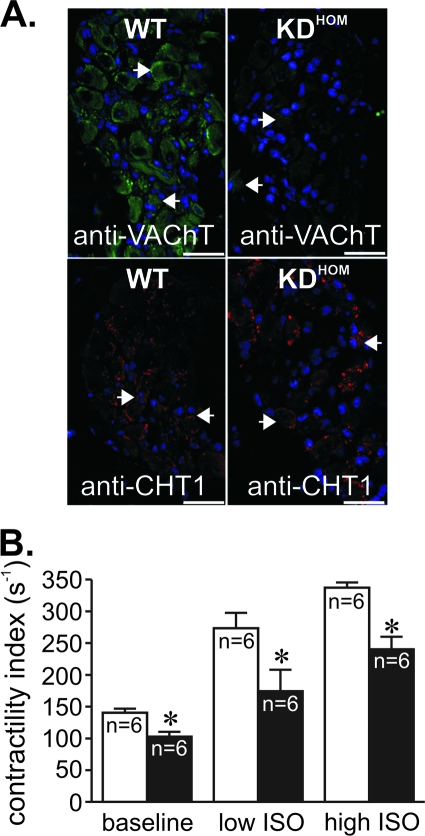
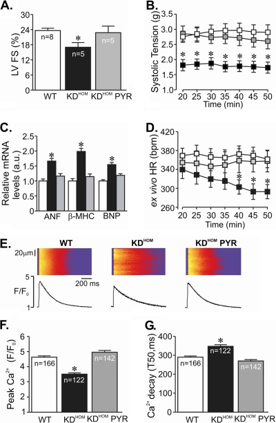
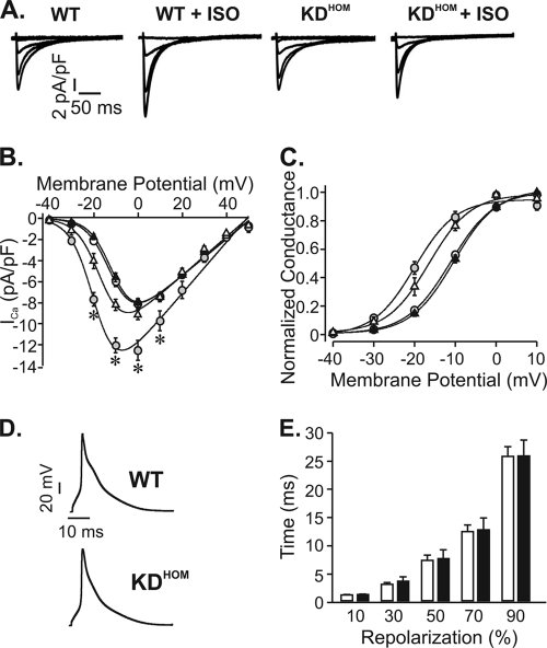
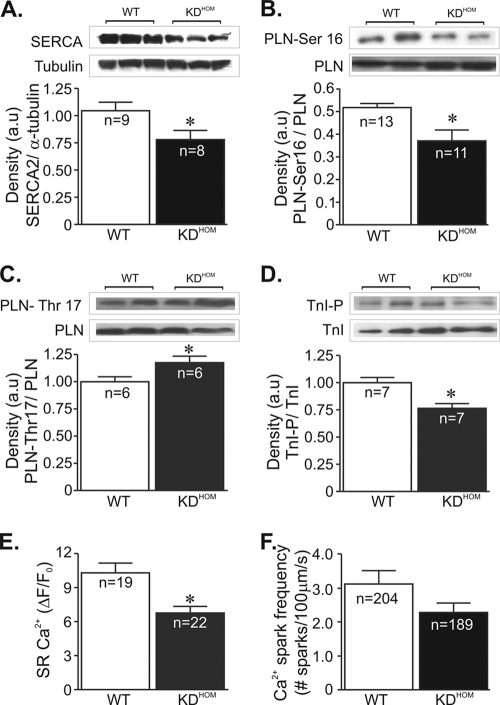
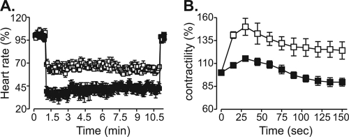
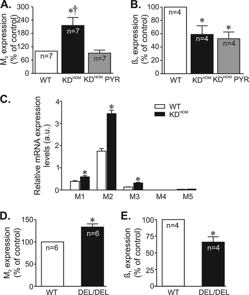
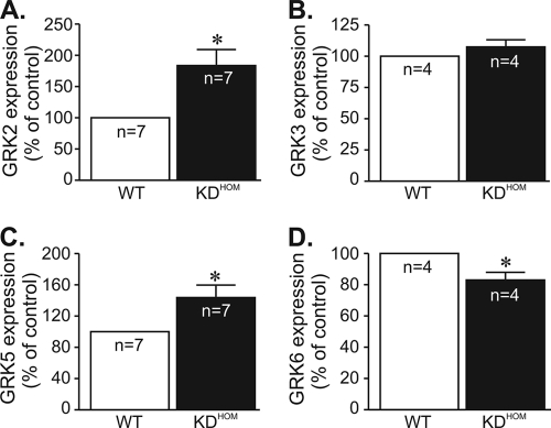
Overwhelming evidence supports the importance of the sympathetic nervous system in heart failure. In contrast, much less is known about the role of failing cholinergic neurotransmission in cardiac disease. By using a unique genetically modified mouse line with reduced expression of the vesicular acetylcholine transporter (VAChT) and consequently decreased release of acetylcholine, we investigated the consequences of altered cholinergic tone for cardiac function. M-mode echocardiography, hemodynamic experiments, analysis of isolated perfused hearts, and measurements of cardiomyocyte contraction indicated that VAChT mutant mice have decreased left ventricle function associated with altered calcium handling. Gene expression was analyzed by quantitative reverse transcriptase PCR and Western blotting, and the results indicated that VAChT mutant mice have profound cardiac remodeling and reactivation of the fetal gene program. This phenotype was attributable to reduced cholinergic tone, since administration of the cholinesterase inhibitor pyridostigmine for 2 weeks reversed the cardiac phenotype in mutant mice. Our findings provide direct evidence that decreased cholinergic neurotransmission and underlying autonomic imbalance cause plastic alterations that contribute to heart dysfunction.


Figures

FIG. 1.
FIG. 1.
VAChT mutant mice present heart failure. (A) VAChT and CHT1 immunoreactivity in neurons of adult WT and VAChT KDHOM mouse intracardiac ganglia. Note the reduction in immunofluorescence for VAChT. There was no decrease in immunoreactivity in nodal ganglia of VAChT KDHOM stained with an antibody against the high-affinity choline transporter (CHT1). Blue labeling corresponds to nuclei stained with DAPI that seem to predominantly label glial cells. Arrowheads indicate some of the neuronal cell bodies in the ganglia. Bar = 20 μm. (B) Left ventricle function (as assessed by contractility index) in VAChT KDHOM mice (black bars) and WT mice (white bars). VAChT mutant mouse hearts had decreased contractility indexes and lower absolute responses to ISO. n, number of mice; *, P value of <0.05 in comparison with the WT.
FIG. 2.
FIG. 2.
Pyridostigmine treatment prevented cardiac dysfunction in VAChT KDHOM mice. (A) Echocardiography analysis of left ventricle fractional shortening (FS) in VAChT mutant mice (black bars) and WT mice (white bars). Note the significant improvement in left ventricle performance after pyridostigmine treatment (gray bars). n, number of mice. (B) Time course of systolic tension in isolated perfused hearts from WT (white squares; 15 hearts) and VAChT KDHOM (black squares; 16 hearts) mice. Pyridostigmine treatment improved systolic tension of hearts from mutant mice (gray squares; 4 hearts). (C) Gene expression of stress cardiac markers in cardiomyocytes from VAChT KDHOM mice and WT mice. The bar graph shows data from at least 5 independent experiments. a.u., arbitrary units. (D) The HR measured in ex vivo beating hearts from VAChT KDHOM mutants (black squares) was lower than that of WT hearts (white squares) after 40 min of perfusion. Pyridostigmine treatment for 2 weeks (gray squares) prevented the observed changes in HR. (E) (Top) Representative confocal images of electrically stimulated intracellular Ca2+ transient recordings in ventricular myocytes. (Bottom) Ca2+ transient line-scan profile. (F) Significant reduction in peak Ca2+ transient amplitude observed in freshly isolated adult VAChT KDHOM ventricular cardiomyocytes compared to the amplitude for WT mice. Pyridostigmine treatment significantly prevented intracellular Ca2+ dysfunction in VAChT KDHOM mice. n, number of ventricular cardiomyocytes analyzed. (G) Ca2+ transient kinetics of decay in VAChT KDHOM cardiomyocytes and WT mice. Note that the observed changes in myocytes from VAChT KDHOM mice can be prevented by 2 weeks of pyridostigmine treatment. *, P value of <0.05 in comparison with WT and KDHOM PYR mice. T50, time from peak Ca2+ transient to 50% decay.
FIG. 3.
FIG. 3.
Electrical properties of VAChT KDHOM cardiomyocytes. (A) Sample ICa currents recorded from depolarizations from −40 mV to 0 mV. ISO (100 nmol/liter) significantly increased the magnitude of ICa in WT cells but not in VAChT KDHOM ventricular myocytes. (B) Average I-V relationships for ICa current density recorded from WT mice (open circles), WT mice with ISO (gray circles), VAChT KDHOM mice (black triangles), and VAChT KDHOM mice with ISO (open triangles). (C) Steady-state activation curves. (D) Sample action potential recordings from WT and VAChT KDHOM ventricular cardiomyocytes. (E) Action potential durations at 10, 30, 50, 70, and 90% repolarization. Fifteen cells from each experimental group were used. *, P value of <0.05 in comparison with WT mice.
FIG. 4.
FIG. 4.
Ca2+ signaling components in VAChT KDHOM hearts. In panels A to D, the top image is a representative Western blot and the bottom shows an average densitometry graph (n, number of heart samples analyzed). (A) SERCA2 levels in VAChT KDHOM hearts were observed to be significantly reduced compared to levels in WT hearts. Phosphorylated-PLN levels at the protein kinase A (PKA)-dependent site (Ser-16) were lower in VAChT mutant hearts (B), while phospho-Thr-17-PLN levels were increased in these hearts (C). (D) Phospho-Ser-23/24-troponin I levels were decreased in VAChT mutant hearts relative to WT hearts. Tubulin expression levels were used as a loading control. (E) The SR Ca2+ content in ventricular cardiomyocytes from VAChT mutants was significantly reduced compared to that in WT cells. n, number of cells. (F) Ca2+ spark frequency showed a tendency to be lower in VAChT KDHOM cardiomyocytes than in WT mice, but the difference was not statistically different. n, number of cells. *, P < 0.05.
FIG. 5.
FIG. 5.
VAChT KDHOM hearts present an altered GPCR response. (A) Percent decrease in beating frequency for WT (white squares; n = 4) and VAChT KDHOM (black squares; n = 4) hearts in response to 13 μmol/liter ACh. (B) Time course of mean percent force increase in WT (n = 5) and VAChT KDHOM (n = 5) perfused hearts in response to 10 μmol/liter ISO.
FIG. 6.
FIG. 6.
Effect of VAChT deficiency on M2 muscarinic and β1-adrenergic receptor message levels. M2 muscarinic and β1-adrenergic receptor message levels were determined by qPCR. n, number of heart samples. (A) M2 mRNA levels were significantly increased in VAChT KDHOM hearts. (B) Significant reduction in β1-adrenergic receptor mRNA levels was observed in VAChT KDHOM hearts. Pyridostigmine treatment prevented the increase in M2 mRNA levels without affecting β1-adrenergic receptor mRNA expression levels in VAChT KDHOM hearts. (C) Determination of M1 to M5 mRNA expression levels by real-time quantitative PCR in ventricular cardiomyocytes from WT and VAChT KDHOM mice. Significant levels of M1 to M3 transcripts were detected in cardiomyocytes from WT mice. M1, M2, and M3 muscarinic receptor transcript levels were upregulated in VAChT KDHOM cardiomyocytes. The bar graph represents data from at least 6 independent experiments. *, P < 0.05. (D and E) Increased M2 and reduced β1-adrenergic receptor transcript messages are observed in hearts of VAChT knockout mice. *, P value of <0.05 in comparison with the WT; †, P value of <0.05 in comparison with KDHOM mice treated with PYR. n, number of heart samples analyzed.
FIG. 7.
FIG. 7.
GRK message expression levels are altered in VAChT mutant hearts. Determination of GRK2 (A), GRK3 (B), GRK5 (C), and GRK6 (D) mRNA expression by real-time quantitative PCR. n, number of hearts analyzed. *, P < 0.05.

References

    1. Amorim, D. S., H. J. Dargie, K. Heer, M. Brown, D. Jenner, E. G. Olsen, P. Richardson, and J. F. Goodwin. 1981. Is there autonomic impairment in congestive (dilated) cardiomyopathy? Lancet i:525-527. - PubMed
    1. Bartholomeu, J. B., A. S. Vanzelli, N. P. Rolim, J. C. Ferreira, L. R. Bechara, L. Y. Tanaka, K. T. Rosa, M. M. Alves, A. Medeiros, K. C. Mattos, M. A. Coelho, M. C. Irigoyen, E. M. Krieger, J. E. Krieger, C. E. Negrao, P. R. Ramires, S. Guatimosim, and P. C. Brum. 2008. Intracellular mechanisms of specific beta-adrenoceptor antagonists involved in improved cardiac function and survival in a genetic model of heart failure. J. Mol. Cell. Cardiol. 45:240-249. - PubMed
    1. Bers, D. M. 2006. Altered cardiac myocyte Ca regulation in heart failure. Physiology (Bethesda) 21:380-387. - PubMed
    1. Bers, D. M. 2002. Cardiac excitation-contraction coupling. Nature 415:198-205. - PubMed
    1. Brandon, E. P., W. Lin, K. A. D'Amour, D. P. Pizzo, B. Dominguez, Y. Sugiura, S. Thode, C. P. Ko, L. J. Thal, F. H. Gage, and K. F. Lee. 2003. Aberrant patterning of neuromuscular synapses in choline acetyltransferase-deficient mice. J. Neurosci. 23:539-549. - PMC - PubMed

Publication types

MeSH terms

Substances