. 2010 Apr;30(7):1783-99.
doi: 10.1128/MCB.01003-09. Epub 2010 Feb 1.

Establishment of extracellular signal-regulated kinase 1/2 bistability and sustained activation through Sprouty 2 and its relevance for epithelial function

Affiliations

Establishment of extracellular signal-regulated kinase 1/2 bistability and sustained activation through Sprouty 2 and its relevance for epithelial function

Weimin Liu et al. Mol Cell Biol. 2010 Apr.

Abstract

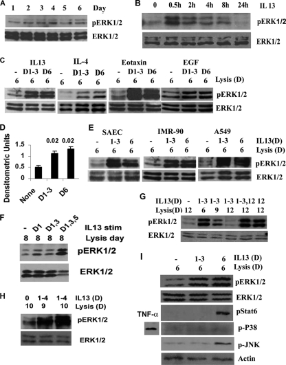
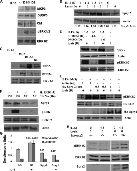
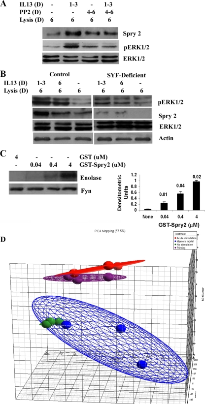
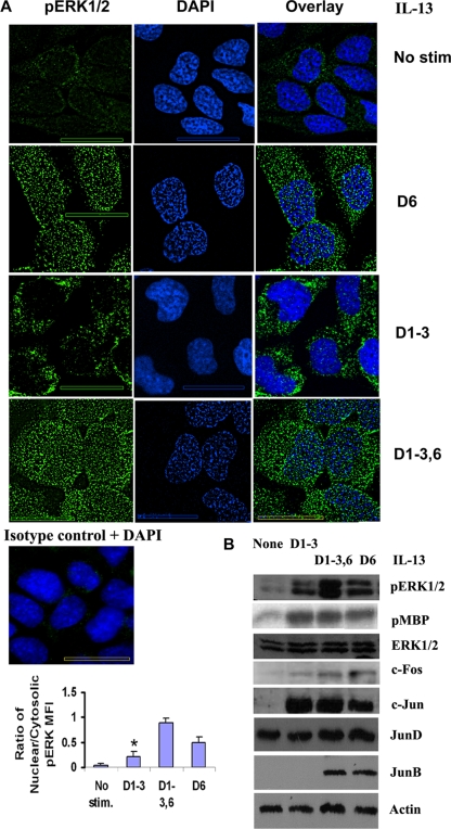
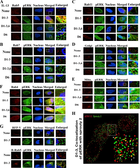
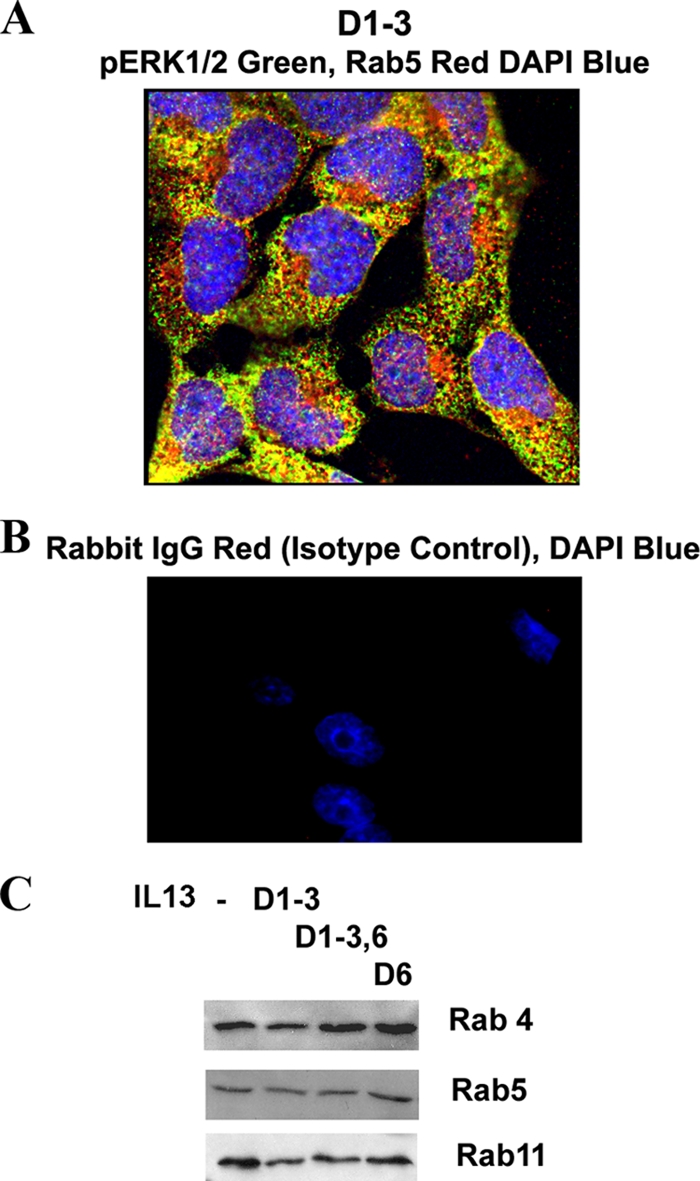
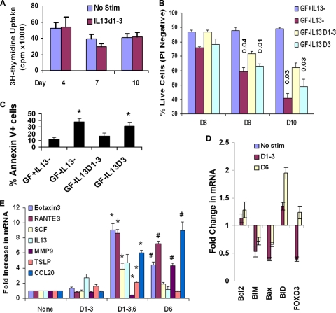
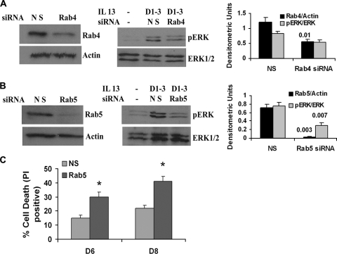
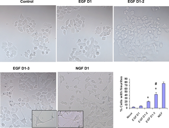
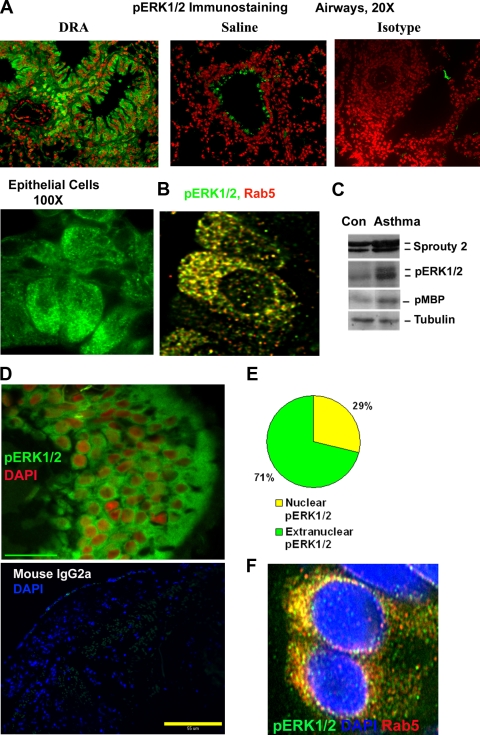
Our objective was to establish an experimental model of a self-sustained and bistable extracellular signal-regulated kinase 1/2 (ERK1/2) signaling process. A single stimulation of cells with cytokines causes rapid ERK1/2 activation, which returns to baseline in 4 h. Repeated stimulation leads to sustained activation of ERK1/2 but not Jun N-terminal protein kinase (JNK), p38, or STAT6. The ERK1/2 activation lasts for 3 to 7 days and depends upon a positive-feedback mechanism involving Sprouty 2. Overexpression of Sprouty 2 induces, and its genetic deletion abrogates, ERK1/2 bistability. Sprouty 2 directly activates Fyn kinase, which then induces ERK1/2 activation. A genome-wide microarray analysis shows that the bistable phospho-ERK1/2 (pERK1/2) does not induce a high level of gene transcription. This is due to its nuclear exclusion and compartmentalization to Rab5+ endosomes. Cells with sustained endosomal pERK1/2 manifest resistance against growth factor withdrawal-induced cell death. They are primed for heightened cytokine production. Epithelial cells from cases of human asthma and from a mouse model of chronic asthma manifest increased pERK1/2, which is associated with Rab5+ endosomes. The increase in pERK1/2 was associated with a simultaneous increase in Sprouty 2 expression in these tissues. Thus, we have developed a cellular model of sustained ERK1/2 activation, which may provide a mechanistic understanding of self-sustained biological processes in chronic illnesses such as asthma.


Figures

FIG. 1.
FIG. 1.
Development of ERK1/2 bistability in human cells. (A) Basal phosphorylation of ERK1/2 in cultured airway epithelial cell line BEAS-2B studied over a period of 6 consecutive days without any stimulation. Following Western blotting with pERK1/2, the membrane was reprobed with an anti-ERK1/2 antibody (n = 3). (B) Kinetics of pERK1/2 in BEAS-2B cells stimulated with IL-13 (20 ng/ml), as measured by Western blotting (n = 3). (C) BEAS-2B cells were stimulated with IL-13 (10 ng/ml), IL-4 (10 ng/ml), eotaxin (10 ng/ml), or EGF (100 ng/ml) separately for 1 h daily for 3 consecutive days. The cells were washed 3 times after each stimulation and cultured without stimulation for 3 additional days after the last stimulation (samples labeled D1-3). One set of controls was stimulated and washed with buffer (−), and another set of controls was stimulated on day 6 with IL-13 or the other cytokines 1 h before lysis (D6). All these samples were lysed on day 6 and Western blotted for pERK1/2 (n = 3 to 6). (D) Densitometric analysis of pERK1/2 expression after repeated IL-13 stimulation as presented in panel C, top (n = 6). Statistical significance (P values) is shown above the bars. (E) Small airway primary epithelial cells (SAEC), the lung fibroblast cell line IMR-90, and the lung alveolar carcinoma cell line A549 were treated with IL-13 as described for panel D and Western blotted for pERK1/2 (n = 3). (F) BEAS-2B cells were stimulated with IL-13 on alternate days and then lysed on day 8 (n = 3). (G) BEAS-2B cells were stimulated with IL-13 for 3 consecutive days (days 1 to 3) and then lysed on the indicated days and Western blotted for pERK1/2. Selected stimulated or nonstimulated samples were also stimulated on day 12 before lysis (n = 3). (H) Effect of 4 days of stimulation. BEAS-2B cells were stimulated with buffer or IL-13 for 1 h daily for 4 consecutive days and Western blotted on the indicated lysis days (n = 3). (I) BEAS-2B cells were stimulated with IL-13 as described for panel C. The cell lysate was Western blotted using antibodies against pERK1/2, ERK1/2, pSTAT6, p-p38 MAPK, and pJNK. For p-p38, an aliquot of cells was additionally stimulated with TNF-α as a positive control. The pJNK membrane was reprobed with an anti-β-actin antibody to demonstrate protein loading (n = 4).
FIG. 2.
FIG. 2.
Role of Sprouty 2 in sustained ERK1/2 activation. (A) Expression of dual-specific phosphatases MKP3 and DUSP3 and receptor-associated ubiquitin ligase Cbl in the repeated-stimulation model. BEAS-2B cells were stimulated repeatedly on days 1 to 3 or only once on day 6 and then Western blotted. The membranes were reprobed for ERK1/2 to demonstrate protein loading (n = 3). (B) Sprouty 2 expression after repeated stimulation. BEAS-2B cells were stimulated with IL-13 for the indicated number of days and then lysed and Western blotted on day 6. The membrane was reprobed for β-actin (n = 4). (C) Mnk1 phosphorylation. BEAS-2B epithelial cells were stimulated with IL-13 as described for Fig. 1C and then Western blotted for pERK1/2 and pMnk1. The membrane was reprobed with an anti-ERK1/2 antibody (n = 3). (D) Effect of a MEK inhibitor on Sprouty 2 expression. BEAS-2B cells were treated with IL-13 as described for Fig. 1C and then incubated with the MEK1/2 inhibitor PD98059 or the diluent DMSO on days 4 to 6. The cell lysate was Western blotted for Sprouty 2 and pERK1/2. The membranes were reprobed with anti-ERK1/2 and anti-β-actin antibodies, respectively (n = 3). (E) Effect of overexpression of Sprouty 2 on pERK1/2. BEAS-2B cells were transfected with two concentrations of a hemagglutinin (HA)-tagged Sprouty 2 expression vector or a control vector and then stimulated with IL-13 as described for Fig. 1C. The cell lysates were Western blotted with anti-pERK1/2 and anti-HA antibodies and reprobed for ERK1/2 and β-actin, respectively (n = 3). (F) Effect of Sprouty 2 knockdown on sustained pERK1/2. BEAS-2B cells were treated with siRNA for Sprouty 2 (SP) or luciferase (nonspecific [NS]) and then stimulated with IL-13 as described for Fig. 1C. The cell lysates were Western blotted for Sprouty 2 and pERK1/2. The membranes were reprobed for actin and ERK1/2, respectively (n = 4). (G) Densitometric analysis of Sprouty 2 and pERK1/2 expression as presented for panel F. Statistical significance (P value) compared to the NS and IL-13 sample is shown above the bars. (H) Effect of Sprouty 2 knockout on pERK1/2. Mouse embryonic fibroblasts with Sprouty 2 homozygous (−/−) and heterozygous (+/−) null deletions were stimulated with IL-13 as described for Fig. 1C. The cell lysates were Western blotted for pERK1/2 and Sprouty 2. The pERK1/2 membrane was reprobed for ERK1/2 (n = 3).
FIG. 3.
FIG. 3.
Role of Src family kinases in sustained ERK1/2 activation and microarray data analyses. (A) Effect of Src family kinase inhibition. BEAS-2B cells were stimulated with IL-13 as described for Fig. 1C and then incubated with the Src family kinase inhibitor PP2 on days 4 to 6. The cell lysates were Western blotted for Sprouty 2 and pERK1/2 (n = 3). (B) Mouse embryonic fibroblasts with homozygous null mutation for Src, Yes, and Fyn (SYF) and control fibroblasts were treated with IL-13 as described for Fig. 1C and then Western blotted for pERK1/2 and Sprouty 2 (n = 4). (C) Effect of recombinant Sprouty 2 on Fyn kinase activation. (Left) Sprouty 2 was expressed as a GST-Sprouty 2 fusion protein, and the purified GST and GST-Sprouty 2 (Spry 2) were used in a Fyn immune complex kinase assay in the presence of [γ-32P]ATP and enolase as a substrate. (Right) Densitometric analysis of phosphorylated enolase bands (from the left panel) from 4 separate experiments. Statistical significance (P values) compared to “none” is shown above the bars. (D) Principal component analysis of the GeneChip data. No stimulation, control cells incubated with buffer instead of IL-13 (green); memory model, cells repeatedly stimulated with IL-13 on days 1 to 3 (blue); acute stimulation, cells stimulated with IL-13 for 30 min on day 6 (red); priming, cells repeatedly stimulated with IL-13 on days 1 to 3 and then restimulated on day 6 (purple).
FIG. 4.
FIG. 4.
Nuclear translocation of pERK1/2. (A, top) BEAS-2B cells were left unstimulated (no stim), stimulated with IL-13 once on day 6 (D6), or stimulated (1 h per day and then washed) repeatedly on days 1 to 3 and then either rested on days 4 to 6 (D1-3) or stimulated again on day 6 (D1-3,6). The cells were then fixed on day 6 and processed for immunofluorescent staining using a mouse anti-pERK1/2 antibody and counterstained with DAPI (4′,6-diamidino-2-phenylindole). Z-stack images (magnification, ×100) were captured and deconvolved with no neighbors using the software Metamorph, v.7. n = 7; scale bar = 10 μm. (Middle) Immunostaining of epithelial cells with a mouse IgG1 isotype antibody (control for the anti-pERK1/2 antibody). The slide was counterstained with DAPI (n = 3). (Bottom) Quantification of the mean fluorescence intensity (MFI) of nuclear and cytosolic pERK1/2 by the software Metamorph, v.7.0. *, P < 0.04 compared to D6 and D1-3,6. n = 7. (B) Kinase activity of pERK1/2 and its effect on AP1 complex proteins. BEAS-2B cells were stimulated with IL-13 as described for panel A. Cells were lysed, and one sample was used in an immune complex kinase assay for ERK1/2 using myelin basic protein (MBP) as a substrate. The incorporation of [γ-32P]ATP into MBP was detected by autoradiography. The membrane was reprobed for ERK1/2 (n = 4). Other samples were Western blotted for pERK1/2, c-Fos, c-Jun, JunB, and JunD. The c-Fos membrane was reprobed for actin (n = 3).
FIG. 5.
FIG. 5.
Endosomal localization of pERK1/2. BEAS-2B cells were stimulated as described for Fig. 4A and then processed for double-immunofluorescent staining for pERK1/2 (green) and the early, late, and recycling endosomal markers Rab5 (A), Rab7 (B), and Rab11 (C) (red), respectively, on day 6. The nucleus was stained blue with DAPI (n = 4). Z-series images (magnification, ×100) were deconvolved with no neighbors using the software Metamorph, v.7, and presented as maximal projection (n = 4). Scale bars = 5 μm. The right panels represent higher magnifications of the boxed areas. (D and E) Immunostaining with Golgi (GM130) (D) and mitochondrial (Mitotracker) (E) markers (n = 4). (F) Colocalization of pERK1/2 with Rab4 (n = 4). (G) Colocalization with red fluorescent protein (RFP)-Rab5. BEAS-2B cells were stimulated with IL-13 as described for Fig. 4A and then transfected with the red fluorescent protein control vector (RFP-V) or RFP-Rab5 fusion construct (R-Rab5) on day 4. The cells were stained for pERK1/2 on day 6 and counterstained with DAPI. Z-series images were deconvolved, and selected images are presented (n = 3). (H) Colocalization of Sprouty 2 with pERK1/2. BEAS-2B cells were stimulated as described for Fig. 4A and then double stained for pERK1/2 (green) and Sprouty 2 (red). Z-series images (magnification, ×100) were 3-dimensionally deconvolved with the point spread function and Metamorph software. The boxed area is enlarged in the bottom right frame. Colocalization (yellow) is shown with arrowheads.
FIG. 6.
FIG. 6.
Subcellular localization of pERK1/2. (A) Colocalization of pERK1/2 with Rab5. An overlay picture of pERK1/2 (green) and Rab5 (red) showing colocalization (yellow) in multiple cells is shown (n = 6). (B) Immunostaining with a rabbit IgG antibody (control for antibodies against Rab4, Rab5, Rab7, Rab11, GM130, and Sprouty 2) and counterstaining with DAPI (n = 3). (C) Expression of Rab4, Rab5, and Rab11 in epithelial cells. BEAS-2B cells were stimulated with IL-13 as described for Fig. 4A and then Western blotted for Rab4, Rab5, and Rab11 (n = 3).
FIG. 7.
FIG. 7.
Biological relevance of sustained ERK1/2 activation. (A) Effect on proliferation. BEAS-2B cells were stimulated with IL-13 as described for Fig. 4A and then cultured in the presence of [3H]thymidine overnight before the conclusion of the culture at the indicated time point. No Stim, no stimulation (n = 3). (B) Effect on cell survival. BEAS-2B cells were stimulated repeatedly with IL-13 (1 h per day) on days 1 to 3 (IL-13 D1-3) or only day 3 (IL-13 D3) and then cultured in basal medium with (GF+) or without (GF−) growth factor-enriched supplement. Cell survival was monitored on days 6, 8, and 10 by staining with propidium iodide (PI). Statistical significance levels compared to the day 1 to 3 samples are shown above the bars (n = 4). (C) Effect on apoptosis. BEAS-2B cells were stimulated with IL-13 as described for Fig. 4A, and cell apoptosis was measured on day 8 by flow cytometry following staining with annexin V and propidium iodide. *, P < 0.04 (n = 3). (D) Real-time PCR for mRNA for selected apoptosis-regulating genes. Fold changes in mRNA compared to that in nonstimulated cells are shown (n = 3). β-Actin and GAPDH (glyceraldehyde-3-phosphate dehydrogenase) genes were used as housekeeping gene controls. (E) Effect on cytokine/protease production. Real-time PCR results for eotaxin 3, RANTES, stem cell factor (SCF), IL-13, MMP9, TSLP, and CCL20 are shown. β-Actin and GAPDH genes were used as housekeeping gene controls. Fold increases in gene expression over that in nonstimulated cells are shown (n = 6). *, P < 0.04; #, P < 0.02.
FIG. 8.
FIG. 8.
(A and B) Effect of Rab4 and Rab5 knockdown on pERK1/2 and cell survival. BEAS-2B cells were stimulated with IL-13 on days 1 to 3 or left untreated and then transfected with Rab4a or Rab5a siRNA on day 4. A nontargeting siRNA that was directed against luciferase was used as a control (NS). Forty-eight hours later cells were divided into two aliquots; one aliquot was Western blotted for Rab4 (A, left), Rab5 (B, left), and pERK (A and B, middle) expression. Bar graphs at the right show densitometric analysis of the corresponding Western blot bands from 3 experiments. Statistical significance (P values) is shown above the bars. (C) One set of cells from the Rab5 knockdown experiment was further cultured for the indicated periods of time in the basic culture medium without the growth factor-rich supplement. At the conclusion the cells were stained with propidium iodide (PI) to determine cell survival. n = 3; *, P < 0.04.
FIG. 9.
FIG. 9.
Neurite growth in PC12 cells. PC12 cells were stimulated once with NGF on day 1 or with increasing frequency with EGF (day 1, days 1 and 2, and days 1 to 3). Images of neurite outgrowth were captured on day 5 using a 10× objective. Neurite outgrowth of 200 cells per culture and from four different cultures was counted for statistical analyses. *, P < 0.01 compared to no stimulation (none); #, P = 0.01 compared to NGF (n = 4).
FIG. 10.
FIG. 10.
Endosomal pERK1/2 in a mouse model of chronic asthma and human asthma. (A) Mice were immunized and exposed to a combination of three allergens, dust mites, ragweed, and Aspergillus (DRA), or saline as described previously (31). Three weeks after the last allergen exposure the mice were examined for pERK1/2 immunostaining (green). Nuclei were stained with DAPI and pseudocolored red for better visualization (n = 6). (B) Z-stack series of an airway epithelium stained with pERK1/2 (green) and Rab5 (red) were deconvolved by the software Metamorph, v.7, with point spread function. An overlay image showing colocalization in yellow is also shown (n = 4). (C) Lung tissue samples from the chronic asthma model (DRA sensitized and exposed) or control (Con; saline) were Western blotted for Sprouty 2 and pERK1/2 (n = 3). Another sample was immunoprecipitated with an anti-ERK1/2 antibody and used in the kinase assay in the presence of myelin basic protein (MBP) as a substrate (n = 3). (D to F) Endosomal pERK1/2 in human asthma. (D) Lung biopsy samples obtained from 6 asthmatic subjects were immunostained for pERK1/2 and counterstained with DAPI (pseudocolored red) as described previously (31). A representative airway epithelial image (magnification, ×100) is shown. The lower panel shows immunostaining with an isotype control antibody. Scale bar = 55 μm. (E) Cells with nuclear staining for pERK1/2 from 100 epithelial cells per sample were counted, and the fraction of epithelial cells with nuclear pERK1/2 is presented (n = 8). (F) Selected biopsy samples were stained for pERK1/2 (green), Rab5 (red), and nuclei (DAPI; blue). Z-stack series were deconvolved by the software Metamorph, v.7, with point spread function. An overlay image showing colocalization in yellow is shown (n = 4).

Similar articles

Cited by

References

    1. Bashor, C. J., N. C. Helman, S. Yan, and W. A. Lim. 2008. Using engineered scaffold interactions to reshape MAP kinase pathway signaling dynamics. Science 319:1539-1543. - PubMed
    1. Bhattacharya, M., N. Ojha, S. Solanki, C. K. Mukhopadhyay, R. Madan, N. Patel, G. Krishnamurthy, S. Kumar, S. K. Basu, and A. Mukhopadhyay. 2006. IL-6 and IL-12 specifically regulate the expression of Rab5 and Rab7 via distinct signaling pathways. EMBO J. 25:2878-2888. - PMC - PubMed
    1. Casci, T., J. Vinós, and M. Freeman. 1999. Sprouty, an intracellular inhibitor of Ras signaling. Cell 96:655-665. - PubMed
    1. Chandramouli, S., C. Y. Yu, P. Yusoff, D. H. Lao, H. F. Leong, K. Mizuno, and G. R. Guy. 2008. Tesk1 interacts with Spry2 to abrogate its inhibition of ERK phosphorylation downstream of receptor tyrosine kinase signaling. J. Biol. Chem. 283:1679-1691. - PubMed
    1. Das, J., M. Ho, J. Zikherman, C. Govern, M. Yang, A. Weiss, A. K. Chakraborty, and J. P. Roose. 2009. Digital signaling and hysteresis characterize ras activation in lymphoid cells. Cell 136:337-351. - PMC - PubMed

Publication types

MeSH terms

Substances