. 2010 Apr 2;285(14):10862-9.
doi: 10.1074/jbc.M109.073171. Epub 2010 Feb 1.

Maspin (SERPINB5) is an obligate intracellular serpin

Affiliations

Maspin (SERPINB5) is an obligate intracellular serpin

Sonia S Y Teoh et al. J Biol Chem. .

Abstract

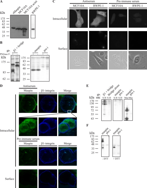
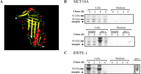
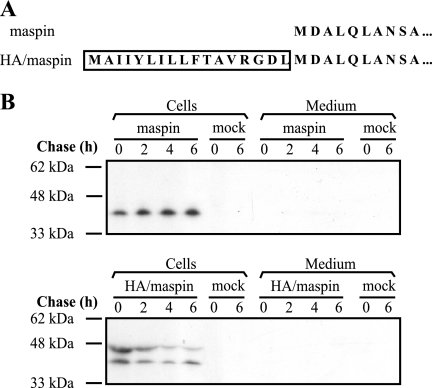
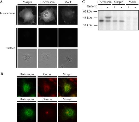
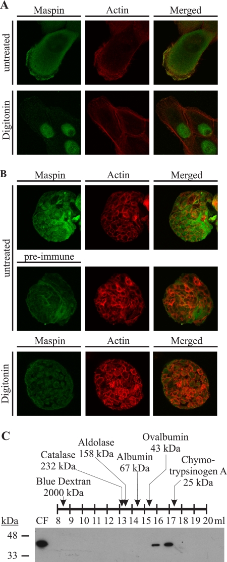
Maspin (SERPINB5) is a tumor suppressor lost in breast and prostate cancer whose molecular function is unknown. It is a non-inhibitory member of the clade B serpins suggested to play a role in a plethora of intracellular and extracellular settings, yet its normal cellular distribution has never been clarified. Here we investigate the distribution of maspin in non-transformed human epithelial cells. By indirect immunofluorescence, maspin has a nucleocytoplasmic distribution in breast (MCF10A) and prostate (RWPE-1) cells and, by immunoblotting and pulse-chase analyses, is neither glycosylated nor secreted. Cell surface biotinylation studies also show that maspin is not present at the cell surface. Differentiation of MCF10A cells into three-dimensional acini results in the redistribution of maspin from the nucleus to the cytoplasm but does not result in secretion. Addition of an efficient conventional signal peptide to maspin directs it into the secretory pathway and results in glycosylation but not secretion. We further show that maspin in the cytoplasm of MCF10A cells is a soluble monomeric protein that is not detectably associated with the cytoskeleton or other extractable components. Taken together, these results suggest that maspin is restricted to an intracellular, possibly nuclear, role in which it influences cell-matrix interactions indirectly. It is probably released only as a consequence of cell damage or necrosis.


Figures

FIGURE 1.
FIGURE 1.
Maspin is expressed by MCF10A and RWPE-1 cells but is absent from the cell surface. A, lysates of MCF10A cells and acini or RWPE-1 cells were separated via 12.5% SDS-PAGE and immunoblotted with recombinant maspin (rMaspin) as a control. The membrane was probed with mouse anti-maspin monoclonal antibody diluted 1:1000 and detected with horseradish peroxidase-conjugated secondary against mouse IgG. RWPE-1 lysates were analyzed on a separate gel. B, maspin in MCF10A cells is not glycosylated. Lysates of 30-min metabolically labeled MCF10A cells were immunoprecipitated (IP) using mouse anti-β1-integrin monoclonal antibody (left panel), rabbit anti-maspin polyclonal antiserum (right panel), or preimmune (pre-i) serum and then either treated with PNGase F or left untreated. Maspin samples were separated by 12.5% SDS-PAGE, and β1-integrin samples were separated by 7.5% SDS-PAGE. Gels were analyzed by fluorography. C, maspin is in the nucleus and cytoplasm and not on the cell surface. MCF10A and RWPE-1 cells were fixed and permeabilized or fixed alone to detect cell surface proteins and then probed with mouse anti-maspin polyclonal antiserum or with the preimmune serum. The primary antibody was detected using goat Alexa 488-conjugated secondary antibodies. Cells were examined by epifluorescence microscopy. Brightfield images of cells examined for cell surface staining are shown. D, maspin is intracellular in each luminal epithelial cell and does not co-localize with β1-integrin. MCF10A cells were grown on Matrigel, and acini were developed for 20 days. Acini were prepared as described for B and examined by confocal microscopy. Images shown are single optical sections. E, MCF10A cells were surface-biotinylated, and lysates were immunoprecipitated using rabbit anti-maspin polyclonal antiserum, preimmune serum, or mouse anti-β1-integrin monoclonal antibody. Immune complexes were collected and analyzed by 10% SDS-PAGE, and the immunoblot (WB) was probed with horseradish peroxidase-conjugated streptavidin (S-H) diluted 1:5000. The blot was stripped and re-probed with mouse anti-maspin monoclonal antibody (Mas) and detected with horseradish peroxidase-conjugated secondary against mouse IgG. Bridge refers to samples immunoprecipitated with bridging antibody alone. F, MCF10A cell lysates were immunoprecipitated, and immune complexes were left nonreduced (without dithiothreitol (−DTT)) or reduced (+DTT) and analyzed via 12.5% SDS-PAGE and immunoblotting with mouse anti-maspin monoclonal antibody followed by horseradish peroxidase-conjugated secondary against mouse IgG.
FIGURE 2.
FIGURE 2.
Maspin is not secreted from MCF10A or RWPE-1 cells. A, ribbon structure of maspin. White arrows denote asparagine residues that may be glycosylated. B and C, pulse-chase experiments were performed on MCF10A cells (B) and the primary prostate cell line, RWPE-1 (C), as indicated under “Experimental Procedures.” Cell lysates and media were collected at each time point and immunoprecipitated with rabbit anti-maspin polyclonal antiserum. Immune complexes were collected, reduced, and analyzed by 10% SDS-PAGE and fluorography. Comparison of antiserum and preimmune (pre-i) serum complexes for MCF10A immunoprecipitation is shown in the middle panel. The preimmune complex from RWPE-1 immunoprecipitation shown in C is from a separate autoradiograph.
FIGURE 3.
FIGURE 3.
Maspin directed into the classical secretory pathway is not released into the extracellular milieu. A, comparison of the N terminus of HA/maspin and maspin. Boxed residues comprise the HA signal peptide. B, COS cells were transfected with maspin (upper panel) and HA/maspin (lower panel) DNA. Forty-eight hours after transfection, cells were used in a pulse-chase experiment as described under “Experimental Procedures.” COS cells transfected without DNA (mock) were included as a negative control.
FIGURE 4.
FIGURE 4.
Glycosylated maspin is retained in the ER. A, COS-1 cells transfected with HA/maspin or maspin were prepared as described in the legend to Fig. 1 and probed with mouse anti-maspin polyclonal antiserum. As a control, COS-1 cells were mock-transfected. The primary antibody was detected with goat anti-mouse IgG conjugated to Alexa 488, and cells were examined by epifluorescence microscopy. The lower panel shows brightfield images of cells examined for cell surface staining. B, COS-1 cells transfected with HA/maspin were fixed, permeabilized, and probed with mouse anti-maspin monoclonal antibody. The primary antibody was detected with goat anti-mouse IgG conjugated to Alexa 488. The ER was marked with Alexa Fluor 594-conjugated concanavalin A (Con A), and the Golgi was marked with rabbit anti-giantin polyclonal antibody detected with goat anti-rabbit IgG conjugated to RITC. Cells were examined by confocal microscopy. Images show single optical sections. C, COS-1 cells were transfected with HA/maspin or maspin DNA or without DNA (Mock). Forty-eight hours after transfection, cells were labeled for 30 min in medium lacking methionine containing 100 μCi of [35S]methionine and then incubated in complete medium for 6 h. Cell lysates and medium were then collected and immunoprecipitated with rabbit anti-maspin polyclonal antiserum. Immune complexes were either treated (+) or left untreated (−) with Endo H. Samples were then reduced and analyzed by 10% SDS-PAGE and fluorography.
FIGURE 5.
FIGURE 5.
Maspin is not stably associated with the cytoskeleton or other cellular proteins and exists as a monomeric serpin. A and B, maspin is a soluble cytoplasmic protein. MCF10A monolayers (A) growing on microscope slides and MCF10A acini (B) were extracted with HMKE buffer containing digitonin and then fixed, permeabilized, and probed with mouse anti-maspin polyclonal antiserum. The primary antibody was detected with goat anti-mouse IgG conjugated to Alexa 488. The actin cytoskeleton was marked by RITC-conjugated phalloidin. Cells were examined by confocal microscopy. Images show single optical sections. C, MCF10A cells were harvested in hypotonic buffer, and gel filtration was performed on the cytosolic fraction (CF) of lysate. Fractions were separated by 12.5% SDS-PAGE, analyzed by immunoblotting for maspin using mouse anti-maspin monoclonal antibody, and detected with horseradish peroxidase-conjugated secondary against mouse IgG. A series of molecular mass standards ranging between 25 and 2000 kDa was assessed, and the fractions in which they appeared are shown. The molecular mass of the eluted maspin was within the predicted monomeric range.

Similar articles

Cited by

References

    1. Silverman G. A., Whisstock J. C., Askew D. J., Pak S. C., Luke C. J., Cataltepe S., Irving J. A., Bird P. I. (2004) Cell. Mol. Life Sci. 61, 301–325 - PMC - PubMed
    1. Zou Z., Anisowicz A., Hendrix M. J., Thor A., Neveu M., Sheng S., Rafidi K., Seftor E., Sager R. (1994) Science 263, 526–529 - PubMed
    1. Gao F., Shi H. Y., Daughty C., Cella N., Zhang M. (2004) Development 131, 1479–1489 - PubMed
    1. Ngamkitidechakul C., Warejcka D. J., Burke J. M., O'Brien W. J., Twining S. S. (2003) J. Biol. Chem. 278, 31796–31806 - PubMed
    1. Sheng S., Carey J., Seftor E. A., Dias L., Hendrix M. J., Sager R. (1996) Proc. Natl. Acad. Sci. U.S.A. 93, 11669–11674 - PMC - PubMed

Publication types

MeSH terms