. 2010 Feb 8;188(3):351-68.
doi: 10.1083/jcb.200911128. Epub 2010 Feb 1.

Mutual regulation of cyclin-dependent kinase and the mitotic exit network

Affiliations

Mutual regulation of cyclin-dependent kinase and the mitotic exit network

Cornelia König et al. J Cell Biol. .

Abstract

The mitotic exit network (MEN) is a spindle pole body (SPB)-associated, GTPase-driven signaling cascade that controls mitotic exit. The inhibitory Bfa1-Bub2 GTPase-activating protein (GAP) only associates with the daughter SPB (dSPB), raising the question as to how the MEN is regulated on the mother SPB (mSPB). Here, we show mutual regulation of cyclin-dependent kinase 1 (Cdk1) and the MEN. In early anaphase Cdk1 becomes recruited to the mSPB depending on the activity of the MEN kinase Cdc15. Conversely, Cdk1 negatively regulates binding of Cdc15 to the mSPB. In addition, Cdk1 phosphorylates the Mob1 protein to inhibit the activity of Dbf2-Mob1 kinase that regulates Cdc14 phosphatase. Our data revise the understanding of the spatial regulation of the MEN. Although MEN activity in the daughter cells is controlled by Bfa1-Bub2, Cdk1 inhibits MEN activity at the mSPB. Consistent with this model, only triple mutants that lack BUB2 and the Cdk1 phosphorylation sites in Mob1 and Cdc15 show mitotic exit defects.


Figures

Figure 1.
Figure 1.
Cdk1 binds to the mSPB in early anaphase. (A) Time-lapse analysis of CDK1-GFP SPC42-eqFP611 mCherry-TUB1 cells grown in low fluorescence medium at 30°C. Consecutive sections were taken every 60 s. Shown are deconvolved and projected images. The yellow arrows show Cdk1-GFP at the plus end of cytoplasmic MTs (Maekawa and Schiebel, 2004). The white arrows highlight Cdk1 at the SPBs. The insets (a and b) are enlargements of the two spindle poles. Bar, 5 µm. (B) Cdk1 associates with mSPB in anaphase. CDK1-3GFP SPC42-eqFP611 cells were synchronized with α-factor at 30°C. n > 130 anaphase cells were analyzed and classified for colocalization of the SPB marker Spc42-eqFP611 and Cdk1-3GFP as indicated. Shown is one representative experiment out of five independent experiments. (C) Cdk1-3GFP associates with SPBs independently of the kinetochore structure. CDK1-3GFP SPC42-RFP ndc10-1 cells were synchronized with α-factor and released into medium at 37°C. After 150 min at 37°C, fixed cells were stained with DAPI. Bar, 5 µm. (D) Cdk1-GFP does not localize to SPBs in clb1Δ clb2Δ cells. α-Factor–synchronized wild-type and clb1Δ Gal1-CLB2 cells were arrested in G1 phase and released in YPAD medium at 30°C to induce Clb2 depletion. SPB localization of Cdk1-GFP of cells in anaphase was quantified for wild-type (wt; n = 119) and clb1Δ Gal1-CLB2 (clb1Δ clb2Δ; n = 70) cells.
Figure 2.
Figure 2.
Activity of the MEN regulates binding of Cdk1 to SPBs. (A) Cdk1 does not associate with SPBs in cells with a misaligned anaphase spindle. KAR9 CDK1-3GFP SPC42-eqFP611 and kar9Δ CDK1-3GFP SPC42-eqFP611 cells grown in YPAD were synchronized with α-factor at 30°C and released at 37°C. The pictures in the right corner show enlargements of SPB signals. (B) Quantification of anaphase cells of experiment A with correctly or misaligned spindles. n > 200 cells per strain. (C) Cdc5 and Tem1 are required for Cdk1 localization to SPBs. α-Factor–synchronized wild-type, Gal1-CDC5, and Gal1-UPL-TEM1 cells were grown in YPD medium at 30°C to deplete Cdc5 and Upl-Tem1. Anaphase cells were analyzed for SPB localization of Cdk1-3GFP. n > 75 cells for each strain. (D) α-Factor–synchronized BFA1 CDK1-3GFP SPC42-eqFP611 and bfa1Δ CDK1-3GFP SPC42-eqFP611 cells were grown in YPAD medium at 30°C and examined in anaphase for colocalization of Cdk1-3GFP and the SPB marker Spc42-eqFP611. The pictures in the right corner show enlargements of SPB signals. (E) Quantification of D as illustrated in the figure. n > 80 anaphase cells were analyzed. Bars, 5 µm.
Figure 3.
Figure 3.
SPB localization of Cdk1 depends on Cdc15 kinase activity. (A and B) CDC15 and cdc15-as1 cells were synchronized with α-factor and released at 30°C into YPAD medium with the PP1 analogue 8 to inhibit activity of cdc15-as1 kinase (D’Aquino et al., 2005). Anaphase cells with a 4–10-µm spindle were examined for the Cdk1-GFP localization. n = 38 for CDC15, n = 43 for cdc15-as1. The arrows in A point toward the Cdk1-GFP signal at mSPBs. (C) Localization of Cdc15-GFP and cdc15-as1-GFP in anaphase. Two representative cells are shown for each strain grown in YPAD at 30°C in the presence of PP1 analogue 8. The first Cdc15-GFP and cdc15-as1-GFP pictures were taken under identical conditions. The second pictures in this row are linear enhancements of the first pictures. Bar, 5 µm. (D) Protein levels of Cdc15-GFP and cdc15-as1-GFP detected with anti-GFP antibody. Anti-Tub2 was used as loading control. (E) The Dbf2–Mob1 kinase complex is not required for SPB association of Cdk1 in anaphase. Synchronized wild-type, mob1-67, and dbf2-2 cells were grown in YPAD at 37°C after the release of the α-factor block. Anaphase cells were examined for SPB localization of Cdk1-GFP. (F) Quantification of E. n > 75 anaphase cells per strain. Bars, 5 µm.
Figure 4.
Figure 4.
Cdc15 and Cdk1 show mutual regulation at the mSPB. (A) CDC14 and td-cdc14 cells harboring CDC15-GFP mCherry-TUB1 were examined for Cdc15-GFP localization in anaphase. The arrows highlight Cdc15-GFP at SPBs. Bar, 5 µm. (B and C) Anaphase cells of CDC14 CDC15-GFP, td-cdc14 CDC15-GFP, and td-cdc14 CDC15-7A-GFP were grown in YPAD and analyzed for GFP signal at SPBs. Quantified relative fluorescent intensities are summarized in box-and-whisker plots: boxes span between the 25th and 75th percentile with a line at the median; whiskers extend from the 10th to 90th percentile. P-values were calculated using unpaired t tests and indicate significant differences between * or ** marked bars. (B) n > 50 anaphase cells per strain. (C) n > 50 for CDC15-GFP cells and n = 24 for CDC15-7A-GFP cells. (D) CDC15 and CDC15-7A cells were grown in SC medium. Cells in anaphase were examined for Cdk1-GFP localization to SPBs. Bar, 5 µm. (E) Quantification of Cdk1-GFP signal at the mSPB. Relative fluorescent intensities in box-and-whisker plots as in B and C. n > 50 cells were analyzed per strain. (F) Cdk1-GFP protein levels measured with anti-GFP antibody and actin as loading control.
Figure 5.
Figure 5.
Phosphorylation of Mob1 by Cdk1 kinase inhibits the MEN. (A) Distribution of the two full consensus sites [S/T-P-x-K/R] (bold) and the five minimal consensus sites [S/T-P] in the Mob1 protein. (B) Phosphorylation of Mob1-2A and Mob1-7A by Cdk1–Clb2 is strongly reduced. Purified GST-Mob1, GST-Mob1-2A, and GST-Mob1-7A were incubated with Cdk1–Clb2 in the presence of γ-[32P]ATP. 32P-Mob1 was determined by autoradiography. The two asterisks indicate a protein band in the Cdk1–Clb2 preparation that is phosphorylated by Cdk1–Clb2. CBB: Coomassie brilliant blue–stained gel. (C) Immunoprecipitated Mob1-6HA was incubated as indicated. Immunoblot with anti-HA antibodies. (D) MOB1-6HA and MOB1-2A-6HA cells were arrested with α-factor in G1 phase (t = 0) and released into a synchronized cell cycle at 30°C. Cells were analyzed for Mob1 phosphorylation and Clb2 and Sic1 protein levels by immunoblotting. Budding index and timing of anaphase are shown below the graphs.
Figure 6.
Figure 6.
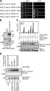
Cdk1 regulates kinase activity of Dbf2–Mob1. (A) Log-phase cells with the indicated phenotypes were serially diluted 10-fold and spotted onto YPD plates. Plates were incubated for 2 d at 23 or 37°C. (B) Active Dbf2–Mob1 complex was incubated without substrate (lane 1), GST (lane 2), and GST-C-Cdc14 (lane 3) in the presence of γ-[32P]ATP. Shown is an autoradiography. (C) Cdk1–Clb2 kinase inhibits the activation of Dbf2–Mob1 kinase by Cdc15 in vitro. GST-Mob1 in a complex with Dbf2 was incubated with Cdk1–Clb2 or Cdc15 in the first and second kinase reaction as indicated in the figure. After the second reaction, Mob1-Dbf2 kinase assays with GST-C-Cdc14 as substrate and anti-Mob1 immunoblots were performed. The top graph shows the specific Dbf2–Mob1 kinase activity. Shown is the outcome of one out of two independent experiments. Both results were identical. (D) Gal1-CLB2-ΔDB cells were arrested with α-factor in G1 phase in YPAR and released into a synchronized cell cycle at 30°C in YPAR. After ∼60 min, galactose (2%) was added. Cells in anaphase were used for immunoprecipitation of TAP-Dbf2 followed by kinase assays using GST-C-Cdc14 as substrate. Phosphorylation was determined by autoradiography and normalized to immunoprecipitated TAP-Dbf2 (immunoblot anti-TAP). Dbf2–Mob1 kinase activity is shown as mean ± SD of three experiments with the activity of wild-type cells set to 1.
Figure 7.
Figure 7.
Cdc14 dephosphorylates Mob1. (A) Cdc14 dephosphorylates Mob1 in vitro. Gal1-CDC20 MOB1-6HA cells were arrested in metaphase through depletion of Cdc20. Immunoprecipitated Mob1-6HA was incubated without any additional protein (lane 1), with GST-Cdc14 (lane 2), or GST-Cdc14-C283S (lane 3) for 1 h at 30°C. Shown is an immunoblot with anti-HA and anti-Cdc14 antibodies. (B) Overexpressed Cdc14 dephosphorylates Mob1 in metaphase. α-Factor–synchronized MOB1-6HA cdc26Δ pMET3-CDC20 (lane 1–3), MOB1-6HA cdc26Δ pMET3-CDC20 Gal1-CDC14 (lane 4–6), and MOB1-6HA cdc26Δ pMET3-CDC20 Gal1-CDC14C283S cells (lane 7–9) were arrested in metaphase through Cdc20 depletion. Cells were shifted to 37°C for 1 h to inactivate the APC. Galactose was then added to induce the Gal1 promoter (t = 0). The percentage of metaphase cells was determined by DAPI staining for each time point. Samples were analyzed with the indicated antibodies. (C) Dephosphorylation of Mob1 is dependent on CDC14. CDC14 MOB1-6HA and td-cdc14 MOB1-6HA cells were synchronized with α-factor at 23°C and released from the G1 block at 37°C. Samples were withdrawn as indicated and analyzed for Mob1 phosphorylation, Clb2, and Sic1 protein levels. A crossreacting band of the anti-Clb2 antibody was used as loading control (LC).
Figure 8.
Figure 8.
bub2Δ CDC15-7A MOB1-2A cells accumulate phenotypes that are consistent with premature mitotic exit and cytokinesis defects. (A) bub2Δ CDC15-7A MOB1-2A cells show enhanced growth defects at elevated temperatures. Serial dilutions of cells with the indicated phenotypes were spotted onto YPD plates. Plates were incubated for 2–3 d at the indicated temperature. (B and C) Defects of bub2Δ CDC15-7A and bub2Δ CDC15-7A MOB1-2A cells. Cells were cultured in YPAD medium at 23°C and analyzed by fluorescence microscopy for Cdc14-GFP and mCherry-Tub1 localization. Bar, 2 µm. (C) Quantification of B. Cells were classified as indicated in the figure (n > 100 cells per strain).
Figure 9.
Figure 9.
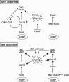
Model for the function of Cdk1 at the mSPB. In early anaphase Cdc14 becomes released from the nucleolus by the FEAR pathway (Stegmeier et al., 2002). Cdc15 and Mob1 become partially dephosphorylated (dashed lines). In addition, Tem1 binds to the mSPB (Molk et al., 2004) and recruits nonphosphorylated Cdc15 to this SPB. Cdc15 directs Cdk1 and Dbf2–Mob1 kinases to the mSPB. We propose that the close vicinity of the proteins at the mSPB leads to phosphorylation of Mob1 and Cdc15 by Cdk1. Cdk1-phosphorylated Cdc15 dissociates from the mSPB restricting Cdk1 at the mSPB (symbolized as inhibition of Cdc15). Phosphorylation of Mob1 by Cdk1 leads to a decrease in Dbf2–Mob1 kinase activity. These events restrict full activation of the MEN at the mSPB. At the dSPB, the Bfa1–Bub2 complex inhibits activation of Tem1 (Bardin et al., 2000; Pereira et al., 2000). In late anaphase the increase in Cdc14 activity and the decrease in Cdk1 activity disrupt the regulation loop between Cdk1 and Cdc15 at the mSPB (thick dashed lines symbolize complete dephosphorylation by Cdc14). This together with the phosphorylation of Bfa1 by Cdc5 polo-like kinase at the dSPB (Hu et al., 2001; Geymonat et al., 2003; Maekawa et al., 2007) allows full activation of the MEN. Dbf2–Mob1 kinase then phosphorylates Cdc14 (Mohl et al., 2009).

Similar articles

Cited by

References

    1. Asakawa K., Yoshida S., Otake F., Toh-e A. 2001. A novel functional domain of Cdc15 kinase is required for its interaction with Tem1 GTPase in Saccharomyces cerevisiae. Genetics. 157:1437–1450 - PMC - PubMed
    1. Azzam R., Chen S.L., Shou W., Mah A.S., Alexandru G., Nasmyth K., Annan R.S., Carr S.A., Deshaies R.J. 2004. Phosphorylation by cyclin B-Cdk underlies release of mitotic exit activator Cdc14 from the nucleolus. Science. 305:516–519 10.1126/science.1099402 - DOI - PubMed
    1. Bailly E., Cabantous S., Sondaz D., Bernadac A., Simon M.N. 2003. Differential cellular localization among mitotic cyclins from Saccharomyces cerevisiae: a new role for the axial budding protein Bud3 in targeting Clb2 to the mother-bud neck. J. Cell Sci. 116:4119–4130 10.1242/jcs.00706 - DOI - PubMed
    1. Bardin A.J., Visintin R., Amon A. 2000. A mechanism for coupling exit from mitosis to partitioning of the nucleus. Cell. 102:21–31 10.1016/S0092-8674(00)00007-6 - DOI - PubMed
    1. Caydasi A.K., Pereira G. 2009. Spindle alignment regulates the dynamic association of checkpoint proteins with yeast spindle pole bodies. Dev. Cell. 16:146–156 10.1016/j.devcel.2008.10.013 - DOI - PubMed

MeSH terms