. 2010 Mar 1;184(5):2297-304.
doi: 10.4049/jimmunol.0903065. Epub 2010 Feb 1.

Nod1 and nod2 are expressed in human and murine renal tubular epithelial cells and participate in renal ischemia reperfusion injury

Affiliations

Nod1 and nod2 are expressed in human and murine renal tubular epithelial cells and participate in renal ischemia reperfusion injury

Alana A Shigeoka et al. J Immunol. .

Abstract

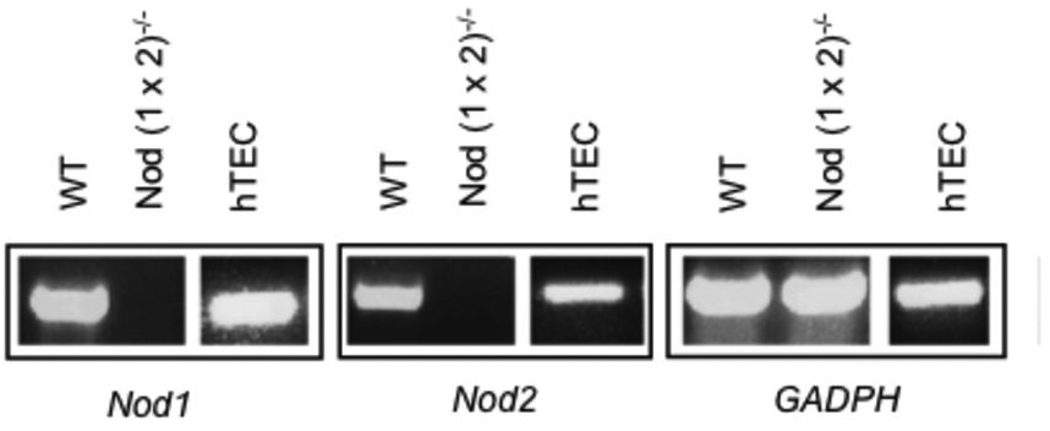
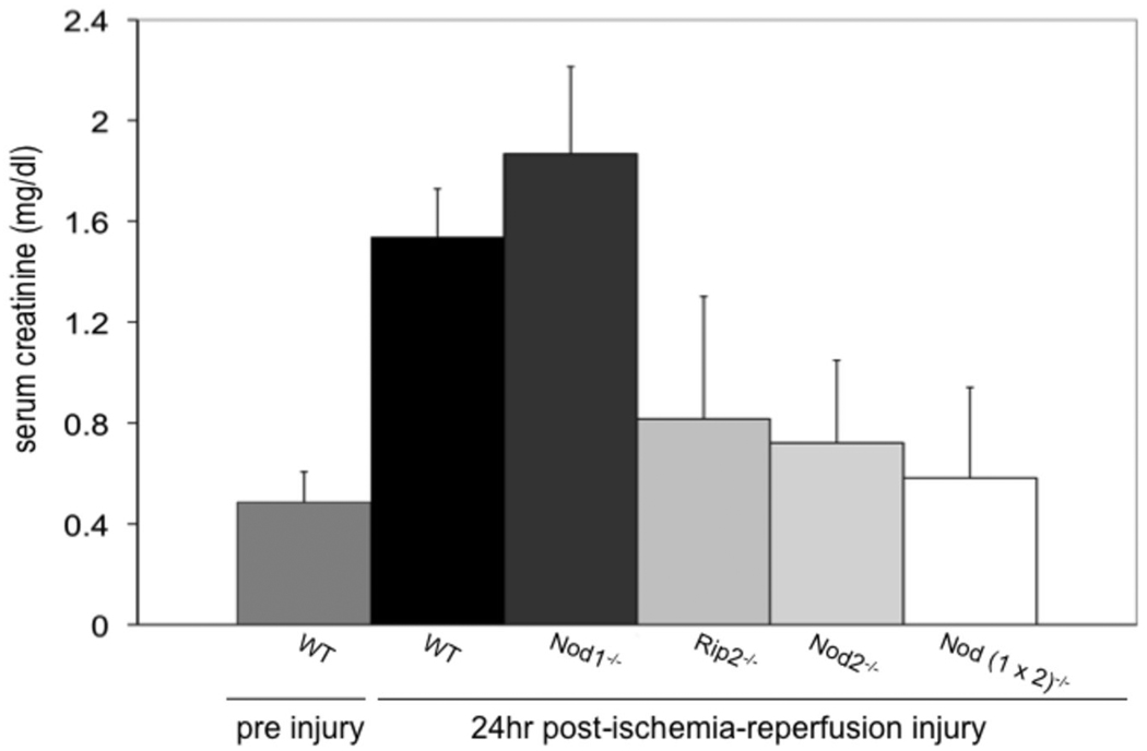
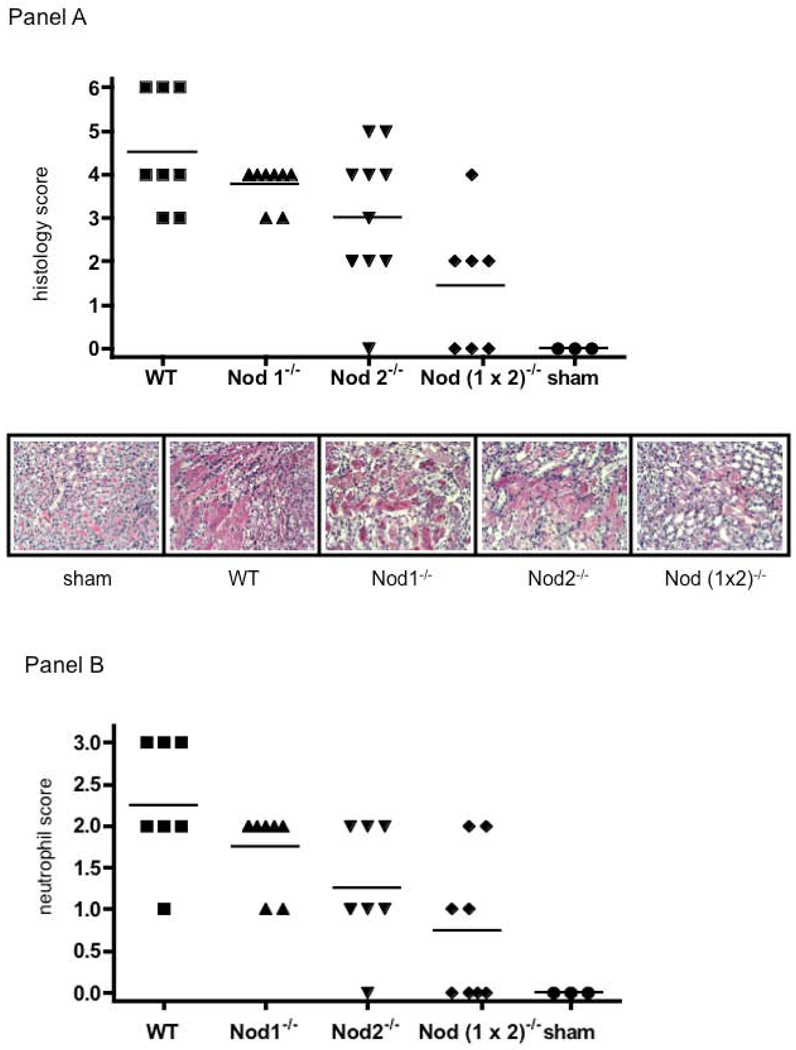
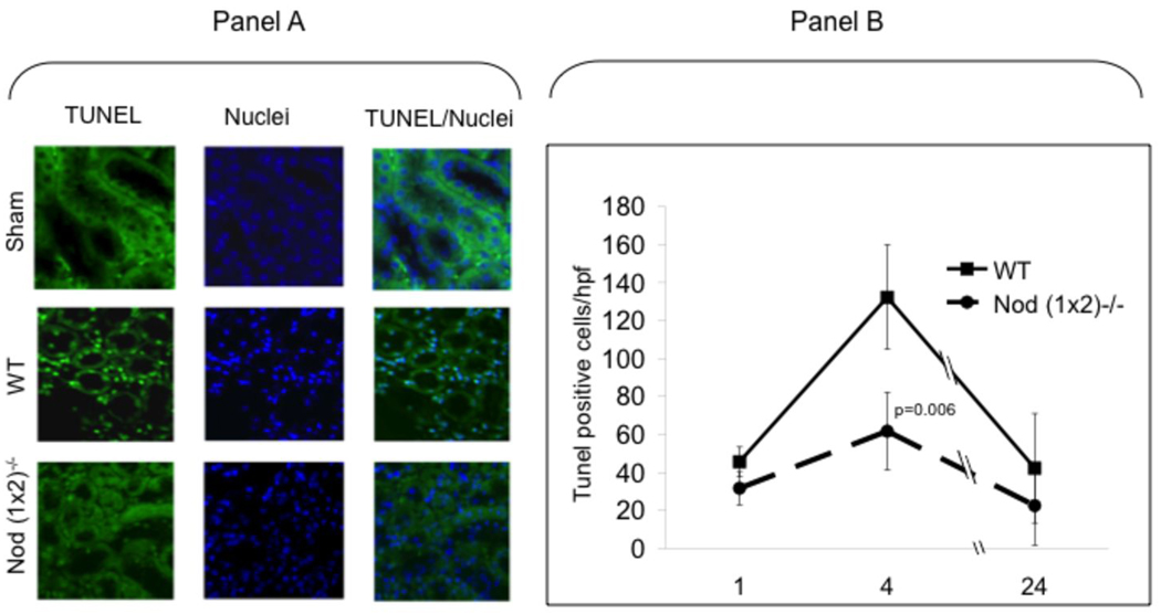
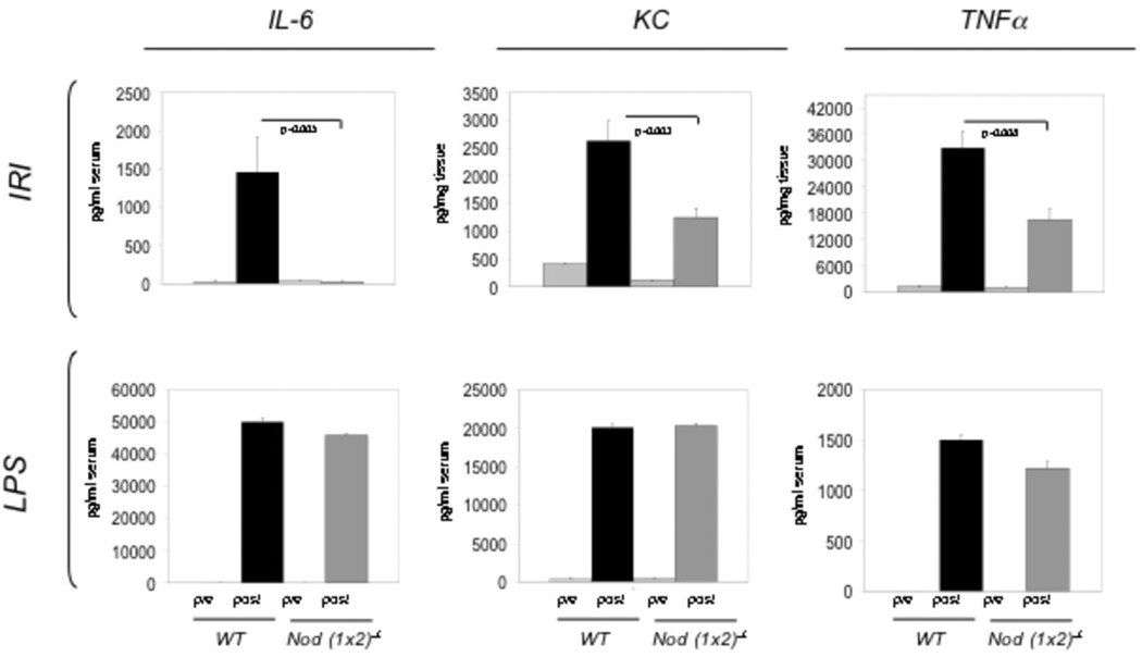
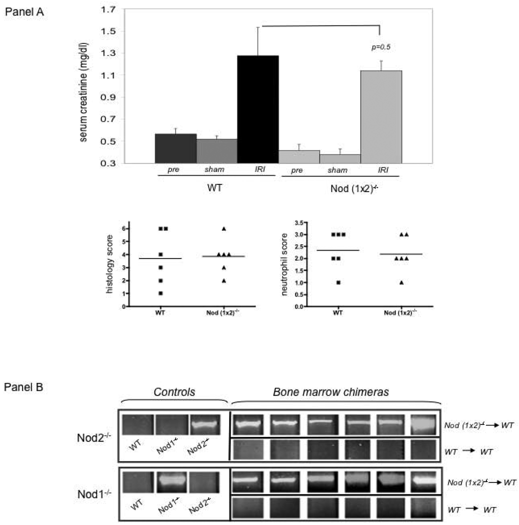
Nucleotide-binding oligomerization domain (Nod) 1 and Nod2 are members of a family of intracellular innate sensors that participate in innate immune responses to pathogens and molecules released during the course of tissue injury, including injury induced by ischemia. Ischemic injury to the kidney is characterized by renal tubular epithelial apoptosis and inflammation. Among the best studied intracellular innate immune receptors known to contribute to apoptosis and inflammation are Nod1 and Nod2. Our study compared and contrasted the effects of renal ischemia in wild-type mice and mice deficient in Nod1, Nod2, Nod(1 x 2), and in their downstream signaling molecule receptor-interacting protein 2. We found that Nod1 and Nod2 were present in renal tubular epithelial cells in both mouse and human kidneys and that the absence of these receptors in mice resulted in protection from kidney ischemia reperfusion injury. Significant protection from kidney injury was seen with a deficiency of Nod2 and receptor-interacting protein 2, and the simultaneous deficiency of Nod1 and Nod2 provided even greater protection. We conclude that the intracellular sensors Nod1 and Nod2 play an important role in the pathogenesis of acute ischemic injury of the kidney, although possibly through different mechanisms.


Figures

Figure 1
Figure 1. Nod1 and Nod2 in renal tubular epithelium of mice and humans
Figure 1 shows three representative panels depicting Nod1 and Nod2 genes as well as a housekeeping gene (GADPH) control (listed as Nod1, Nod2, GADPH below each panel). Each panel illustrates gene expression, detected by semiquantitative PCR in renal tubular epithelium of either WT (C57BL/6) mice, Nod (1×2)−/− mice or human tubular epithelial cells (hTEC) extracted from normal human kidney tissue. The results are representative of 4 mice per group and 3 different human kidneys extracted for renal tubular cells with identical results.
Figure 2
Figure 2. Kidney injury following ischemia/reperfusion injury
WT, Nod deficient and Rip2-deficient mice were subjected to 25 min ischemia/24hr reperfusion and serum obtained at 24 h to detect creatinines from WT (n=11), Nod1−/− (n=11), RIP2−/−, (n=6) Nod2−/− (n=11) and Nod (1 × 2) −/− mice (n=11). Pre injury creatinines were obtained 2 weeks earlier for each group of mice (WT pre injury values are shown in the figure). WT pre injury creatinines did not differ significantly from Nod1−/− (0.3±0.032), for Rip2−/− (0.5±0.059), Nod2−/− (0.35±0.12) and Nod (1×2)−/− (0.43±0.057). Shams had no significant change from baseline pre injury levels (data not shown). Error bars represent SDs of creatinines and statistical significance was determined with two-tailed Student’s t test.
Figure 3
Figure 3
Panel A. Histological evaluation of renal injury after bilateral renal artery clamping. Panel A shows blinded histological scoring of renal injury from WT (n=8), Nod1−/− (n=8), Nod2−/− (n=10-, Nod (1×2)−/− (n=7), and sham (n=3), mice that were subjected to 25min ischemia/24hr reperfusion. The top panel shows the blinded histologic score (see materials and methods for details of scoring) and the bottom panel shows a representative micrograph of each group of mice (PAS, 200×). Means of histological score are as follows; WT (m=4.5); Nod1−/− (m= 3.8, p=0.14); Nod2−/− (m=3.0, p=0.17); Nod (1 × 2)−/− (m=1.4, p=0.04); Sham (m=0). Panel B. Neutrophil infiltration into kidneys of WT versus Nod deficient mice after bilateral renal artery clamping. This figure shows neutrophil infiltration following 25min ischemia/24hr reperfusion in WT (n=8), Nod1−/− (n=8, p=00.11), Nod2−/− (n=8, p=0.013), Nod (1×2)−/− (n=8, p=0.002) and sham (n=3) mice. The top panel shows the blinded score (see materials and methods for details of scoring) of neutrophil infiltration. Statistical significance was determined with a two-tailed Student's t-test.
Figure 4
Figure 4. Renal apoptosis in IR injured kidneys
WT versus Nod (1×2)−/− mice were subjected to ischemia (25min bilateral renal artery clamping) and reperfusion (1hr, 4hr, 24hr). After indicated times of reperfusion, the mice were sacrificed, kidneys removed, fixed and paraffin-embedded. After deparaffinization, the TUNEL assay was performed and images viewed using fluorescent microscopy. Panel A shows representative images of TUNEL and DAPI staining 4hrs after reperfusion. Panel B shows results of blinded counting of TUNEL-positive cells in nine random, non-overlapping, sections (n=4 mice per group). Data are expressed as means ± SD. Statistical significance was determined using a Student’s two-tailed T test.
Figure 5
Figure 5. Cytokine and chemokine expression in WT vs. Nod (1×2)−/− mice
The top row (labeled IRI) shows peak levels of the indicated cytokines/chemokines (24hrs post IRI). IL-6 levels were detected in serum, whereas KC and TNFα were detected in tissue homogenates. The bottom row (labeled LPS) shows peak levels of the indicated cytokines/chemokines after i.p. injection with LPS (10mg/kg). Peak levels of serum IL-6 and KC were noted at 3hrs post-LPS injection, whereas the serum peak for TNFa was seen at 6 hrs post injection. The pre bars represent unmanipulated controls whereas the post bars represent after either IRI or LPS injection. Statistical significance was determined using a Student’s two-tailed T test. The data represent six mice per group and error bars indicate SDs.
Figure 6
Figure 6. Chimeric mice subjected to ischemia/reperfusion injury
WT mice were irradiated, injected with either WT (n=6) or Nod (1 ×2)−/− (n=6) bone marrow 6 weeks earlier and, after engraftment, subjected to 25min ischemia/24 h reperfusion. Panel A shows serum creatinines of mice treated with 25min ischemia/24hr reperfusion as well as blinded histological scores of renal injury and neutrophil infiltration. Pre creatinine values were obtained 2 weeks prior to injury. Sham creatinines were obtained after laparatomy, without IRI. Six mice were treated per group. Error bars represent SDs. Statistical significance was determined by Student T test. Panel B confirms chimeric bone marrow was indeed engrafted in the indicated experimental groups. The top panels show the Nod2 mutant allele (indicating the Nod2−/−) was detected in Nod2−/− control mice, but not in Wt or Nod1−/− control mice. As shown in the WT mice transplanted with Nod (1×2)−/− bone marrow (titled Bone Marrow Chimeras), the Nod2 mutation was highly expressed. The bottom panels show the Nod1 mutant allele is detected in Nod1−/− mice, but not in WT or Nod2−/− mice. The Nod1−/− mutation is also expressed in the WT mice transplanted with the Nod (1×2) −/− bone marrow (Bone Marrow Chimeras).

References

    1. Sansonetti PJ. The innate signaling of dangers and the dangers of innate signaling. Nat Immunol. 2006;7:1237–1242. - PubMed
    1. Franchi L, Warner N, Viani K, Nunez G. Function of Nod-like receptors in microbial recognition and host defense. Immunol Rev. 2009;227:106–128. - PMC - PubMed
    1. Kreig A, Correa RG, Garrison JB, Le Negrate G, Welsh K, Huan Z, Knoefel WT, Reed JC. XIAP mediates NOD signaling via interaction with RIP2. PNAS e pub. 2009 - PMC - PubMed
    1. Shigeoka AA, Holscher TD, King AJ, Hall FW, Kiosses WB, Tobias PS, Mackman N, McKay DB. TLR2 is constitutively expressed within the kidney and participates in ischemic renal injury through both MyD88-dependent and -independent pathways. J Immunol. 2007;178:6252–6258. - PubMed
    1. Zhai Y, Shen XD, O'Connell R, Gao F, Lassman C, Busuttil RW, Cheng G, Kupiec-Weglinski JW. Cutting edge: TLR4 activation mediates liver ischemia/reperfusion inflammatory response via IFN regulatory factor 3-dependent MyD88-independent pathway. J Immunol. 2004;173:7115–7119. - PubMed

Publication types

MeSH terms

LinkOut - more resources