

Abnormalities of visual processing and frontostriatal systems in body dysmorphic disorder
- PMID: 20124119
- PMCID: PMC2853756
- DOI: 10.1001/archgenpsychiatry.2009.190
Abnormalities of visual processing and frontostriatal systems in body dysmorphic disorder
Abstract
Context: Body dysmorphic disorder (BDD) is a psychiatric disorder in which individuals are preoccupied with perceived defects in their appearance, often related to their face. Little is known about its pathophysiology, although early research provides evidence of abnormal visual processing.
Objective: To determine whether patients with BDD have abnormal patterns of brain activation when visually processing their own face with high, low, or normal spatial resolution.
Design: Case-control study.
Setting: A university hospital.
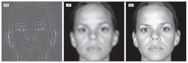
Participants: Seventeen right-handed medication-free subjects with BDD and 16 matched healthy control subjects. Intervention Functional magnetic resonance imaging while viewing photographs of face stimuli. Stimuli were neutral-expression photographs of the patient's own face and a familiar face (control stimuli) that were unaltered, altered to include only high spatial frequency (fine spatial resolution), or altered to include only low spatial frequency (low spatial resolution).
Main outcome measure: Blood oxygen level-dependent signal changes in the BDD and control groups during each stimulus type.
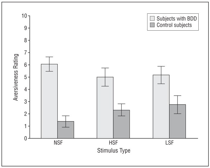
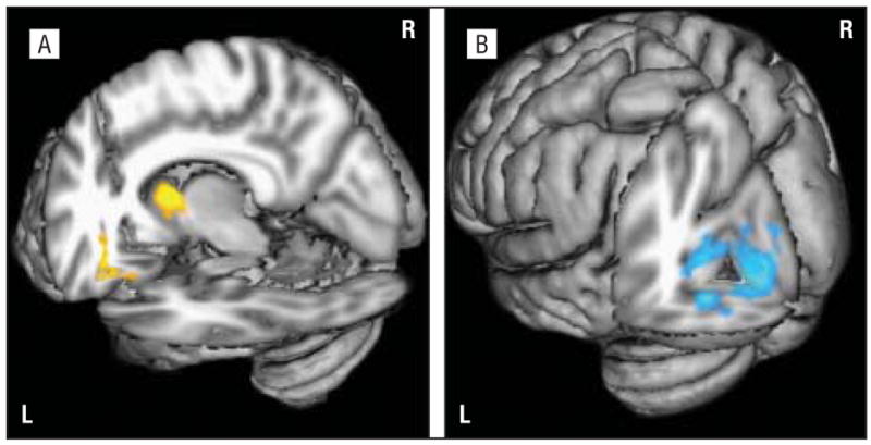
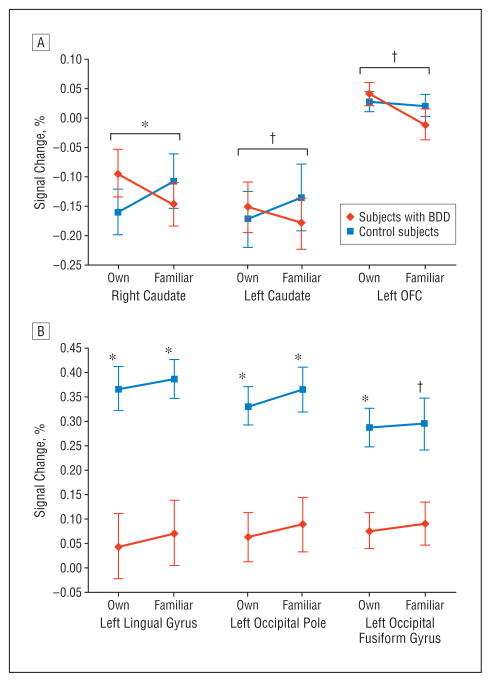
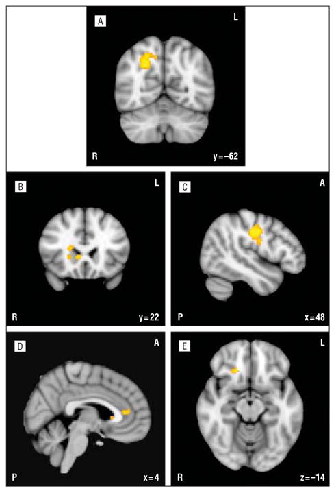
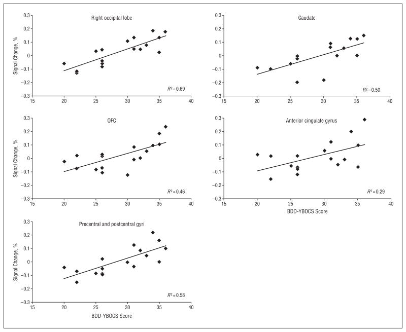
Results: Subjects with BDD showed relative hyperactivity in the left orbitofrontal cortex and bilateral head of the caudate for the unaltered own-face vs familiar-face condition. They showed relative hypoactivity in the left occipital cortex for the low spatial frequency faces. Differences in activity in frontostriatal systems but not visual cortex covaried with aversiveness ratings of the faces. Severity of BDD symptoms correlated with activity in frontostriatal systems and visual cortex.
Conclusions: These results suggest abnormalities in visual processing and frontostriatal systems in BDD. Hypoactivation in the occipital cortex for low spatial frequency faces may indicate either primary visual system abnormalities for configural face elements or top-down modulation of visual processing. Frontostriatal hyperactivity may be associated both with aversion and with symptoms of obsessive thoughts and compulsive behaviors.
Figures
Similar articles
-
Abnormalities of object visual processing in body dysmorphic disorder.Psychol Med. 2011 Nov;41(11):2385-97. doi: 10.1017/S0033291711000572. Epub 2011 Apr 18. Psychol Med. 2011. PMID: 21557897 Free PMC article.
-
Anorexia nervosa and body dysmorphic disorder are associated with abnormalities in processing visual information.Psychol Med. 2015 Jul;45(10):2111-22. doi: 10.1017/S0033291715000045. Epub 2015 Feb 5. Psychol Med. 2015. PMID: 25652023 Free PMC article.
-
Visual information processing of faces in body dysmorphic disorder.Arch Gen Psychiatry. 2007 Dec;64(12):1417-25. doi: 10.1001/archpsyc.64.12.1417. Arch Gen Psychiatry. 2007. PMID: 18056550
-
Visual processing in anorexia nervosa and body dysmorphic disorder: similarities, differences, and future research directions.J Psychiatr Res. 2013 Oct;47(10):1483-91. doi: 10.1016/j.jpsychires.2013.06.003. Epub 2013 Jun 27. J Psychiatr Res. 2013. PMID: 23810196 Free PMC article. Review.
-
A hierarchy of visual processing deficits in body dysmorphic disorder: a conceptual review and empirical investigation.Cogn Neuropsychiatry. 2024 Mar;29(2):116-140. doi: 10.1080/13546805.2024.2326243. Epub 2024 Apr 2. Cogn Neuropsychiatry. 2024. PMID: 38563811 Review.
Cited by
-
Abnormalities of object visual processing in body dysmorphic disorder.Psychol Med. 2011 Nov;41(11):2385-97. doi: 10.1017/S0033291711000572. Epub 2011 Apr 18. Psychol Med. 2011. PMID: 21557897 Free PMC article.
-
Recognizing Body Dysmorphic Disorder (Dysmorphophobia).J Cutan Aesthet Surg. 2015 Jul-Sep;8(3):165-8. doi: 10.4103/0974-2077.167279. J Cutan Aesthet Surg. 2015. PMID: 26644741 Free PMC article.
-
Associations of observer's gender, Body Mass Index and internalization of societal beauty ideals to visual body processing.Psychol Res. 2021 Nov;85(8):3026-3039. doi: 10.1007/s00426-020-01471-5. Epub 2021 Jan 12. Psychol Res. 2021. PMID: 33433640 Free PMC article.
-
Anorexia nervosa and body dysmorphic disorder are associated with abnormalities in processing visual information.Psychol Med. 2015 Jul;45(10):2111-22. doi: 10.1017/S0033291715000045. Epub 2015 Feb 5. Psychol Med. 2015. PMID: 25652023 Free PMC article.
-
Abnormal brain network organization in body dysmorphic disorder.Neuropsychopharmacology. 2013 May;38(6):1130-9. doi: 10.1038/npp.2013.18. Epub 2013 Jan 15. Neuropsychopharmacology. 2013. PMID: 23322186 Free PMC article.
References
-
- Otto MW, Wilhelm S, Cohen LS, Harlow BL. Prevalence of body dysmorphic disorder in a community sample of women. Am J Psychiatry. 2001;158(12):2061–2063. - PubMed
-
- Faravelli C, Salvatori S, Galassi F, Aiazzi L, Drei C, Cabras P. Epidemiology of somatoform disorders: a community survey in Florence. Soc Psychiatry Psychiatr Epidemiol. 1997;32(1):24–29. - PubMed
-
- Rief W, Buhlmann U, Wilhelm S, Borkenhagen A, Brahler E. The prevalence of body dysmorphic disorder: a population-based survey. Psychol Med. 2006;36(6):877–885. - PubMed
-
- Koran LM, Abujaoude E, Large MD, Serpe RT. The prevalence of body dysmorphic disorder in the United States adult population. CNS Spectr. 2008;13(4):316–322. - PubMed
-
- Phillips KA, Diaz SF. Gender differences in body dysmorphic disorder. J Nerv Ment Dis. 1997;185(9):570–577. - PubMed
Publication types
MeSH terms
Grants and funding
LinkOut - more resources
Full Text Sources
Other Literature Sources
