. 2010 Feb 16;121(6):792-803.
doi: 10.1161/CIRCULATIONAHA.109.900928. Epub 2010 Feb 1.

Reduction of AMP-activated protein kinase alpha2 increases endoplasmic reticulum stress and atherosclerosis in vivo

Affiliations

Reduction of AMP-activated protein kinase alpha2 increases endoplasmic reticulum stress and atherosclerosis in vivo

Yunzhou Dong et al. Circulation. .

Abstract

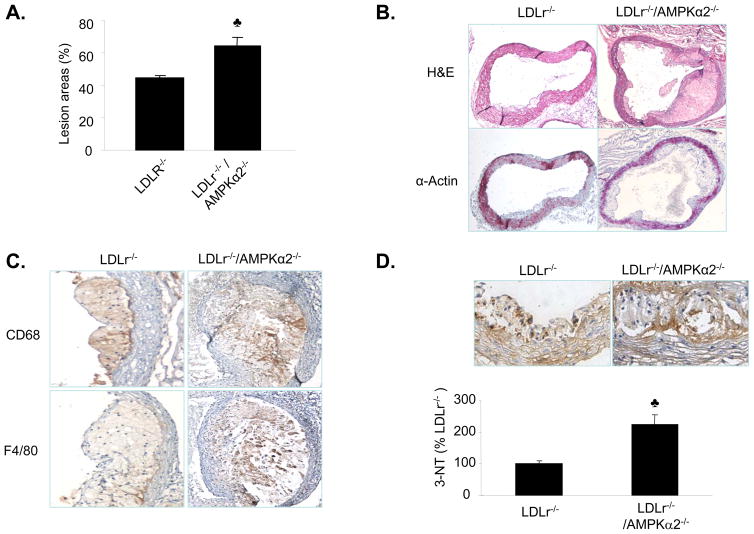
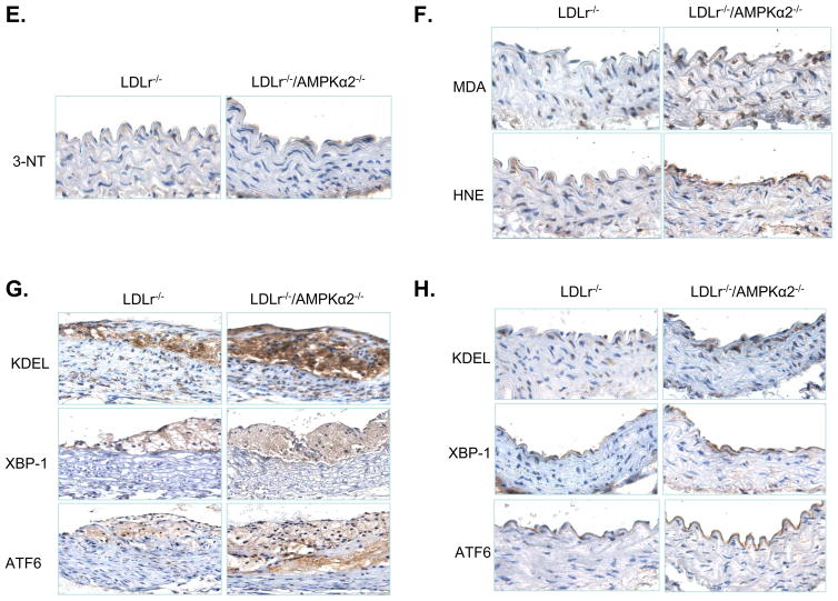
Background: Aberrant endoplasmic reticulum (ER) stress is associated with several cardiovascular diseases, including atherosclerosis. The mechanism by which aberrant ER stress develops is poorly understood. This study investigated whether dysfunction of AMP-activated protein kinase (AMPK) causes aberrant ER stress and atherosclerosis in vivo.

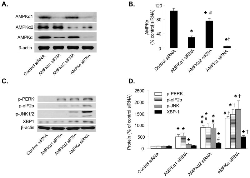
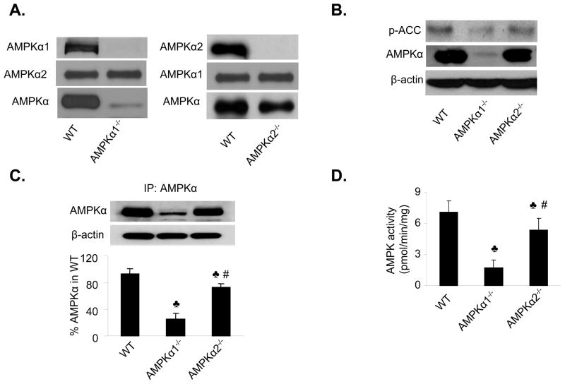
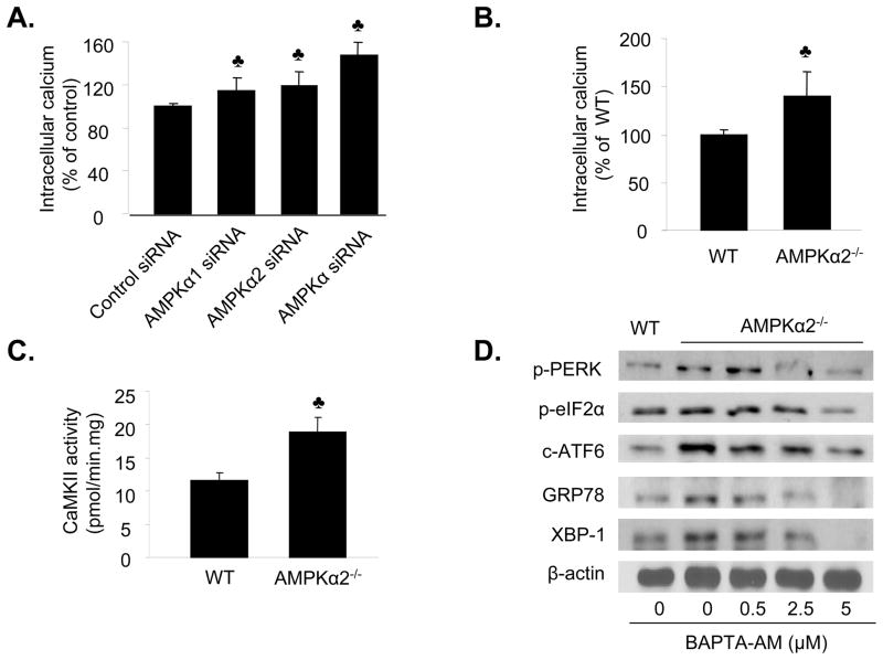
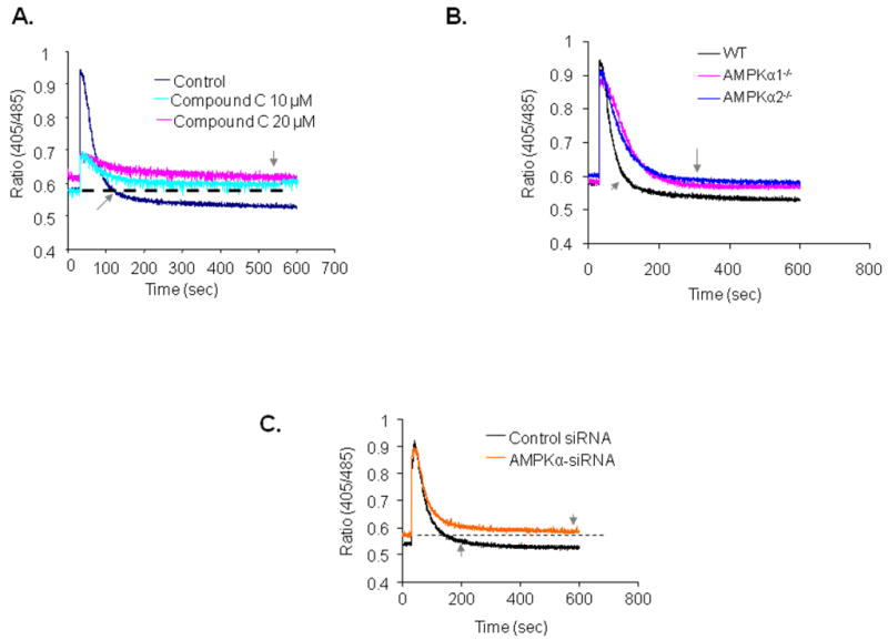
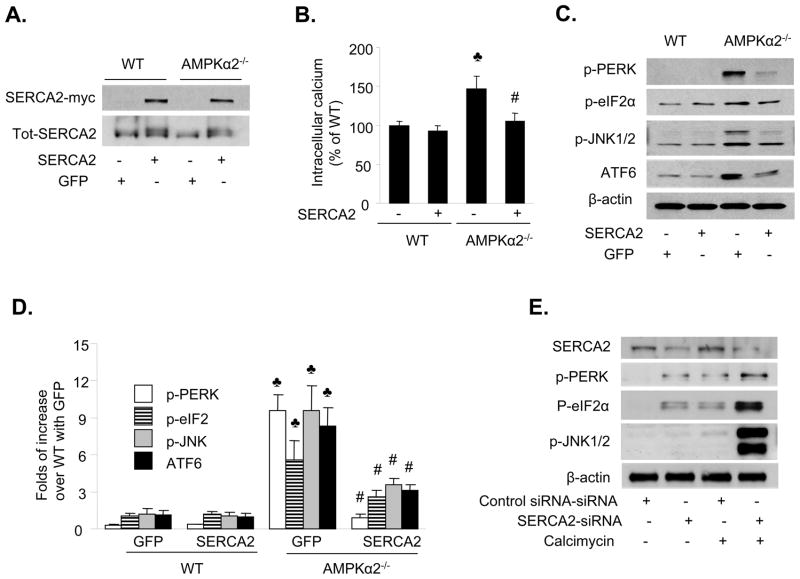
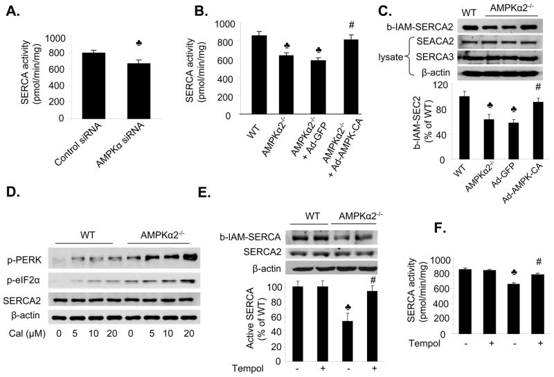
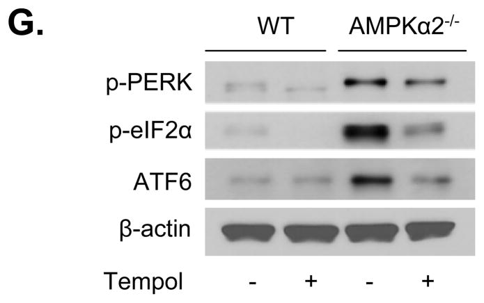
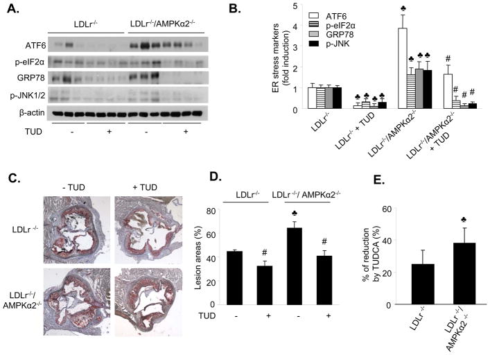
Methods and results: Human umbilical vein endothelial cells and mouse aortic endothelial cells from AMPK-deficient mice were used to assess the level of ER stress with Western blotting. Reduction of AMPKalpha2 expression significantly increased the level of ER stress in human umbilical vein endothelial cells. In addition, mouse aortic endothelial cells from AMPKalpha2 knockout (AMPKalpha2(-/-)) mice had higher expression of markers of ER stress and increased levels of intracellular Ca2+. These phenotypes were abolished by adenovirally overexpressing constitutively active AMPK mutants (Ad-AMPK-CA) or by transfecting sarcoendoplasmic reticulum calcium ATPase (SERCA). Inhibition of SERCA induced ER stress in endothelial cells. Furthermore, reduction of AMPKalpha expression suppressed SERCA activity. In addition, SERCA activity was significantly reduced concomitantly with increased oxidation of SERCA in mouse aortic endothelial cells from AMPKalpha2(-/-) mice. Both of these phenotypes were abolished by adenovirally overexpressing Ad-AMPK-CA. Furthermore, Tempol, which restored SERCA activity and decreased oxidized SERCA levels, markedly reduced the level of ER stress in mouse aortic endothelial cells from AMPKalpha2(-/-) mice. Finally, oral administration of tauroursodeoxycholic acid, a chemical chaperone that inhibits ER stress, significantly reduced both ER stress and aortic lesion development in low-density lipoprotein receptor- and AMPKalpha2-deficient mice.

Conclusions: These results suggest that AMPK functions as a physiological suppressor of ER stress by maintaining SERCA activity and intracellular Ca2+ homeostasis.


Conflict of interest statement

Conflict of Interest Disclosures

None

Figures

Figure 1
Figure 1. Reduction of AMPKα2 expression induces ER stress in endothelial cells
A&B. Western blot analysis of AMPKα2 in HUVEC. The blot is representative of at least three blots from three independent experiments; p<0.05, AMPKα1 or α2 or AMPKα siRNA vs. control siRNA, respectively; #p<0.05, AMPKα2 siRNA vs. AMPKα1 siRNA; p<0.05, AMPKα siRNA vs. AMPKα1 or α2 siRNA. n=4. C&D. Genetic inhibition of AMPKα2 induces ER stress in HUVEC. p<0.05 vs. control siRNA; #p<0.05 vs. AMPKα1 siRNA, †p<0.05 vs. AMPKα1 or AMPK α2 siRNA; n=4.
Figure 2
Figure 2. AMPKα2-dependent ER stress in isolated mouse aortic endothelial cells
A. Characterization of MAEC from WT, AMPKα1 KO, and AMPKα2 KO. AMPKα, AMPKα1, and AMPKα2 were detected by using the specific antibodies in western blots. The blot is a representative of 6 blots from 6 individual experiments. B. Effects of AMPKα1 deletion and AMPKα2 deletion on the levels of phospho-ACC at Ser79 in MAEC. The blot is a representative of three blots obtained from three individual experiments. C. Distribution of AMPKα1 and AMPKα2 in isolated of MAEC. AMPKα was first immunoprecipitated from MAEC of WT, AMPKα1, and AMPKα2 followed by western blot analysis of AMPK. The blot is a representative of four blots from four independent experiments. p<0.05 vs. WT; #p<0.05 vs. AMPKα1−/−; D. Assays of AMPK activity in MAEC from WT, AMPKα1−/− or AMPKα2−/− primary cultured endothelial cells. P<0.05 vs. WT; #p<0.05 vs. AMPKα1−/−; n=4. E&F. Increased levels of ER stress in MAEC from AMPKα1−/− or AMPKα2−/− mice. P<0.05 vs. WT; #p<0.05 vs. AMPKα1−/−; n=8. G&H. Adenoviral overexpression of Ad-AMPK-CA alleviates ER stress in MAEC from AMPKα2−/− mice. p<0.05 vs. WT; #P<0.05 vs. AMPKα2−/− or AMPKα2−/− infected with Ad-GFP; n=5.
Figure 3
Figure 3. AMPKα deletion causes a calcium-dependent ER stress response in endothelial cells
A. Genetic inhibition of AMPK increases the intracellular Ca2+ levels in HUVEC. p<0.05 vs. control siRNA; n=6. B. Elevation of intracellular Ca2+ in cultured MAEC from AMPKα2−/− mice. p<0.05 vs. WT; n=8. C. Increased activity of Ca2+–dependent CAMKII in MAEC from AMPKα2−/− mice. p<0.05 vs. WT; n=4. D. Reduction of ER stress by BAPTA in MAEC isolated from AMPKα2−/− mice. The blot is a representative of four blots from four independent experiments.
Figure 4
Figure 4. AMPK inhibition impairs ionomycin-induced Ca2+ release and return transients
After being loaded with Indo-1/AM dye, the cells were stimulated with ionomycin (10 μM) which induces Ca2+ release from the ER. The small arrow indicates the timing to return to basal Ca2+ levels. A. Effects of Compound C on ionomycin-induced Ca2+ release and intracellular Ca2+ stores. B. Defective Ca2+ release and stores in MAEC derived from AMPKα1−/− mice (red), AMPKα2−/− mice (blue) and WT mice (black), n=3. C. Genetic inhibition of AMPK impairs ionomycin-induced Ca2+ release and intracellular Ca2+ stores. HUVEC were transfected with control siRNA or AMPK-specific siRNA. The ratiometric Ca2+ dynamics were monitored 36 h after the siRNA transfections with AMPK-siRNA (orange) or control siRNA (black), n=3.
Figure 5
Figure 5. SERCA-mediated ER stress in MAEC from AMPKα2−/− mice
A. Transfection of SERCA2b into MAEC from WT and AMPKα2−/− mice. The blot is representative of four blots obtained from four independent experiments. B. Transfection of SERCA2b into MAEC from AMPKα2−/− mice normalizes intracellular Ca2+ levels. p<0.05 vs. WT; #p<0.05 vs. AMPKα2−/− alone. C&D. Overexpression of SERCA2 suppresses ER stress in MAEC from AMPKα2−/− mice. The blot is representative of three blots obtained from three independent experiments. p<0.05 vs. WT + Ad-GFP; #p<0.05 vs. AMPKα2−/− + Ad-GFP. E. Reduction of SERCA2 expression by siRNA induces ER stress in HUVEC. After the siRNA transfection, the cells were exposed to calcimycin (5 μM) for 1 h. ER stress markers were monitored using western blotting. The blot is a representative of three blots obtained from three independent experiments.
Figure 6
Figure 6. AMPKα2 deletion inhibits SERCA activity but increases its oxidation status in endothelial cells
A. Reduction of AMPK expression with AMPKα-specific siRNA decreases SERCA activity; p<0.05 vs. control siRNA; n=4. B. Adenoviral overexpression of AMPK-CA normalizes SERCA activity in MAEC from AMPKα2−/− mice. p<0.05 vs. WT, n=4; #p<0.05 vs. AMPKα2−/− +Ad-GFP; n=4; C. Adenoviral overexpression of AMPK-CA prevents the oxidation of SERCA in MAEC from AMPKα2−/− mice. ♣p<0.05 vs. WT; # p<0.05 vs. Ad-GFP; n=4. D. Increased ER stress in response to calcimycin in MAEC from AMPKα2−/− mice. MAEC from WT and AMPKα2−/− mice were exposed to calcimycin at the indicated concentrations for 2 h. Both ER stress markers and SERCA2 expression were monitored using western blotting, as described in the Materials and Methods. The blot is representative of three blots from three independent experiments. E. Anti-oxidant Tempol prevents SERCA from oxidation in MAEC from AMPKα2−/− mice. p<0.05 vs. WT; #p<0.05 vs. AMPKα2−/−; n=4. F. The antioxidant Tempol normalizes SERCA activity in MAEC from AMPKα2−/− mice. MAEC from WT and AMPKα2−/−mice were treated with Tempol (10 μM) for 16 h. SERCA activity was monitored as described in the Materials and Methods. p<0.05 vs. WT; #p<0.05 vs. AMPKα2−/−; n=4. G. Tempol supplementation reduces ER stress in MAEC from AMPKα2−/− mice. ER stress markers were monitored in MAEC from WT or AMPKα2−/− treated with Tempol (10 μM) for 16 h. The blot is representative of four blots obtained from four independent experiments;
Figure 7
Figure 7. Increased atherosclerosis in the aortic roots of LDLr−/−/AMPKα2−/− mice
A. Atherosclerotic lesion size in the aortic root in LDLr−/− and LDLr−/−/AMPKα2−/− mice, as determined by Oil Red O staining. The lesion area is expressed as a percentage of the total analyzed area of the aortic root. P<0.05 vs. LDLr−/−, n=5 or 6 for each group. B. Histological features of an atherosclerotic lesion in the aortic arch of male LDLr−/− and LDL−/−/AMPKα2−/− mice. Cross sections of the aortic arch from LDLr−/− and LDLr−/−/AMPKα2−/− mice were stained with hematoxylin and eosin (upper panel), or with an antibody against α-smooth muscle actin (bottom). C. Immunohistochemical staining for CD68 (upper) and F4/80 (low) in the aortic roots. D. Immunohistochemical staining with an antibody against 3-NT in the aortic roots. P<0.05 vs. LDLr−/−, n=5 or 6 for each group. E. Immunohistochemical staining with an antibody against 3-NT in the aortic arches. F. Immunohistochemical staining for malondialdehyde (MDA) and HNE. G. Immunohistochemical staining for ER stress markers (ATF6, KDEL and XBP-1) in the aortic roots of LDLr−/− and LDLr−/−/AMPKα2−/− mice. H. Immunohistochemical staining of ER stress markers (ATF6, KDEL and XBP-1) in the non-atherosclerotic aortic arches of LDLr−/− and LDLr−/−/AMPKα2−/− mice.
Figure 8
Figure 8. Chronic administration of TUDCA reduces ER stress and aortic lesions in mice deficient for both LDLr−/− and AMPKα2
LDLr−/− and LDLr−/−/AMPKα2−/− mice were fed a high fat diet for 10 weeks, with or without TUDCA treatment. Each group contained six to eight mice. A&B. Chronic administration of TUDCA attenuates ER stress in vivo. p<0.05 vs. LDLr−/−; #p<0.05 vs. LDLr−/−/AMPKα2−/−; C&D. Chronic administration of TUDCA suppresses aortic lesions in LDLr−/−and LDLr/AMPKα2−/− mice. p<0.05 LDLr−/−/AMPKα2−/− vs. LDLr−/−; #p<0.05 LDLr−/− vs. LDLr−/−+TUD; LDLr−/−/AMPKα2−/− vs LDLr−/−/AMPKα2−/−+TUD, respectively; n=6–8. E. Comparison of the effects of TUDCA in LDLr−/− and LDLr−/−/AMPKα2−/− mice, p<0.05 vs. LDLr−/−, n=6–8.

References

    1. Lin JH, Walter P, Yen TS. Endoplasmic reticulum stress in disease pathogenesis. Annu Rev Pathol. 2008;3:399–425. - PMC - PubMed
    1. Xu C, Bailly-Maitre B, Reed JC. Endoplasmic reticulum stress: cell life and death decisions. J Clin Invest. 2005;115:2656–2664. - PMC - PubMed
    1. Marciniak SJ, Ron D. Endoplasmic reticulum stress signaling in disease. Physiol Rev. 2006;86:1133–1149. - PubMed
    1. Ozcan U, Cao Q, Yilmaz E, Lee AH, Iwakoshi NN, Ozdelen E, Tuncman G, Gorgun C, Glimcher LH, Hotamisligil GS. Endoplasmic reticulum stress links obesity, insulin action, and type 2 diabetes. Science. 2004;306:457–461. - PubMed
    1. Wellen KE, Hotamisligil GS. Inflammation, stress, and diabetes. J Clin Invest. 2005;115:1111–1119. - PMC - PubMed

Publication types

MeSH terms

Substances