. 2010 Jul;25(7):2107-19.
doi: 10.1093/ndt/gfq006. Epub 2010 Feb 1.

Differential expression of claudin tight junction proteins in the human cortical nephron

Affiliations

Differential expression of claudin tight junction proteins in the human cortical nephron

Adam Kirk et al. Nephrol Dial Transplant. 2010 Jul.

Abstract

Background: In renal tubules, paracellular permeability is tightly controlled to facilitate solute absorption and urinary concentration and is regulated by tight junctions, which incorporate claudin proteins. There is very limited information confirming the localization of these proteins in the human renal cortex. Most data is inferred from mouse, bovine and rabbit studies and differences exist between mouse and other species.

Methods: A survey of claudin staining was performed on human kidney cortex embedded in glycolmethacrylate resin to enhance tissue morphology and facilitate the cutting of 2 microm serial sections.

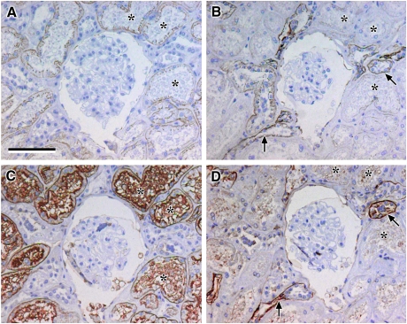
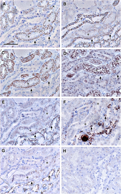
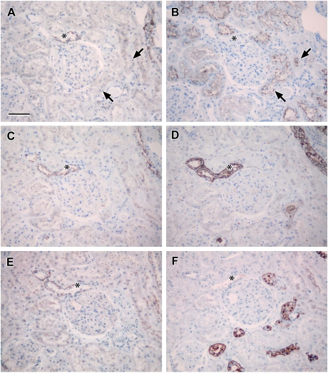
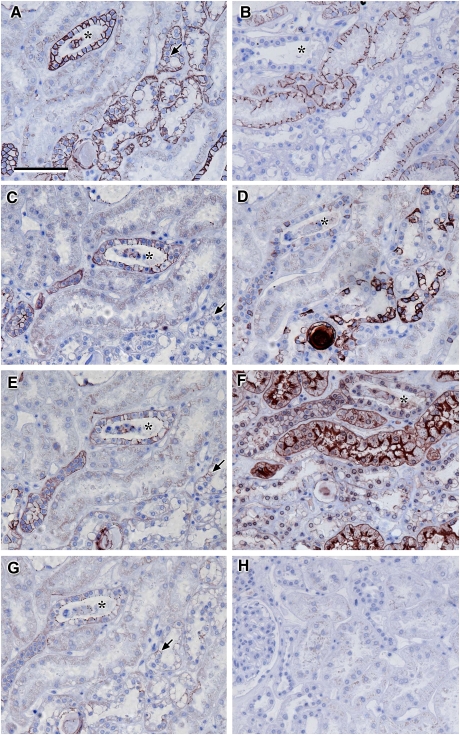
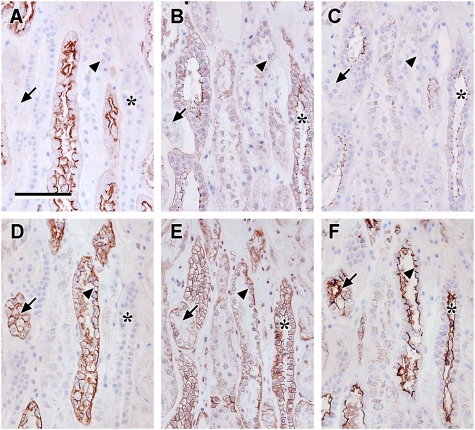
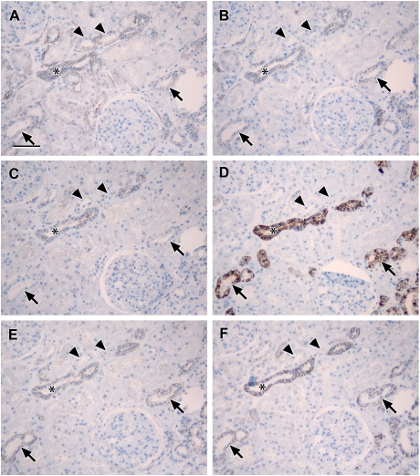
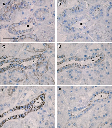
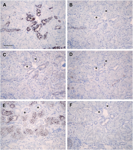
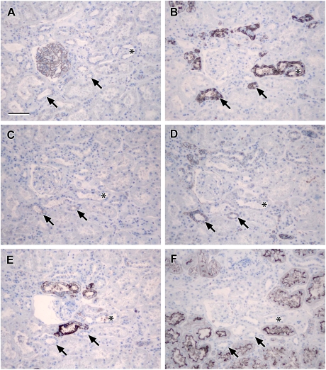
Results: Claudin-2, -10 and -11 antibodies labelled renal tubular epithelial cells, correlating with Lotus tetragonolobus and N-cadherin positive proximal tubules. Claudin-3, -10, -11 and -16 antibodies strongly stained a population of tubules that were positive for Tamm Horsfall protein on adjacent sections, confirming expression in the thick ascending limb of the Loop of Henle. Claudin-3, -4 and -8 antibodies reacted with tubules that correlated with the distal nephron markers, E-cadherin, epithelial membrane antigen and Dolichos biflorus and claudin-3, -4, -7 and -8 with the distal tubule marker, calbindin, and the collecting duct marker, aquaporin-2. Claudin-14 was localized in distal convoluted tubules, correlating positively with calbindin but negatively with aquaporin-2, whereas claudin-1 staining was identified in the parietal epithelium of Bowman's capsule, distal convoluted tubule and collecting duct. Cellular and tight junction localization of claudin staining in renal tubules was heterogeneous and is discussed.

Conclusions: Complex variation in the expression of human claudins likely determines paracellular permeability in the kidney. Altered claudin expression may influence pathologies involving abnormalities of absorption.


Figures

Fig. 1
Fig. 1
Localization of N- and E-cadherin: photomicrographs showing serial sections of human renal cortical tissue immunohistochemically stained for N-cadherin (A), E-cadherin (B), L. tetragonolobus (C) and D. biflorus (D) [note that anatomically similar proximal structures were recognized by N-cadherin antibody and L. tetragonolobus where E-cadherin staining was weak or absent (asterisks, A–C); strong E-cadherin was seen in coincident structures that were negative for N-cadherin and L. tetragonolobus, but positive for D. biflorus (arrows, B and D), indicating strong expression in the distal nephron (TAL or DCT) or collecting duct; lectins L. tetragonolobus and D. biflorus stained in a different cellular pattern to the N- and E-cadherin antibodies, as they label carbohydrates present on the surface of the RTECs whereas the cadherins stain the lateral cell borders; scale bar 100 µm].
Fig. 2
Fig. 2
Localization of claudin-2, -10 and -11: photomicrographs showing serial sections of human renal cortical tissue stained for N-cadherin (A), E-cadherin (B), claudin-2 (C), L. tetragonolobus (D), claudin-10 (E), D. biflorus (F) and claudin-11 (G); representative negative control (H) [N-cadherin and claudin-2 stained strongly positive in L. tetragonolobus positive proximal tubules which showed faint discrete junctional staining for claudin-10 and E-cadherin, faint cytoplasmic staining for claudin-11 and negative for D. biflorus (asterisks, A–G); in addition, strongly positive claudin-10 and -11 staining coincided with strong E-cadherin staining in tubules with a subpopulation of cells intensely stained with D. biflorus suggestive of TAL (arrows); scale bar 100 µm].
Fig. 3
Fig. 3
Localization of claudin-10 and -11 compared to claudin-16, THP and calbindin: photomicrographs showing serial sections of human renal cortical tissue immunohistochemically stained for claudin-10 (A), L. tetragonolobus (B), claudin-11 (C), THP (D), claudin-16 (E) and calbindin (F) [very faint discrete claudin-10 staining was seen in junctional areas of tubules coincident with L. tetragonolobus (arrows in A and B); robust claudin-10, -11 and -16 staining (asterisks in A, C and E) was seen in tubules coincident with those stained by THP, but not L. tetragonolobus or calbindin (asterisks in B, D and F); taken together, these data indicate that claudins-10, -11 and -16 are expressed in the TAL; scale bar 100 µm].
Fig. 4
Fig. 4
Localization of claudin-3, -4 and -8: photomicrographs showing serial sections of human renal cortical tissue stained for E-cadherin (A), N-cadherin (B), claudin-8 (C), D. biflorus (D), claudin-3 (E), L. tetragonolobus (F) and claudin-4 (G); representative negative control (H) [discrete tight junction-type claudin-3, -4 and -8 staining coincided with strong positive E-cadherin staining (asterisks, A–G), suggesting a distal nephron or collecting duct distribution; note that the staining was in anatomically different tubules to N-cadherin (B) and L. tetragonolobus (F) positive tubules; claudin-3 also appeared to stain tubules that were coincident with strong E-cadherin and D. biflorus, revealing possible TAL location (arrows in A, C, E and G); scale bar 100 µm].
Fig. 5
Fig. 5
Localization of claudin-3, -4 and -8 compared to THP: photomicrographs showing serial sections of human renal cortical tissue stained for L. tetragonolobus (A), claudin-8 (B), claudin-4 (C), THP (D), claudin-3 (E) and EMA (F) [claudin-8, -4 and -3 stained tubules that were coincident with similar ones that were positive for EMA and negative for L. tetragonolobus (asterisks in A–C, E and F), confirming the expression of these claudins in the distal nephron (TAL or DCT) or collecting duct; THP staining was coincident with a subpopulation of EMA positive tubules that was positive for claudin-3 and not claudin-4 or -8 or L. tetragonolobus, indicating that claudin-3 is expressed in the TAL (arrows and arrowheads in A–F); scale bar 100 µm].
Fig. 6
Fig. 6
Localization of claudin-3, -4 -7 and -8 compared to aquaporin-2 and calbindin: photomicrographs showing serial sections of human renal cortical tissue stained for claudin-3 (A), claudin-4 (B), aquaporin-2 (C), calbindin (D), claudin-8 (E) and claudin-7 (F) [claudin-3, -4, -8 and -7 stained tubules that were coincident with similar ones that were positive for aquaporin-2 and calbindin (asterisks in A–F) but were also localized in calbindin positive, aquaporin-2 negative tubules (arrowheads in A–F); claudin-4, -7 and -8 showed similar staining patterns, whereas claudin-3 strongly stained additional tubules which were negative for claudin-4, -7, -8, aquaporin-2 and calbindin (arrows in A–F); scale bar 100 µm].
Fig. 8
Fig. 8
High-magnification images comparing claudin localization with ZO-1 and occludin: photomicrographs showing serial sections of human renal cortical tissue stained for claudin-14 (A) and ZO-1 (B) with a second sequence of serial sections showing staining of claudin-3 (C), -4 (D) and -7 (E) with occludin (F) [claudin-14 was seen in basolateral locations in tubules corresponding to those with discrete tight junction staining for ZO-1 (A and B, square symbol marks corresponding tubule); in serial sections from the same tissue sample, claudin-4 was observed to be mostly in discrete tight junction staining patterns (D), whereas claudin-3 and -7 showed strong basolateral cell membrane staining in addition to tight junction staining (C and E, asterisks mark corresponding tubule); in corresponding tubules, occludin was stained in a discrete pattern consistent with tight junction localization (F); scale bar 50 µm].
Fig. 7
Fig. 7
Localization of claudin-14: photomicrographs showing serial sections of human renal cortical tissue stained for calbindin (A), aquaporin-2 (B), claudin-14 (C), THP (D), L. tetragonolobus (E) and ZO-1 (F) [claudin-14 stained tubules that were coincident with similar ones that were positive for calbindin (asterisks in A and C) and negative for aquaporin-2, THP and L. tetragonolobus (asterisks in B, D and E), confirming the expression of claudin-14 in the distal tubule; basolateral claudin-14 staining corresponded with tubules showing discrete junctional ZO-1 staining (asterisks in C and F); scale bar 100 µm].
Fig. 9
Fig. 9
Localization of claudin-1: photomicrographs showing serial sections of human renal cortical tissue stained for ZO-1 (A), calbindin (B), aquaporin-2 (C), claudin-1 (D), THP (E) and L. tetragonolobus (F) [claudin-1-, calbindin- and aquaporin-2-stained coincident tubules that were negative for THP and L. tetragonolobus (arrowheads in B–F); there were some calbindin positive tubules that were negative for claudin-1, THP and aquaporin-2 (asterisks in B–E); claudin-1 basolateral membrane location, corresponding with tubules showing discrete junctional ZO-1 staining (arrowheads in A and D); robust ZO-1 staining was seen in glomerular epithelial cells (A); scale bar 100 µm].

Similar articles

Cited by

References

    1. Rector FC., Jr Sodium, bicarbonate, and chloride absorption by the proximal tubule. Am J Physiol. 1983;244:F461–F471. - PubMed
    1. Berry CA, Rector FC., Jr Mechanism of proximal NaCl reabsorption in the proximal tubule of the mammalian kidney. Semin Nephrol. 1991;11:86–97. - PubMed
    1. Aronson PS, Giebisch G. Mechanisms of chloride transport in the proximal tubule. Am J Physiol. 1997;273:F179–F192. - PubMed
    1. Shah M, Quigley R, Baum M. Maturation of rabbit proximal straight tubule chloride/base exchange. Am J Physiol. 1998;274:F883–F888. - PMC - PubMed
    1. Kwon O, Nelson WJ, Sibley R, et al. Backleak, tight junctions, and cell–cell adhesion in postischemic injury to the renal allograft. J Clin Invest. 1998;101:2054–2064. - PMC - PubMed

Publication types