

Adult T-cell leukemia/lymphoma development in HTLV-1-infected humanized SCID mice
- PMID: 20124219
- PMCID: PMC2852366
- DOI: 10.1182/blood-2009-10-246959
Adult T-cell leukemia/lymphoma development in HTLV-1-infected humanized SCID mice
Retraction in
-
Banerjee P, Tripp A, Lairmore MD, Crawford L, Sieburg M, Ramos JC, Harrington W Jr, Beilke MA, Feuer G. Adult T-cell leukemia/lymphoma development in HTLV-1-infected humanized SCID mice. Blood. 2010;115(13):2640-2648.Blood. 2014 Jul 10;124(2):305. doi: 10.1182/blood-2014-05-577882. Epub 2014 Jun 10. Blood. 2014. PMID: 24916510 Free PMC article. No abstract available.
Abstract
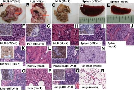
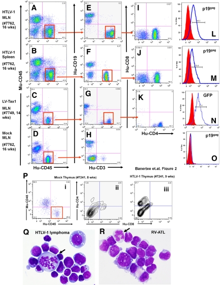
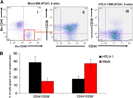
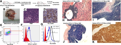
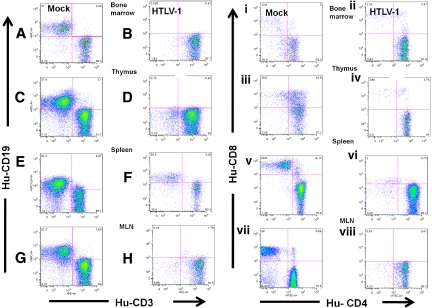
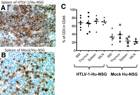
The molecular and genetic factors induced by human T-lymphotropic virus type-1 (HTLV-1) that initiate adult T-cell leukemia/lymphoma (ATLL) remain unclear, in part from the lack of an animal model that accurately recapitulates leukemogenesis. HTLV-1-infected humanized nonobese diabetic severe combined immunodeficiency (HU-NOD/SCID) mice were generated by inoculation of NOD/SCID mice with CD34(+) hematopoietic progenitor and stem cells (CD34(+) HP/HSCs) infected ex vivo with HTLV-1. HTLV-1-HU-NOD/SCID mice exclusively developed CD4(+) T-cell lymphomas with characteristics similar to ATLL and elevated proliferation of infected human stem cells (CD34(+)CD38(-)) in the bone marrow were observed in mice developing malignancies. Purified CD34(+) HP/HSCs from HTLV-1-infected patient peripheral blood mononuclear cells revealed proviral integrations suggesting viral infection of human bone marrow-derived stem cells. NOD/SCID mice reconstituted with CD34(+) HP/HSCs transduced with a lentivirus vector expressing the HTLV-1 oncoprotein (Tax1) also developed CD4(+) lymphomas. The recapitulation of a CD4(+) T-cell lymphoma in HU-NOD/SCID mice suggests that HSCs provide a viral reservoir in vivo and act as cellular targets for cell transformation in humans. This animal model of ATLL will provide an important tool for the identification of molecular and cellular events that control the initiation and progression of the lymphoma and potential therapeutic targets to block tumor development.
Figures
Similar articles
-
Human T-cell leukemia virus type 1 tax oncoprotein suppression of multilineage hematopoiesis of CD34+ cells in vitro.J Virol. 2003 Nov;77(22):12152-64. doi: 10.1128/jvi.77.22.12152-12164.2003. J Virol. 2003. PMID: 14581552 Free PMC article.
-
An animal model of adult T-cell leukemia: humanized mice with HTLV-1-specific immunity.Blood. 2014 Jan 16;123(3):346-55. doi: 10.1182/blood-2013-06-508861. Epub 2013 Nov 6. Blood. 2014. PMID: 24196073
-
Human T-cell leukemia virus infection of human hematopoietic progenitor cells: maintenance of virus infection during differentiation in vitro and in vivo.J Virol. 1996 Jun;70(6):4038-44. doi: 10.1128/JVI.70.6.4038-4044.1996. J Virol. 1996. PMID: 8648741 Free PMC article.
-
Prevention of human T-cell lymphotropic virus type 1 infection and adult T-cell leukemia/lymphoma.Recent Results Cancer Res. 2014;193:211-25. doi: 10.1007/978-3-642-38965-8_12. Recent Results Cancer Res. 2014. PMID: 24008301 Review.
-
NF-κB signaling mechanisms in HTLV-1-induced adult T-cell leukemia/lymphoma.FEBS J. 2018 Sep;285(18):3324-3336. doi: 10.1111/febs.14492. Epub 2018 May 14. FEBS J. 2018. PMID: 29722927 Free PMC article. Review.
Cited by
-
Animal models on HTLV-1 and related viruses: what did we learn?Front Microbiol. 2012 Sep 21;3:333. doi: 10.3389/fmicb.2012.00333. eCollection 2012. Front Microbiol. 2012. PMID: 23049525 Free PMC article.
-
The utility of the new generation of humanized mice to study HIV-1 infection: transmission, prevention, pathogenesis, and treatment.Retrovirology. 2011 Aug 11;8:65. doi: 10.1186/1742-4690-8-65. Retrovirology. 2011. PMID: 21835012 Free PMC article. Review.
-
Animal Models Utilized in HTLV-1 Research.Virology (Auckl). 2013 Nov 18;4:49-59. doi: 10.4137/VRT.S12140. eCollection 2013. Virology (Auckl). 2013. PMID: 25512694 Free PMC article. Review.
-
Controversies in targeted therapy of adult T cell leukemia/lymphoma: ON target or OFF target effects?Viruses. 2011 Jun;3(6):750-69. doi: 10.3390/v3060750. Epub 2011 Jun 14. Viruses. 2011. PMID: 21994752 Free PMC article. Review.
-
Humanized Rag1-/- γc-/- mice support multilineage hematopoiesis and are susceptible to HIV-1 infection via systemic and vaginal routes.PLoS One. 2011;6(6):e20169. doi: 10.1371/journal.pone.0020169. Epub 2011 Jun 14. PLoS One. 2011. PMID: 21695116 Free PMC article.
References
-
- Matsuoka M. Human T-cell leukemia virus type I and adult T-cell leukemia. Oncogene. 2003;22(33):5131–5140. - PubMed
-
- Gessain A, Mahieux R. A virus called HTLV-1. Epidemiological aspects [in French]. Presse Med. 2000;29(40):2233–2239. - PubMed
-
- Gessain A, Mahieux R. Epidemiology, origin and genetic diversity of HTLV-1 retrovirus and STLV-1 simian affiliated retrovirus [in French]. Bull Soc Pathol Exot. 2000;93(3):163–171. - PubMed
-
- Hino S. Milk-borne transmission of HTLV-I as a major route in the endemic cycle. Acta Paediatr Jpn. 1989;31(4):428–435. - PubMed
Publication types
MeSH terms
Grants and funding
LinkOut - more resources
Full Text Sources
Other Literature Sources
Molecular Biology Databases
Research Materials
Miscellaneous
