

Autophagy in unicellular eukaryotes
- PMID: 20124347
- PMCID: PMC2817228
- DOI: 10.1098/rstb.2009.0237
Autophagy in unicellular eukaryotes
Abstract
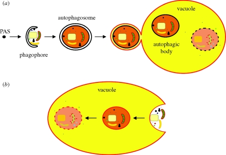
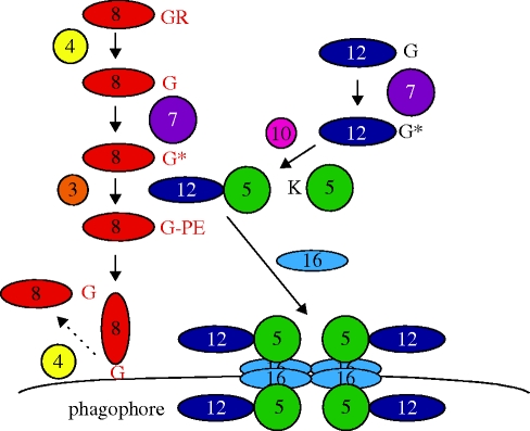
Cells need a constant supply of precursors to enable the production of macromolecules to sustain growth and survival. Unlike metazoans, unicellular eukaryotes depend exclusively on the extracellular medium for this supply. When environmental nutrients become depleted, existing cytoplasmic components will be catabolized by (macro)autophagy in order to re-use building blocks and to support ATP production. In many cases, autophagy takes care of cellular housekeeping to sustain cellular viability. Autophagy encompasses a multitude of related and often highly specific processes that are implicated in both biogenetic and catabolic processes. Recent data indicate that in some unicellular eukaryotes that undergo profound differentiation during their life cycle (e.g. kinetoplastid parasites and amoebes), autophagy is essential for the developmental change that allows the cell to adapt to a new host or form spores. This review summarizes the knowledge on the molecular mechanisms of autophagy as well as the cytoplasm-to-vacuole-targeting pathway, pexophagy, mitophagy, ER-phagy, ribophagy and piecemeal microautophagy of the nucleus, all highly selective forms of autophagy that have first been uncovered in yeast species. Additionally, a detailed analysis will be presented on the state of knowledge on autophagy in non-yeast unicellular eukaryotes with emphasis on the role of this process in differentiation.
Figures
References
-
- Alvarez V. E., Kosec G., Sant'Anna C., Turk V., Cazzulo J. J., Turk B.2008Autophagy is involved in nutritional stress response and differentiation in Trypanosoma cruzi. J. Biol. Chem. 283, 3454–3464 (doi:10.1074/jbc.M708474200) - DOI - PubMed
-
- Barrett M. P., Burchmore R. J., Stich A., Lazzari J. O., Frasch A. C., Cazzulo J. J., Krishna S.2003The trypanosomiases. The Lancet 362, 1469–1480 (doi:10.1016/S0140-6736(03)14694-6) - DOI - PubMed
-
- Bates P. A., Rogers M. E.2004New insights into the developmental biology and transmission mechanisms of Leishmania. Curr. Mol. Med. 4, 601–609 (doi:10.2174/1566524043360285) - DOI - PubMed
-
- Baxter B. K., Abeliovich H., Zhang X., Stirling A. G., Burlingame A. L., Goldfarb D. S.2005Atg19p ubiquitination and the cytoplasm to vacuole trafficking pathway in yeast. J. Biol. Chem. 280, 39 067–39 076 (doi:10.1074/jbc.M508064200) - DOI - PubMed
-
- Bellu A. R., Komori M., van der Klei I. J., Kiel J. A. K. W., Veenhuis M.2001Peroxisome biogenesis and selective degradation converge at Pex14p. J. Biol. Chem. 276, 44 570–44 574 (doi:10.1074/jbc.M107599200) - DOI - PubMed
Publication types
MeSH terms
Substances
LinkOut - more resources
Full Text Sources
Molecular Biology Databases