. 2010 Jun;9(6):1243-59.
doi: 10.1074/mcp.M900387-MCP200. Epub 2010 Feb 2.

Identification and validation of novel spinophilin-associated proteins in rodent striatum using an enhanced ex vivo shotgun proteomics approach

Affiliations

Identification and validation of novel spinophilin-associated proteins in rodent striatum using an enhanced ex vivo shotgun proteomics approach

Anthony J Baucum 2nd et al. Mol Cell Proteomics. 2010 Jun.

Abstract

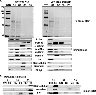
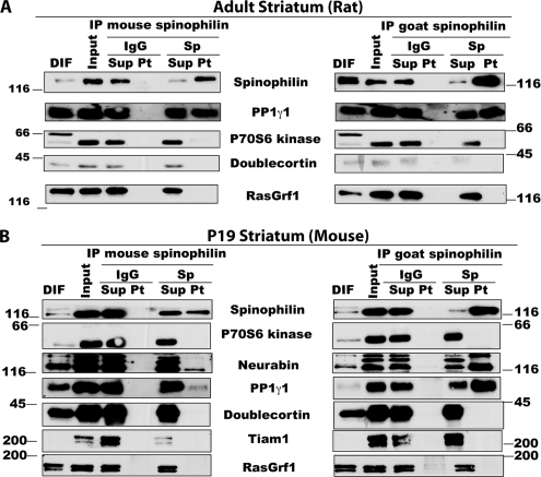
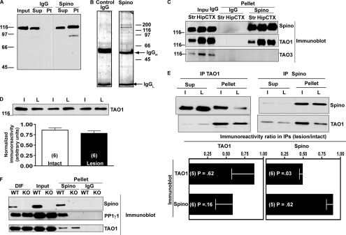
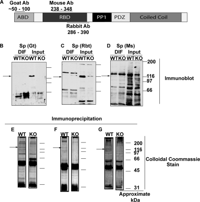
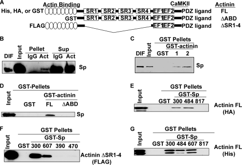
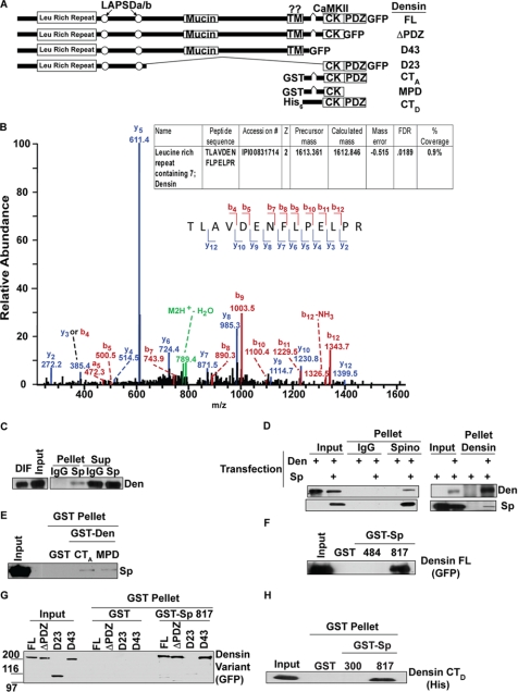
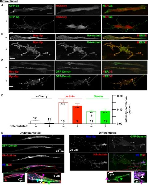
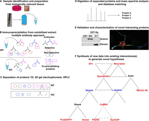
Spinophilin regulates excitatory postsynaptic function and morphology during development by virtue of its interactions with filamentous actin, protein phosphatase 1, and a plethora of additional signaling proteins. To provide insight into the roles of spinophilin in mature brain, we characterized the spinophilin interactome in subcellular fractions solubilized from adult rodent striatum by using a shotgun proteomics approach to identify proteins in spinophilin immune complexes. Initial analyses of samples generated using a mouse spinophilin antibody detected 23 proteins that were not present in an IgG control sample; however, 12 of these proteins were detected in complexes isolated from spinophilin knock-out tissue. A second screen using two different spinophilin antibodies and either knock-out or IgG controls identified a total of 125 proteins. The probability of each protein being specifically associated with spinophilin in each sample was calculated, and proteins were ranked according to a chi(2) analysis of the probabilities from analyses of multiple samples. Spinophilin and the known associated proteins neurabin and multiple isoforms of protein phosphatase 1 were specifically detected. Multiple, novel, spinophilin-associated proteins (myosin Va, calcium/calmodulin-dependent protein kinase II, neurofilament light polypeptide, postsynaptic density 95, alpha-actinin, and densin) were then shown to interact with GST fusion proteins containing fragments of spinophilin. Additional biochemical and transfected cell imaging studies showed that alpha-actinin and densin directly interact with residues 151-300 and 446-817, respectively, of spinophilin. Taken together, we have developed a multi-antibody, shotgun proteomics approach to characterize protein interactomes in native tissues, delineating the importance of knock-out tissue controls and providing novel insights into the nature and function of the spinophilin interactome in mature striatum.


Figures

Fig. 1.
Fig. 1.
Subcellular fractionation of striatal proteins. A, distribution of proteins between subcellular fractions prepared at different ionic strengths using increasingly stringent detergents. Equivalent volumes of each fraction were fractionated by SDS-PAGE and transferred to nitrocellulose membrane. The membranes were stained for total protein using Ponceau S (top) and then Western blotted for the indicated proteins (lower panels). B, association of PP1γ1 and neurabin with spinophilin immune complexes isolated from each soluble extract. Western blots of proteins present in immune complexes isolated using a goat spinophilin or a control IgG (Sp and IgG, respectively) are shown. STD, protein standard (in kDa); TH, tyrosine hydroxylase.
Fig. 2.
Fig. 2.
Characterization of previously identified SpAPs in striatum. A, immunoprecipitation of adult rat striatal spinophilin using multiple antibodies and Western blot analysis for known SpAPs. B, immunoprecipitation of P19 mouse striatal spinophilin using multiple antibodies and Western blot analysis for known SpAPs. IP, immunoprecipitate; DIF, detergent-insoluble fraction; Sup, supernatant; Pt, pellet.
Fig. 3.
Fig. 3.
Nonspecific association of TAO1 with spinophilin immune complexes isolated using mouse spinophilin antibody. A, the mouse spinophilin antibody recognizes a single, strong band and one faint band at a lower molecular weight in rat striatum and in samples immunoprecipitated using a rabbit spinophilin antibody. B, multiple proteins detected in spinophilin immune complexes isolated from rat striatum using the mouse spinophilin antibody are absent from IgG control samples. The gels were stained with colloidal Coomassie Blue. C, TAO1 is strongly detected by Western blotting immune complexes, but not in IgG controls, isolated using the mouse spinophilin antibody from rat striatum (Str), hippocampus (Hip), and cortex (CTX). TAO3, which is structurally similar to TAO1, is not detected in any spinophilin immunoprecipitates. D, total levels of TAO1 as detected by Western blot are unchanged in 6-OHDA-lesioned rats. E, apparent reciprocal co-immunoprecipitation of TAO1 and spinophilin using a mouse spinophilin antibody for immunoprecipitation and Western blotting. The apparent interaction is decreased in 6-OHDA-lesioned rats. The ratios of lesion/intact from normalized values (the mean ± standard error of the mean (error bars)) are compared with a theoretical value of 1 using column statistics. F, use of spinophilin KO striatum demonstrates that association of TAO1, but not PP1γ1, with spinophilin immune complexes isolated using the mouse antibody is nonspecific. L, lesioned; I, intact; Sup, supernatant; Pt, pellet; Spino, spinophilin; IPs, immunoprecipitates; DIF, detergent-insoluble fraction; IgGH, IgG heavy; IgGL, IgG light.
Fig. 4.
Fig. 4.
Isolation of immune complexes from WT and spinophilin KO striatum using three spinophilin antibodies. A, domain structure of spinophilin showing the approximate location of epitopes used to raise the commercially available goat, mouse, and rabbit spinophilin antibodies. B–D, each panel shows an immunoblot of a detergent-insoluble and -soluble fractions (DIF and Input, respectively) from striatum of WT and spinophilin KO mice. B, goat (Gt) antibody. C, rabbit (Rbt) antibody. D, mouse (Ms) antibody. E–G, each panel shows a colloidal Coomassie stain of the spinophilin immune complexes isolated from a Triton-soluble fraction of WT and KO striatum. E, goat (Gt) antibody. F, rabbit (Rbt) antibody. G, mouse (Ms) antibody. Ab, antibody; RBD, receptor binding domain.
Fig. 5.
Fig. 5.
Validation of putative novel SpAPs using GST-spinophilin co-sedimentation assays. A, schematic illustrating the overlapping fragments of spinophilin included in three GST-Sp fusion proteins, spanning the entire amino acid sequence. B, CaMKIIα, CaMKIIβ, myosin Va, NF-L, PSD-95, α-actinin, and densin specifically associate with different GST-Sp protein fragments. GAPDH did not precipitate with GST or with any of the GST-Sp constructs. TIF, Triton-insoluble fraction.
Fig. 6.
Fig. 6.
Characterization of interaction between spinophilin and α-actinin. A, domain structure of α-actinin and the constructs used in these experiments. (SR1–4, spectrin repeats; EF1/2, calcium-binding EF hand domains; ΔABD, deleted actin binding domain). B, spinophilin specifically co-immunoprecipitates with an α-actinin antibody compared with the IgG control (Pellets). C, spinophilin in a detergent-soluble (1% Triton X-100 and 1% deoxycholate fraction) mouse whole brain extract specifically associates with GST-α-actinin-1-FL and GST-α-actinin-2-FL in a co-sedimentation assay. D, Myc-spinophilin expressed in HEK293 cells specifically associates with GST-α-actinin-2, but not GST-α-actinin-2-ΔABD or GST, in a co-sedimentation assay. E, HA-α-actinin-2-FL expressed in HEK293 cells specifically associates with GST-Sp constructs containing residues 1–300 or 151–484, but not residues 446–817, in a co-sedimentation assay. F, FLAG-α-actinin-2-ΔSR1–4 specifically associates with GST-Sp(1–300) and GST-Sp(151–607), but not GST-Sp(286–390) or GST-Sp(427–470) (300, 607, 390, and 470, respectively), in a co-sedimentation assay. G, purified His-α-actinin-2 associates with GST-Sp(300), GST-Sp(484), and GST-Sp(607) but not GST alone or GST-Sp(817). DIF, detergent-insoluble fraction; Sup, supernatant; Act, actinin.
Fig. 7.
Fig. 7.
Characterization of interaction between spinophilin and densin. A, domain structure of densin and the constructs used for these experiments (LAPSDa/b, leucine-rich repeat and PDZ-specific domains a and b; Mucin, mucin homology domain; TM, transmembrane-like domain; CK, CaMKII binding region). B, MS/MS, collisionally induced dissociation, m/z spectra from the tryptic peptide matching densin is shown. The strong y-5 ion corresponds to the fragmentation after the proline residue. The inset table shows the IPI accession number, charge state of the peptide (z), precursor mass (in Da), calculated mass (in Da), mass error (in Da), and the percent coverage. C, densin is enriched in spinophilin immune complexes from striatum (Pellet) compared with the IgG control. D, GFP-densin-FL expressed in HEK293 cells co-precipitates with the spinophilin immune complex (Pellet) only when spinophilin is co-expressed; furthermore, Myc-spinophilin co-precipitates with the densin immune complex only when densin is co-expressed. E, Myc-spinophilin expressed in HEK293 cells specifically binds to GST-densin-CTA and GST-densin-MPD in a co-sedimentation assay. F, densin-FL expressed in HEK293 cells specifically associates with GST-Sp(817), but not GST-Sp(484) or GST, in a co-sedimentation assay. G, GST-Sp(817) specifically binds to densin-FL, densin-ΔPDZ, and densin-D43, but not densin-D23, in a co-sedimentation assay. H, bacterially expressed His-densin-CTD specifically binds to GST-Sp(817). DIF, detergent-insoluble fraction; Den, densin.
Fig. 8.
Fig. 8.
Localization of spinophilin and novel SpAPs in STHdh+/Hdh+ striatal cells. A, co-expression of GFP-spinophilin and mCherry shows spinophilin localization in cortical regions of undifferentiated cells and in the neurite-like projections of differentiated cells. Scale bars, 20 μm. B, co-expression of Myc-spinophilin and HA-α-actinin-2 reveals partial co-localization in both undifferentiated and differentiated cells. Scale bars, 20 μm. C, co-expression of Myc-spinophilin and GFP-densin reveals partial co-localization in both undifferentiated and differentiated cells. Scale bars, 20 μm. D, ICQ analysis comparing the distribution of spinophilin with the distribution of mCherry, HA-α-actinin-2, or GFP-densin in undifferentiated (−) and differentiated (+) striatal cells. ICQ values (maximum theoretical range, −0.5 to +0.5) are shown as the mean ± standard error of the mean (error bars). Significant differences from corresponding mCherry-spinophilin ICQ scores are indicated as follows: ***, p < 0.001; and **, p < 0.01. Co-localization of densin-FL with spinophilin is significantly weaker than co-localization of α-actinin-2 with spinophilin in undifferentiated cells (#, p < 0.05). E, localization of Myc-spinophilin, GFP-densin, and HA-α-actinin-2 in triple transfected undifferentiated and differentiated cells. The small panels below show magnified images of the indicated regions of these cells containing colored arrows to point out regions of the cells containing different co-localization patterns: green, GFP-densin alone; red, HA-α-actinin-2 alone; magenta, co-localization of HA-α-actinin-2 with spinophilin; cyan, co-localization of densin with spinophilin; yellow, co-localization of HA-α-actinin-2 with densin; white, co-localization of all three proteins. Scale bar, 5 or 20 μm as noted.
Fig. 9.
Fig. 9.
Methodological work flow for identification, validation, and characterization of novel SpAPs. A, homogenize a biologically relevant tissue under conditions optimized to solubilize protein(s) of interest with least disruption to protein-protein interactions. B, immunoprecipitate protein of interest using multiple antibodies from WT tissue using a KO tissue control. C, separate precipitated complexes by SDS-PAGE. D, separate enzymatic digests of proteins by HPLC for MS and MS/MS analysis and match the mass spectra to an appropriate protein database. E, validate and further characterize novel interactions using biochemical and co-localization approaches. These studies map the interaction domain and allow for development of novel tools (e.g. fragments or mutated proteins) that may be used to disrupt specific interactions in intact cells. F, the spinophilin interactome characterized in the present studies. Spinophilin directly interacts with α-actinin and densin (in blue). Both of these proteins interact with additional partners, many of which were also identified in our current study (red). Solid lines indicate known direct interactions, whereas dashed lines connect protein pairs that have been previously shown to exist in the same multiprotein complexes (, , –90).

References

    1. Kim E., Sheng M. (2004) PDZ domain proteins of synapses. Nat. Rev. Neurosci 5, 771–781 - PubMed
    1. Kennedy M. B., Beale H. C., Carlisle H. J., Washburn L. R. (2005) Integration of biochemical signalling in spines. Nat. Rev. Neurosci 6, 423–434 - PubMed
    1. Dindot S. V., Antalffy B. A., Bhattacharjee M. B., Beaudet A. L. (2008) The Angelman syndrome ubiquitin ligase localizes to the synapse and nucleus, and maternal deficiency results in abnormal dendritic spine morphology. Hum. Mol. Genet 17, 111–118 - PubMed
    1. Ingham C. A., Hood S. H., Arbuthnott G. W. (1989) Spine density on neostriatal neurones changes with 6-hydroxydopamine lesions and with age. Brain Res 503, 334–338 - PubMed
    1. Ingham C. A., Hood S. H., van Maldegem B., Weenink A., Arbuthnott G. W. (1993) Morphological changes in the rat neostriatum after unilateral 6-hydroxydopamine injections into the nigrostriatal pathway. Exp. Brain Res 93, 17–27 - PubMed

Publication types

MeSH terms