. 2010 Mar 1;123(Pt 5):660-70.
doi: 10.1242/jcs.057984. Epub 2010 Feb 2.

Human Timeless and Tipin stabilize replication forks and facilitate sister-chromatid cohesion

Affiliations

Human Timeless and Tipin stabilize replication forks and facilitate sister-chromatid cohesion

Adam R Leman et al. J Cell Sci. .

Abstract

The Timeless-Tipin protein complex has been reported to be important for replication checkpoint and normal DNA replication processes. However, the precise mechanisms by which Timeless-Tipin preserves genomic integrity are largely unclear. Here, we describe the roles of Timeless-Tipin in replication fork stabilization and sister chromatid cohesion. We show in human cells that Timeless is recruited to replication origin regions and dissociate from them as replication proceeds. Cdc45, which is known to be required for replication fork progression, shows similar patterns of origin association to those of Timeless. Depletion of Timeless-Tipin causes chromosome fragmentation and defects in damage repair in response to fork collapse, suggesting that it is required for replication fork maintenance under stress. We also demonstrate that depletion of Timeless-Tipin impairs sister chromatid cohesion and causes a defect in mitotic progression. Consistently, Timeless-Tipin co-purifies with cohesin subunits and is required for their stable association with chromatin during S phase. Timeless associates with the cohesion-promoting DNA helicase ChlR1, which, when overexpressed, partially alleviates the cohesion defect of cells depleted of Timeless-Tipin. These results suggest that Timeless-Tipin functions as a replication fork stabilizer that couples DNA replication with sister chromatid cohesion established at replication forks.


Figures
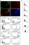
Fig. 1.
Fig. 1.
Timeless is recruited to replication forks. (A) HeLa cells were grown overnight on coverslips. In situ extraction of soluble proteins by Triton X-100 was performed before fixation and immunostaining of cells. Timeless and PCNA localization was determined using affinity-purified anti-timeless (Tim, Green) and anti-PCNA (PCNA, Red) antibodies. DNA was costained with DAPI (4′,6′-diamidino-2-phenylindole). The merged image of timeless and PCNA is shown (Merge). Representative images of repeat experiments are shown. (B,C) HeLa cells were synchronized in very early S phase and released into fresh medium to allow cells to progress through the cell cycle. Cells were collected at the indicated times and processed for ChIP. Precipitated DNA recovered from antibody-containing beads was used to monitor the association of timeless (B) and Cdc45 (C) at MYC (c-Myc), HBB (β-globin), MCM4 and ACTG1 (G-actin) loci. The replication origins (origin: 0 kb) and their proximal positions (11 kb for MYC; 31-kb for HBB) were analyzed at MYC and HBB loci. Percentage precipitated DNA over input DNA is shown at each time-point. Data are from at least three independent experiments, and error bars represent s.d. (D) DNA content of cells used in ChIP experiments in B determined by flow cytometry analysis.
Fig. 2.
Fig. 2.
Timeless is required for replication fork stabilization. (A) HeLa cells were transiently transfected with the indicated siRNAs (Luc, luciferase; Tim, timeless; Tip, tipin). Levels of timeless (Tim), tipin (Tip) and tubulin (Tub), 24 hours after the transfection are shown, as monitored by western blotting (WB). siRNA depletion of timeless also caused a reduction of the tipin level, and vice versa. Representative results of repeat experiments are shown. (B,C) HeLa cells were transfected with timeless siRNA, then treated with 5 mM HU for 5 hours (B) or 10 μM CPT (C) for 3 hours. Cells were washed and harvested at the indicated times. Cells were counted, and 1×10⁶ cells were embedded in an agarose plug for chromosomal DNA preparation. Chromosomal DNA in plugs was separated by PFGE and visualized using ethidium bromide staining. Downregulation of timeless induced strong accumulation of subchromosomal DNA fragments in response to both HU and CPT treatment. Quantification of DNA damage was shown as relative intensity of fragmented DNA by setting the minimum intensity (no drug in control siRNA cells) to 1. Sp and Sc indicate Genomic DNA from S. pombe and S. cerevisiae as size markers, respectively. Representative results of repeat experiments are shown.
Fig. 3.
Fig. 3.
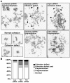
The timeless-tipin complex is involved in sister chromatid cohesion. (A) HeLa cells were transfected with luciferase (Luc), timeless (Tim) or tipin (Tip) siRNA, grown for an additional 30 hours, treated with colcemid for 90 minutes, incubated in hypotonic buffer, and fixed. Fixed cells were dropped onto a glass slide from a height of 0.75 m and stained with DAPI. Representative images of cells with fragmented chromosomes and cells with cohesion defects are shown. (B) The frequency of different phenotypes shown in A was determined and expressed as percentage of total metaphase cells. At least 100 metaphase cells were counted for each experiment. Average percentages of three independent experiments are shown.
Fig. 4.
Fig. 4.
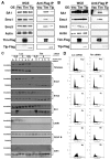
Timeless is involved in stable maintenance of cohesin subunits on chromatin. (A,B) HEK293 cells were transfected with pcDNA3-3FLAG (Vec), pcDNA3-timeless-3FLAG (Tim), or pcDNA-tipin-3FLAG (Tip) and grown for 48 hours. Cell extracts were prepared from these cells in the absence (A) or presence (B) of DNaseI. FLAG-tagged proteins were immunoprecipitated from cell extracts with anti-FLAG antibody. Immunoprecipitates were probed with the indicated antibodies to determine interacting proteins. Representative results of repeat experiments are shown. WCE, whole cell extract; anti-FLAG IP, immunoprecipitated fraction; OE, overexpressed protein. (C) HEK293 cells were transfected with the indicated siRNA, synchronized at the G1-S transition, and released into the fresh medium to allow cells to progress through the cell cycle. Cells were collected at the indicated times and fractionated into Triton-X-100-soluble and - insoluble fractions for protein analyses. Levels of the indicated proteins were determined by western blotting. Representative results of repeat experiments are shown. A, asynchronous. (D) DNA content of cells used in fractionation experiments in C was determined by flow cytometry.
Fig. 5.
Fig. 5.
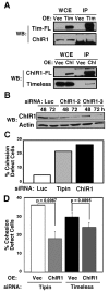
ChlR1 and timeless-tipin cooperate in establishment of sister chromatid cohesion. (A) ChlR1 associates with timeless. HEK293 cells were transfected with pcDNA3-3FLAG (Vec), pcDNA3-timeless-3FLAG (Tim) or pcDNA3-hChlR1-3FLAG (Chl) and grown for 48 hours. Immunoprecipitation (IP) was performed with the anti-FLAG antibody. Associated proteins were examined by western blotting using the indicated antibodies. Representative results of repeat experiments are shown. WCE, whole cell extract; IP, immunoprecipitated fraction; WB, protein detected by western blotting; OE, overexpressed protein. Representative results of repeat experiments are shown. (B) HeLa cells were transiently transfected with the indicated siRNAs. Cells were collected 48 or 72 hours after transfection, and levels of ChlR1 were monitored by western blotting, using antibodies against ChlR1. Luc, Luciferase. (C) Chromosome spread analysis was performed as described in Fig. 3. Cells treated with tipin siRNA and ChlR1-2 siRNA showed significant cohesion defects. (D) ChlR1 overexpression partially suppressed cohesion defects of timeless or tipin-depleted cells. HeLa cells stably overexpressing ChlR1 or vector alone (Vec) were transfected with the indicated siRNA. Cohesion was evaluated by chromosome spread analysis. At least 50 metaphase cells were counted for each experiment. Error bars correspond to the s.d. obtained from three independent experiments. OE, overexpression.
Fig. 6.
Fig. 6.
Depletion of timeless impairs mitotic progression. (A) HeLa cells transfected with timeless siRNA showed a slight reduction in the number of mitotic cells compared with cells transfected with luciferase siRNA. At least 300 cells were counted from each experiment. Error bars correspond to the s.d. obtained from three independent experiments. (B) Quantification of each mitotic stage from HeLa cells transfected with timeless siRNA or luciferase siRNA. At least 50 mitotic cells were counted from each experiment. Error bars indicate s.d. from three independent experiments. (C) BubR1 is activated in HeLa cells transfected with Timeless siRNA. Whole-cell extracts from HeLa cells treated with luciferase (Luc) siRNA, timeless (Tim) siRNA and nocadazole (Noc) were examined by western blotting using the anti-BubR1 antibody. Western blotting of actin is shown as loading control. Representative results of repeat experiments are shown.
Fig. 7.
Fig. 7.
S-phase stress-response mechanisms. The timeless-tipin fork protection complex (FPCTim-Tip) is involved in the ATR-Chk1-dependent replication checkpoint. FPCTim-Tip has checkpoint-independent functions that are important for fork protection, DNA repair, and sister chromatid cohesion. In this model, FPCTim-Tip stabilizes replication forks in a configuration that is recognized by replication checkpoint sensors. In addition, FPCTim-Tip cooperates with ChlR1 to promote proper establishment of sister chromatid cohesion.

References

    1. Aguilera A., Gomez-Gonzalez B. (2008). Genome instability: a mechanistic view of its causes and consequences. Nat. Rev. Genet. 9, 204-217 - PubMed
    1. Ansbach A. B., Noguchi C., Klansek I. W., Heidlebaugh M., Nakamura T. M., Noguchi E. (2008). RFCCtf18 and the Swi1-Swi3 complex function in separate and redundant pathways required for the stabilization of replication forks to facilitate sister chromatid cohesion in Schizosaccharomyces pombe. Mol. Biol. Cell 19, 595-607 - PMC - PubMed
    1. Aparicio T., Guillou E., Coloma J., Montoya G., Mendez J. (2009). The human GINS complex associates with Cdc45 and MCM and is essential for DNA replication. Nucleic Acids Res. 37, 2087-2095 - PMC - PubMed
    1. Barnes J. W., Tischkau S. A., Barnes J. A., Mitchell J. W., Burgoon P. W., Hickok J. R., Gillette M. U. (2003). Requirement of mammalian Timeless for circadian rhythmicity. Science 302, 439-442 - PubMed
    1. Bartek J., Lukas J. (2007). DNA damage checkpoints: from initiation to recovery or adaptation. Curr. Opin. Cell Biol. 19, 238-245 - PubMed

Publication types

MeSH terms