. 2010 Jun;24(6):1879-92.
doi: 10.1096/fj.09-144659. Epub 2010 Feb 2.

Granulin epithelin precursor: a bone morphogenic protein 2-inducible growth factor that activates Erk1/2 signaling and JunB transcription factor in chondrogenesis

Affiliations

Granulin epithelin precursor: a bone morphogenic protein 2-inducible growth factor that activates Erk1/2 signaling and JunB transcription factor in chondrogenesis

Jian Q Feng et al. FASEB J. 2010 Jun.

Abstract

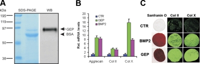
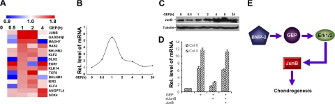
Granulin epithelin precursor (GEP) has been implicated in development, tissue regeneration, tumorigenesis, and inflammation. Herein we report that GEP stimulates chondrocyte differentiation from mesenchymal stem cells in vitro and endochondral ossification ex vivo, and GEP-knockdown mice display skeleton defects. Similar to bone morphogenic protein (BMP) 2, application of the recombinant GEP accelerates rabbit cartilage repair in vivo. GEP is a key downstream molecule of BMP2, and it is required for BMP2-mediated chondrocyte differentiation. We also show that GEP activates chondrocyte differentiation through Erk1/2 signaling and that JunB transcription factor is one of key downstream molecules of GEP in chondrocyte differentiation. Collectively, these findings reveal a novel critical role of GEP growth factor in chondrocyte differentiation and the molecular events both in vivo and in vitro.


Figures

Figure 1.
Figure 1.
Expression of GEP during chondrogenesis both in vitro and in vivo. A, B) Expressions of GEP and collagen X were examined in the course of chondrogenesis of a micromass culture of C3H10T1/2 cells. Micromass cultures of C3H10T1/2 cells were stimulated by BMP2 protein at various time points, as indicated, and the mRNA levels of GEP (A) and collagen type X (Col X; B) were assayed using real-time PCR. Units are arbitrary; the normalized values were calibrated against the d 0 time point, here given the value of 1. C) GEP immunostaining assay shows that GEP (arrows) is first detected in the late condensation stage (E12.5) and then at the differentiation stage (E13.5) and continues during entire chondrogenic developmental stages in both proliferating and hypertrophic zones (from E17.5 to 1 mo old). Scale bar = 100 μm.
Figure 2.
Figure 2.
GEP stimulates chondrogenesis in vitro. A) Characterization of recombinant GEP protein. A purified recombinant human GEP (10 ng/μl in 0.2% BSA) was either stained with Coomassie Brilliant Blue R-250 (left) or detected by Western blotting with a polyclonal anti-GEP antibody (right). B) Comparisons of GEP and BMP2 in stimulations of chondrogenesis of murine BMSCs. BMSC aggregate cultures were incubated in the absence (CTR) or presence of either 100 ng/ml BMP2 or 100 ng/ml GEP for 2 wk, followed by quantitative measurements of aggrecan, collagen II (Col II), and collagen X (Col X) using real-time PCR. Units are arbitrary; normalized values were calibrated against controls, here given the value of 1. C) Comparable potency of GEP and BMP2 in stimulation of chondrogenesis and activation of collagen II and collagen X expressions. hMSC aggregate cultures were incubated in the absence (CTR) or presence of 100 ng/ml BMP2 or GEP for 3 wk followed by Safranin O staining (left, red) or immunostains of collagen II (middle, green) or collagen X (right, green).
Figure 3.
Figure 3.
GEP stimulates chondrocyte hypertrophy, mineralization, and endochondral bone growth. A, B) Safranin O/Fast Green staining of metatarsal bones. Metatarsals from 14.5-d-old mouse embryos were cultured in the absence (CTR) or presence of 100 ng/ml GEP for 5 d and stained with Safranin O/Fast Green, shown in low-power (A) and high-power (B) microphotographs. C) Alizarin red/Alcian blue staining of metatarsals. Explants were fixed and processed for staining. A representative photograph of an explanted metatarsal is presented. D) Percentage changes in total (T) and mineralization (M) length of metatarsal bones. Percentage changes in bone length were calculated as (length at d 5 − length at d 0)/length at d 0. *P < 0.05 vs. control.
Figure 4.
Figure 4.
GEP knockdown (KD) leads to a sharp reduction of chondrogenic markers. A) Immunostaining of GEP in the growth plate of the femur from 3-wk-old wild-type (WT) (top) and conditional KD (bottom) mice crossing between siGEP transgenic mice and Sox2 Cre mice. B) Reduction in thickness of the growth plate stained with Safranin O stain in GEP KD (top) compared with control (bottom). C) Reduction of BrdU (reflection of cell proliferation) in the GEP KD proliferation zone (top) compared with control (bottom). D) Reduction of alkaline phosphatase mRNA in the GEP KD growth plate (top) compared with control (bottom). E) Sharp reduction of collagen II mRNA in the GEP KD growth plate (top) compared with control (bottom). F) Collagen X is largely undetectable in the GEP KD hypertrophic zone (top) compared with control (bottom).
Figure 5.
Figure 5.
GEP accelerates cartilage repair. AC) Articular cartilage and subchondral bone sections stained with Safranin O/Fast Green. There is a lack of cartilage repair in the trauma region of the control group that was implanted with a collagen sponge only (A; arrow). Implantations of the collagen sponge containing a recombinant BMP2 protein accelerates high-quality hyaline-appearing repair as reflected by well-integrated and organized new tissues stained positive with Safranin O (B; pink, arrow). Implantations of the collagen sponge containing the equivalent GEP display a similar hyaline-like repairing response (C; pink, arrow). D) Quantitation of tissue repair. Collagen sponge with BMP2 or GEP accelerates cartilage repair.
Figure 6.
Figure 6.
GEP is a downstream molecule of BMP2 and is required for BMP2 stimulation of chondrogenesis. A) Effects of growth factors and cytokines on GEP mRNA in chondrocytes by real-time PCR. Expression of GEP mRNA was normalized against 18S rRNA. ***P < 0.001. B) siGEP, a GEP siRNA construct, reduces 80% of endogenous GEP mRNA. C3H10T1/2 cells were transfected with either pSuper (CTR) or siGEP, and level of GEP mRNA was measured by real-time PCR. C) Suppression of GEP by siRNAs inhibits BMP2-induced chondrogenesis. Micromass cultures of C3H10T1/2 cells transfected with either pSuper (CTR) or siGEP were used to test whether BMP2 (100 ng/ml)-induced chondrogenesis is GEP dependent. Expressions of marker genes, as indicated, were determined by real-time PCR.
Figure 7.
Figure 7.
BMP2 and its mediators, Smads, activate the GEP-specific reporter genes. A) BMP2 activates GEP-specific reporter genes in RCS cells. Indicated segments from the 5′-flanking region of the GEP gene were linked to an simian virus 40 promoter and a DNA segment encoding luciferase. Numbers indicate distances in nucleotides from first nucleotide of intron 1. Indicated reporter gene and a pSVgal internal control plasmid were transfected into RCS cells in the presence or absence of 100 ng/ml BMP2 for 48 h, and β-galactosidase and luciferase activities were determined. Luciferase activities were normalized to β-galactosidase activities. B) BMP2 activates the minimal promoter of GEP gene (−275 to −51). RCS cells were transfected with either an internal deletion or a negative control reporter construct, as indicated above, and luciferase and β-galactosidase assays were performed. C) BMP2-activated SMAD4 binds to the GEP promoter (ChIP). RCS cells treated with or without 100 ng/ml BMP2 for 12 h were cross-linked by formaldehyde treatment and lysed. Cell lysates were subjected to immunoprecipitation with control IgG or anti-Smad4. Purified DNA from the cell lysate (input DNA serves as a positive control) and DNA recovered from immunoprecipitation were amplified by PCR using specific primers for GEP minimal promoter. D) BMP2 downstream transcription factors (Smads) activate the GEP-specific reporter genes. GEP-specific reporter construct −1575GEPluc was transfected into RCS cells together with the indicated Smad expression plasmids (i.e., Smad1, Smad4, and Smad5), as well as a pSVgal internal control plasmid. At 48 h after transfection, cultures were harvested and processed as described in A.
Figure 8.
Figure 8.
GEP activates chondrogenesis through the Erk1/2 signal. A) GEP activates Akt and Erk1/2 pathways in chondrocytes. Human C28I2 chondrocytes were starved for 24 h and treated with 50 ng/ml GEP at various time points, as indicated, and cell lysates were analyzed using the PathScan Multiplex Western Cocktail I. Left panel: long-time exposure (5 min). Right panel: short-time exposure (30 s). B) U0126 abolishes GEP-induced type II and type X collagen expression during chondrogenesis. Micromass cultures of C3H10T1/2 cells were cultured in the absence (CTR) or presence of 100 ng/ml GEP for 5 d for initial induction of chondrogenesis, then 0.01% DMSO (GEP) or 1 μM U0126 in 0.01% DMSO (GEP+U0126) was added to the GEP-pretreated groups and incubated for additional 7 d, and expression levels of type II (Col II) and type X (Col X) collagen were analyzed by real-time PCR. Units are arbitrary; normalized values were calibrated against controls, here given the value of 1.
Figure 9.
Figure 9.
JunB is an early responsive gene of GEP and required in GEP-induced chondrogenesis. A) Genome-wide DNA chip analysis for isolating GEP-responsive genes. Total RNA was isolated from human C28I2 chondrocytes treated with 50 ng/ml GEP for various time points, as indicated, and analyzed by microarray analysis. Several up-regulated genes after GEP treatment were determined by hierarchical clustering. B, C) Induction of JunB by GEP was examined in a micromass culture of C3H10T1/2 cells. Micromass cultures of C3H10T1/2 progenitor cells were incubated in the presence of 100 ng/ml recombinant GEP for various time points, as indicated, and cells were harvested followed by real-time PCR and Western blotting for measurements of JunB mRNA (B) and protein (C), respectively. D) GEP-mediated chondrogenesis is largely dependent on JunB. Micromass cultures of C3H10T1/2 cells transfected with pSuper-JunB encoding a siRNA against JunB, JunB expression plasmid, or combinations, as indicated, were incubated with 100 ng/ml GEP for 7 d, and expression of type II (Col II) and type X (Col X) collagen was analyzed by real-time PCR. Units are arbitrary; normalized values were calibrated against controls, here given the value of 1. E) Proposed model to explain the role and regulation of GEP in chondrogenesis.

Similar articles

Cited by

References

    1. Colnot C. Cellular and molecular interactions regulating skeletogenesis. J Cell Biochem. 2005;95:688–697. - PubMed
    1. Franz-Odendaal T A, Vickaryous M K. Skeletal elements in the vertebrate eye and adnexa: morphological and developmental perspectives. Dev Dyn. 2006;235:1244–1255. - PubMed
    1. Goldring M B, Tsuchimochi K, Ijiri K. The control of chondrogenesis. J Cell Biochem. 2006;97:33–44. - PubMed
    1. Ornitz D M, Marie P J. FGF signaling pathways in endochondral and intramembranous bone development and human genetic disease. Genes Dev. 2002;16:1446–1465. - PubMed
    1. Tuan R S. Cellular signaling in developmental chondrogenesis: N-cadherin, Wnts, and BMP-2. J Bone Joint Surg Am. 2003;85A:137–141. - PubMed

Publication types

MeSH terms