Case Reports
. 2010 Apr;3(2):169-78.
doi: 10.1161/CIRCGENETICS.109.908905. Epub 2010 Feb 2.

Chylomicronemia with low postheparin lipoprotein lipase levels in the setting of GPIHBP1 defects

Affiliations
Case Reports

Chylomicronemia with low postheparin lipoprotein lipase levels in the setting of GPIHBP1 defects

Remco Franssen et al. Circ Cardiovasc Genet. 2010 Apr.

Abstract

Background: Recent studies in mice have established that an endothelial cell protein, glycosylphosphatidylinositol-anchored high-density lipoprotein-binding protein 1 (GPIHBP1), is essential for the lipolytic processing of triglyceride-rich lipoproteins.

Methods and results: We report the discovery of a homozygous missense mutation in GPIHBP1 in a young boy with severe chylomicronemia. The mutation, p.C65Y, replaces a conserved cysteine in the GPIHBP1 lymphocyte antigen 6 domain with a tyrosine and is predicted to perturb protein structure by interfering with the formation of a disulfide bond. Studies with transfected Chinese hamster ovary cells showed that GPIHBP1-C65Y reaches the cell surface but has lost the ability to bind lipoprotein lipase (LPL). When the GPIHBP1-C65Y homozygote was given an intravenous bolus of heparin, only trace amounts of LPL entered the plasma. We also observed very low levels of LPL in the postheparin plasma of a subject with chylomicronemia who was homozygous for a different GPIHBP1 mutation (p.Q115P). When the GPIHBP1-Q115P homozygote was given a 6-hour infusion of heparin, a significant amount of LPL appeared in the plasma, resulting in a fall in the plasma triglyceride levels from 1780 to 120 mg/dL.

Conclusions: We identified a novel GPIHBP1 missense mutation (p.C65Y) associated with defective LPL binding in a young boy with severe chylomicronemia. We also show that homozygosity for the C65Y or Q115P mutations is associated with low levels of LPL in the postheparin plasma, demonstrating that GPIHBP1 is important for plasma triglyceride metabolism in humans.


Figures

Figure 1
Figure 1. Identification of a homozygous mutation in GPIHBP1 (p.C65Y) in a young boy with chylomicronemia
A. DNA sequence of exon 3 of GPIHBP1 from a normolipidemic control subject (wild-type) and the p.C65Y homozygote. Nucleotide and amino acid sequences are shown above each chromatogram. The arrow indicates the nucleotide substitution (c.194G>A); this mutation creates an RsaI site. B. RsaI digestion confirming the C65Y mutation. A 330-bp fragment of GPIHBP1 was amplified from genomic DNA with primers 5′-CATCTGAGCAGTGGGTGCTGG-3′ and 5′-AGGTGGCTCTGCAGGGCTC-3′. RsaI cleaves the amplified DNA fragment from the C65Y homozygote. C. Distribution of triglycerides within the lipoproteins of the proband (C65Y), his parents, and his siblings. Plasma lipoproteins were size-fractionated by Fast Protein Liquid Chromatography with a Superose 6 HR column. This study showed that nearly all of the triglycerides in the proband’s plasma were in large lipoprotein particles. D. Pedigree. The proband, indicated by the black arrow, is homozygous for the cysteine-to-tyrosine substitution at residue 65. Subjects with open symbols were not available for analysis. C65Y heterozygotes are denoted by half-filled symbols; symbols with small darkened circles indicate individuals without any GPIHBP1 mutation. Parental consanguinity is indicated by a double line. Crossed over means deceased.
Figure 2
Figure 2. Detection of the GPIHBP1-C65Y mutant at the cell surface
CHO pgsA-745 cells were electroporated with an empty vector or an expression vector for wild-type human GPIHBP1, GPIHBP1-C65Y, GPIHBP1-C65A, GPIHBP1-Q115P, or GPIHBP1-G56R. All constructs contained an amino-terminal S-protein tag. The expression of GPIHBP1 was determined in nonpermeabilized cells (A) and in permeabilized cells (B) by immunofluorescence microscopy with a FITC-conjugated goat antiserum against the S-protein tag. Cell nuclei were visualized with DAPI (blue).
Figure 3
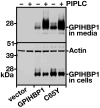
Figure 3. Release of the GPIHBP1-C65Y mutant from the surface of cells with a phosphatidylinositol-specific phospholipase C (PIPLC)
CHO pgsA-745 cells were electroporated with empty vector, an expression vector for wild-type human GPIHBP1, or an expression vector for GPIHBP1-C65Y. 24 h later, the cells were incubated with PIPLC (5 U/ml) in serum-free medium for 1 h at 37° C. The amounts of GPIHBP1 in the cell culture media and the cell lysates were assessed by western blotting with a goat antibody against the S-protein tag. Actin was used as a loading control.
Figure 4
Figure 4. Assessing the amounts of GPIHBP1 at the surface of cells relative to the total amounts of GPIHBP1 in the cell with a goat antibody against the S-protein tag and a mouse monoclonal antibody against human GPIHBP1
(A) Western blot analysis of GPIHBP1 at the cell surface. CHO pgsA-745 cells were electroporated with an empty vector or an expression vector encoding wild-type human GPIHBP1, GPIHBP1-C65Y, GPIHBP1-Q115P, or GPIHBP1-G56R. All constructs contained an amino-terminal S-protein tag. The next day, the cells were incubated for 2 h at 4° C with a goat antiserum against the S-protein tag. After washing the cells six times in ice-cold PBS, cell extracts were prepared for western blotting with a IR680-conjugated donkey antibody against goat IgG and a mouse monoclonal antibody against human GPIHBP1. The mouse monoclonal antibody was detected with an IR800-conjugated donkey antibody against mouse IgG. (B) Quantification of the western blot. The signal corresponding to the goat anti-S-protein tag IgG was normalized to the signal for the monoclonal antibody against human GPIHBP1 and expressed relative to the ratio for wild-type GPIHBP1 (set at 100%).
Figure 5
Figure 5. Assessing the binding of human V5-tagged LPL to GPIHBP1-C65Y
CHO pgsA-745 cells were electroporated with empty vector, an expression vector for wild-type human GPIHBP1, GPIHBP1-C65Y, GPIHBP1-Q115P, or GPIHBP1-G56R. 24 h later, the cells were incubated for 2 h at 4° C with human V5-tagged LPL with or without heparin (500 U/ml). At the end of the incubation, the cells were washed six times with ice-cold PBS, and cell extracts were prepared for western blotting. The amount of LPL bound to the cells was determined with a mouse monoclonal against the V5 tag; the levels of GPIHBP1 expression were assessed with a goat antibody against the S-protein tag. Actin was used as a loading control.
Figure 6
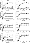
Figure 6. The appearance of LPL and HL in the plasma after heparin
A–B. LPL in the plasma after an intravenous bolus of heparin in the subject homozygous for a C65Y mutation in GPIHBP1, his heterozygous parents, and four normolipidemic controls. LPL activity (A) and mass (B) in postheparin plasma were measured during an 18-min sampling period after an intravenous injection of heparin (50 U/kg body weight). Proband (▲); father (○) mother (◇); and controls (■). For the controls, data are expressed as mean ± SEM. C–D. LPL in the plasma after an intravenous bolus of heparin in the subject homozygous for a Q115P mutation in GPIHBP1 and normolipidemic controls. LPL activity (C) and mass (D) in postheparin plasma was measured during an 18-min sampling period after an intravenous injection of heparin (50 U/kg body weight). Proband (▲); and controls (■). For the controls (n = 4), data are expressed as mean ± SEM. E–F. HL in the plasma after an intravenous bolus of heparin in the subject homozygous for a C65Y mutation in GPIHBP1, his heterozygous parents, and four normolipidemic controls. HL activity (E) and mass (F) in postheparin plasma were measured during an 18-min sampling period after an intravenous injection of heparin (50 U/kg body weight). Proband (▲); father (○); mother (◇); and controls (■). For the controls, data are expressed as mean ± SEM. G–H. HL in the plasma after an intravenous bolus of heparin in the subject homozygous for a Q115P mutation in GPIHBP1, and normolipidemic controls. HL activity (G) and mass (H) in postheparin plasma was measured during an 18-min sampling period after an intravenous injection of heparin (50 U/kg body weight). Proband (▲); and controls (■). For the (n = 4), data are expressed as mean ± SEM.
Figure 7
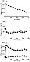
Figure 7. LPL mass and triglycerides in the plasma during a continuous infusion of heparin in the Q115P homozygote and controls
We analyzed LPL mass and triglyceride levels in multiple samples taken every 30 min. (A) Plasma triglycerides in the Q115P homozygote. (B) Plasma triglycerides in normolipidemic control subjects. For the experiments shown in Panels A and B, an inhibitor of lipolysis, tetrahydrolipstatin, was added to the blood after they were collected. Thus, the fall in plasma triglycerides observed in Panel A could not be ascribed to continuing lipolysis occurring in the tube ex vivo. (C) Plasma LPL mass measurements in the normolipidemic controls and the Q115 P homozygote. Proband (▲); and controls (■). For the controls (n = 7), data are expressed as mean ± SEM.
Figure 8
Figure 8. Model for the Ly6 domain of human GPIHBP1 based on the known structure of CD59
The amino acid sequence of GPIHBP1 was aligned to NCBI conserved domain cd000117 with the Conserved Domain Database Search Service, v2.17 (http://www.ncbi.nlm.nih.gov/Structure/cdd/cdd.shtml). Using the NMR structure of CD59 (PDB code 1CDR) as a template, 20 models of GPIHBP1 structure were built with the loop model procedure of MODELER 9v6., The model with the best DOPE (Discrete Optimized Protein Energy) score was selected. Human GPIHBP1 has two putative N-linked glycosylation sites (Asn-78, Asn-82). The locations of the 10 cysteines and the predicted disulfide bonds (C65–C89, C68–C77, C83–C110, C114–C130, and C131–C136) are shown.

Similar articles

Cited by

References

    1. Wang H, Eckel RH. Lipoprotein Lipase: from gene to obesity. Am J Physiol Endocrinol Metab. 2009;297:E271–E288. - PubMed
    1. Williams KJ. Molecular processes that handle - and mishandle - dietary lipids. J Clin Invest. 2008;118:3247–3259. - PMC - PubMed
    1. Young SG, Davies BS, Fong LG, Gin P, Weinstein MM, Bensadoun A, Beigneux AP. GPIHBP1: an endothelial cell molecule important for the lipolytic processing of chylomicrons. Curr Opin Lipidol. 2007;18:389–396. - PMC - PubMed
    1. Beigneux AP, Davies BS, Gin P, Weinstein MM, Farber E, Qiao X, Peale F, Bunting S, Walzem RL, Wong JS, Blaner WS, Ding ZM, Melford K, Wongsiriroj N, Shu X, De Sauvage FJ, Ryan RO, Fong LG, Bensadoun A, Young SG. Glycosylphosphatidylinositol-anchored high-density lipoprotein-binding protein 1 plays a critical role in the lipolytic processing of chylomicrons. Cell Metab. 2007;5:279–291. - PMC - PubMed
    1. Beigneux AP, Davies BS, Bensadoun A, Fong LG, Young SG. GPIHBP1, a GPI-anchored protein required for the lipolytic processing of triglyceride-rich lipoproteins. J Lipid Res. 2009;50 (Suppl):S57–S62. - PMC - PubMed

Publication types