. 2010 Feb;9(2):499-509.
doi: 10.1158/1535-7163.MCT-09-0664. Epub 2010 Feb 2.

Hormonal regulation and distinct functions of semaphorin-3B and semaphorin-3F in ovarian cancer

Affiliations

Hormonal regulation and distinct functions of semaphorin-3B and semaphorin-3F in ovarian cancer

Doina Joseph et al. Mol Cancer Ther. 2010 Feb.

Abstract

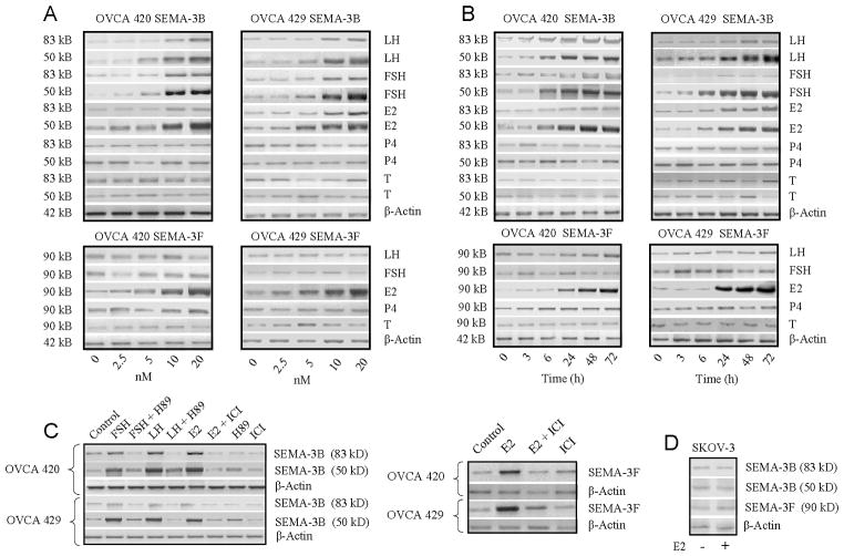
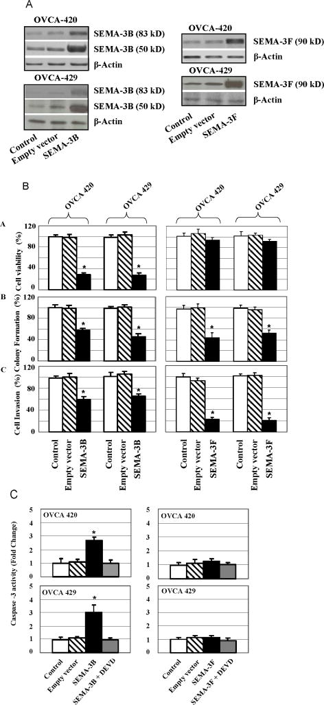
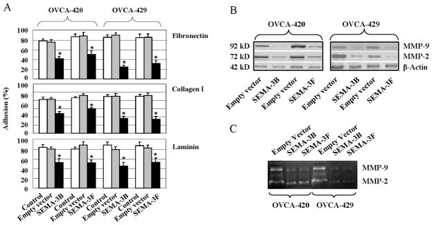
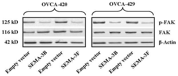
Semaphorins comprise a family of molecules that influence neuronal growth and guidance. Class-3 semaphorins, semaphorin-3B (SEMA3B) and semaphorin-3F (SEMA3F), illustrate their effects by forming a complex with neuropilins (NP-1 or NP-2) and plexins. We examined the status and regulation of semaphorins and their receptors in human ovarian cancer cells. A significantly reduced expression of SEMA3B (83 kDa), SEMA3F (90 kDa), and plexin-A3 was observed in ovarian cancer cell lines when compared with normal human ovarian surface epithelial cells. The expression of NP-1, NP-2, and plexin-A1 was not altered in human ovarian surface epithelial and ovarian cancer cells. The decreased expression of SEMA3B, SEMA3F, and plexin-A3 was confirmed in stage 3 ovarian tumors. The treatment of ovarian cancer cells with luteinizing hormone, follicle-stimulating hormone, and estrogen induced a significant upregulation of SEMA3B, whereas SEMA3F was upregulated only by estrogen. Cotreatment of cell lines with a hormone and its specific antagonist blocked the effect of the hormone. Ectopic expression of SEMA3B or SEMA3F reduced soft-agar colony formation, adhesion, and cell invasion of ovarian cancer cell cultures. Forced expression of SEMA3B, but not SEMA3F, inhibited viability of ovarian cancer cells. Overexpression of SEMA3B and SEMA3F reduced focal adhesion kinase phosphorylation and matrix metalloproteinase-2 and matrix metalloproteinase-9 expression in ovarian cancer cells. Forced expression of SEMA3F, but not SEMA3B in ovarian cancer cells, significantly inhibited endothelial cell tube formation. Collectively, our results suggest that the loss of SEMA3 expression could be a hallmark of cancer progression. Furthermore, gonadotropin- and/or estrogen-mediated maintenance of SEMA3 expression could control ovarian cancer angiogenesis and metastasis.


Figures

Figure 1
Figure 1
Down-regulation of SEMA3B, SEMA3F and plexin A-3, but not NP-1, NP-2 and plexinA1 in ovarian cancer cells. (A) Normal immortalized human ovarian surface epithelial (HOSE) cells (HOSE 6–8, and HOSE 642), and ovarian cancer (OVCA) cells (OVCA 420 and OVCA 429) were evaluated by Western blot for the expression of SEMA3B, SEMA3F, plexin-A1, plexin A-3, NP-1 and NP-2. Cells and conditioned media were harvested and 20 μg of protein from the whole-cell extract or conditioned media was loaded in each lane. The blot was probed with the indicated antibody. β -Actin was used as a loading control. (B) Mouse E16 cerebellum and (C) rat brain lysates were used as positive controls for SEMA3B and SEMA3F respectively. Lysates probed with only secondary antibody (rabbit IgG) were used as negative controls. (D) Suppression of SEMA3B, SEMA3F, and plexin A-3 expression in G3 ovarian tumors. Total cellular RNA (1 μg) was isolated from epithelial ovarian tumor tissues of different grades varying from G0 to G3. Semi- quantitative RT-PCR was used to compare SEMA3B, SEMA3F and plexin A-3 mRNA expression in control normal ovarian tissue (Normal, N=3), benign tumor (G0, N=3), borderline tumor (G1, N=3), and high-grade tumor (G3, N=3). SEMA3B, and SEMA3F, and plexin A-3 expression was significantly lower in G3 ovarian tissues.
Figure 2
Figure 2
Time and dose dependent up-regulation of SEMA3B, and SEMA3F expression in ovarian cancer cells. (A) Effect of reproductive hormones on the expression of SEMA3B and SEMA3F was studied in two ovarian cancer (OVCA 420 and OVCA 429) cell lines. Cells were cultured with indicated doses of Follicle-stimulating hormone (FSH), luteinizing hormone (LH), estradiol (E2), progesterone (P4) or testosterone (T), for 24 h. Expression of SEMA3B and SEMA3F in the lysates of OVCA cell lines was evaluated by Western blots. (B) To study the time-dependent effect, cells were treated with indicated hormones (10 nmol/L each) for 3, 6, 24, 48 and 72 hours. Cells were harvested and 20 μg of protein from the whole-cell extract was loaded in each lane. The blots were probed with anti-human SEMA3B or SEMA3F antibodies. β-Actin was used as a loading control. (C) To confirm the specificity of FSH, LH, and estradiol, 2 × 10⁵ cells per T-25 flask were cultured with 10 nmol/L FSH, 10 nmol/L LH, or 10 nmol/L estradiol alone or with the protein kinase A inhibitor H-89 at 0.1 mmol/L (for FSH and LH), or 0.1 mmol/L ICI 182,780 (for estrogen) for 72 h. Control cells were treated with vehicle alone. After 72 h, cells were harvested and 20 μg of protein from the whole-cell extract was loaded in each lane. The blots were probed with anti-human SEMA3B or SEMA3F antibodies. β-Actin was used as a loading control. (D) To study the expression of SEMA3B and SEMA3F, SKOV-3, estrogen non-responsive cells were cultured with estradiol (10 nmol/L) for 24 h. Control cells were treated with ethanol (final concentration of ethanol in the medium was 0.1%). Protein was extracted from cell cultures and loaded in each lane and blots were probed with SEMA3B and SEMA3F. β-Actin was used as a loading control.
Figure 3
Figure 3
Effect of SEMA3B and SEMA3F overexpression on ovarian cancer cells malignant phenotypes. Effect of SEMA3B and SEMA3F overexpression on the viability, colony formation, and migration of ovarian cancer (OVCA) cells was investigated in two ovarian cancer cell lines. The cell lines (OVCA-420 and OVCA-429) were transfected with a vector containing SEMA3B or SEMA3F. Control cells were transfected with respective empty vectors. Second control was cells without transfection. (A) Protein from transfected OVCA cell lines was extracted and analyzed for the expression of SEMA3B and SEMA3F by Western blot. A significant difference in the protein levels in transfected cell lines and those observed in control cell cultures is shown in the figure. (BA) After 3 days of transfection, cell viability was measured by MTS assay and viability of transfected cells was expressed as percentage growth compared with that of control cells (100%). The data shown represent the mean±SEM of three independent experiments. Statistically significant decreases in cell growth compared to those seen in control cells are indicated by *P<0.05. (BB) SEMA3B and SEMA3F transfected and control cells were cultured on soft agar, and 2 weeks later, colonies were counted. The value shown is the percentage compared with that of empty vector transfected control cells (100%). The data shown represent the mean±SEM of three independent experiments. Statistically significant changes in colony formation, as compared to those seen in control cells, are indicated by *P<0.05. (BC) OVCA cells transfected with SEMA3B and SEMA3F were plated on Matrigel. After 22 h, cells that migrated through the Matrigel were counted. The data shown represent the mean±SEM of three independent experiments. Statistically significant changes in cell invasion, as compared to those seen in control cells, are indicated by *P<0.05. (C) Activation of caspase-3 by overexpression of SEMA3B, and SEMA3F in OVCA cell lines. Three days following transfection, OVCA (OVCA 420 and OVCA 429) cells were harvested and analyzed for caspase-3 activity by measuring the cleavage of pNA-labeled caspase-3–specific substrate, DEVD/pNA. The results were calculated as the mean±SEM of three experiments. Statistical significance of difference between empty vector transfected cells and SEMA3B or SEMA3F transfected group is indicated with asterisk.
Figure 4
Figure 4
Inhibition of OVCA cell adhesion and metastasis by ectopic expression of SEMA3B and SEMA3F. (A) Effect of SEMA3B and SEMA3F overexpression on adhesion to ECM components (fibronectin, collagen type 1, and laminin), was investigated in two ovarian cancer cell lines. SEMA and mock transfected cells (1 × 10⁴ cells/well) were transferred in wells coated with ECM components and then incubated for 90 min. After gentle washing with warm PBS, cell adhesion was determined by measuring absorbance at 590 nm. Data are representative of three independent experiments performed in triplicate, and presented are the mean±SEM. *P < 0.05 (Mock transfected versus SEMA-3 transfected cells). (B) Restoration of SEMA3B and SEMA3F expression in OVCA cells decreases MMP-2 and MMP-9 protein expression. Total cellular protein extracts were extracted as described in Materials and Methods. Representative picture from three independent experiments is shown. β-Actin serves as loading control. (C) The gelatinolytic activity of MMP-2 and -9 in CM of ovarian cancer cells. Ovarian cancer cells overexpressing SEMA3B or SEMA3F or mock empty vector transfected cells were serum starved for 12 h, and then incubated in the serum-free culture medium for 24 h. Conditioned media were collected and then subjected to gelatin zymography.
Figure 5
Figure 5
Ectopic expression of SEMA3B and SEMA3F inhibited phosphorylated FAK expression of OVCA cells. Two OVCA cell lines were transfected with SEMA3B and SEMA3F as described in Materials and Methods. Three days after transfection cell lysates were prepared for immunoblotting using antibody against FAK and p-FAK.
Figure 6
Figure 6
SEMA3F inhibits in vitro capillary-like tube formation. The effect of SEMA3B or SEMA3F on capillary-like network (tube) formation in HUVEC cells was evaluated by plating HUVEC cells (5 × 10⁴ per well) on growth factor–reduced Matrigel in growth media generated from OVCA 429 cells expressing SEMA3B or SEMA3F. Representative micrographs of the network of tubes formed are shown. The pictures shown are representative of three independent experiments, magnification, ×100.

Similar articles

Cited by

References

    1. Comeau MR, Johnson R, DuBose RF, et al. A poxvirus-encoded semaphorin induces cytokine production from monocytes and binds to a novel cellular semaphorin receptor, VESPR. Immunity. 1998;8:473–82. - PubMed
    1. Tamagnone L, Artigiani S, Chen H, et al. Plexins are a large family of receptors for transmembrane, secreted, and GPI-anchored semaphorins in vertebrates. Cell. 1999;99:71–80. - PubMed
    1. Winberg ML, Noordermeer JN, Tamagnone L, et al. Plexin A is a neuronal semaphorin receptor that controls axon guidance. Cell. 1998;95:903–16. - PubMed
    1. Luo Y, Raible D, Raper JA. Collapsin, a protein in brain that induces the collapse and paralysis of neuronal growth cones. Cell. 1993;75:217–27. - PubMed
    1. Kolodkin AL, Matthes DJ, Goodman CS. The semaphorin genes encode a family of transmembrane and secreted growth cone guidance molecules. Cell. 1993;75:1389–99. - PubMed

Publication types