. 2010 Feb 15;70(4):1356-66.
doi: 10.1158/0008-5472.CAN-09-2178. Epub 2010 Feb 2.

The neurofibromatosis type 1 tumor suppressor controls cell growth by regulating signal transducer and activator of transcription-3 activity in vitro and in vivo

Affiliations

The neurofibromatosis type 1 tumor suppressor controls cell growth by regulating signal transducer and activator of transcription-3 activity in vitro and in vivo

Sutapa Banerjee et al. Cancer Res. .

Abstract

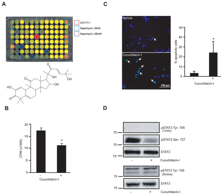
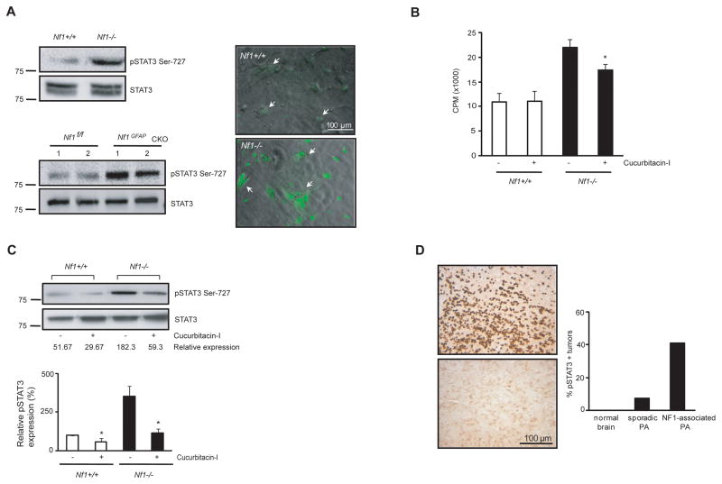
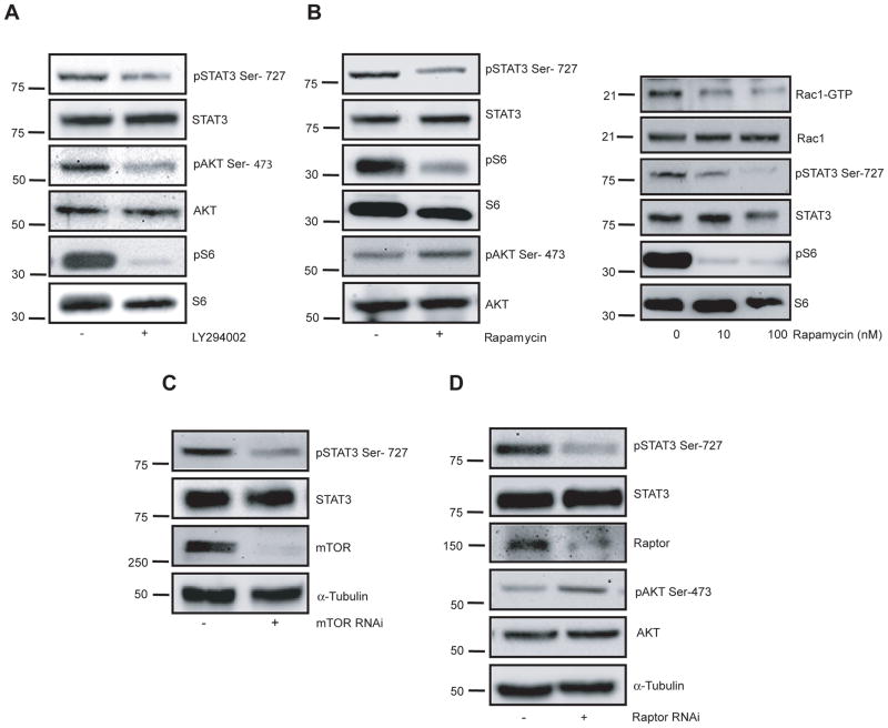
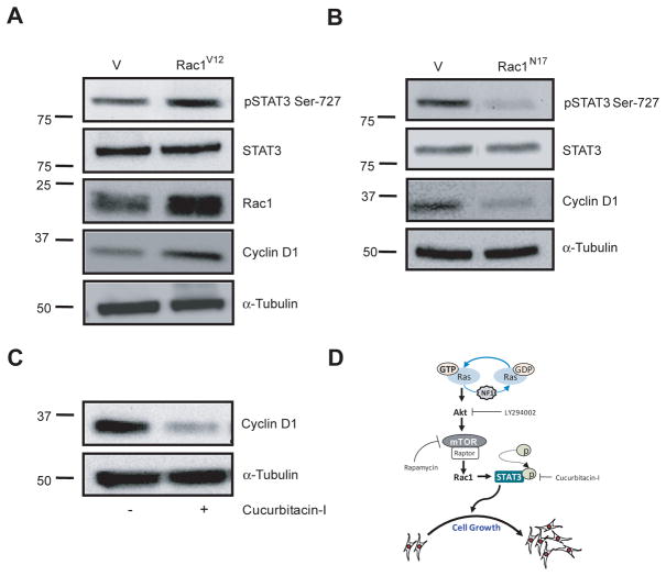
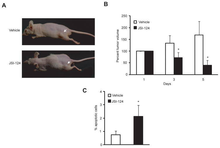
Neurofibromatosis type 1 (NF1) is a common cancer predisposition syndrome in which affected individuals develop benign and malignant nerve tumors. The NF1 gene product neurofibromin negatively regulates Ras and mammalian target of rapamycin (mTOR) signaling, prompting clinical trials to evaluate the ability of Ras and mTOR pathway inhibitors to arrest NF1-associated tumor growth. To discover other downstream targets of neurofibromin, we performed an unbiased cell-based high-throughput chemical library screen using NF1-deficient malignant peripheral nerve sheath tumor (MPNST) cells. We identified the natural product, cucurbitacin-I (JSI-124), which inhibited NF1-deficient cell growth by inducing apoptosis. We further showed that signal transducer and activator of transcription-3 (STAT3), the target of cucurbitacin-I inhibition, was hyperactivated in NF1-deficient primary astrocytes and neural stem cells, mouse glioma cells, and human MPNST cells through Ser(727) phosphorylation, leading to increased cyclin D1 expression. STAT3 was regulated in NF1-deficient cells of murine and human origin in a TORC1- and Rac1-dependent manner. Finally, cucurbitacin-I inhibited the growth of NF1-deficient MPNST cells in vivo. In summary, we used a chemical genetics approach to reveal STAT3 as a novel neurofibromin/mTOR pathway signaling molecule, define its action and regulation, and establish STAT3 as a tractable target for future NF1-associated cancer therapy studies.


Conflict of interest statement

Potential Conflicts of interest: None

Figures

Figure 1
Figure 1. Cucurbitacin-I inhibits NF1-deficient cell growth
A, Top, ST88-14 cells were treated with the various compounds in all wells of the 96-well plates, except in columns 1 and 12, where ST88-14 cells were treated either with DMSO (0.25%) or with two concentrations of rapamycin (10 nM and 100 nM). Bottom, The chemical structure of Cucurbitacin-I is shown. B, Cucurbitacin-I treatment reduced ST88-14 cell growth by 30% as measured by [3H]-thymidine incorporation (p<0.001). C, Representative TUNEL labeling photomicrographs demonstrate increased numbers of apoptotic ST88-14 cells following Cucurbitacin-I treatment (arrows denote TUNEL-positive cells). The percentages of TUNEL-labeled cells were presented as the mean ± SD of three fields from different separate experiments. Asterisks (*) denote statistically significant differences from vehicle-treated ST88-14 cells. Magnification=20X. Scale bar=100μm. D, Top, STAT3 Ser-727 phosphorylation is reduced by 10 fold in ST88-14 cells following treatment with Cucurbitacin-I. STAT3 Tyr-705 phosphorylation was not detected following 1 min exposure. Bottom, No changes in STAT3 Tyr-705 phosphorylation were observed. Total STAT3 protein expression was used as a protein loading control.
Figure 2
Figure 2. Cucurbitacin-I inhibits STAT3 activation in Nf1-deficient astrocytes
A, Top, STAT3 Ser-727 phosphorylation is increased 3-fold in Nf1−/ relative to WT astrocytes. Bottom, Nf1GFAPCKO mouse brains have increased STAT3 Ser-727 phosphorylation compared to WT (Nf1flox/flox) mouse brains. STAT3 expression was used as a protein loading control. Right, Increased nuclear STAT3 Ser-727 phosphorylation is observed in Nf1−/− relative to WT astrocytes by immunocytochemistry. Magnification=20X. Scale bar=100μm. B, Cucurbitacin-I treatment reduces Nf1−/− astrocyte proliferation as assessed by [3H]-thymidine incorporation. Asterisks (*) denote statistically significant differences from vehicle-treated Nf1−/− astrocytes. C, STAT3 Ser-727 phosphorylation was decreased following Cucurbitacin-I treatment in Nf1−/− astrocytes (quantitation shown below). D, Representative photomicrographs of human glioma TMA sections stained with STAT3 Ser-727 phospho-antibody. The number of phospho-STAT3 Ser-727-immunopositive specimens in each group is shown in the graph.
Figure 3
Figure 3. STAT3 activation is regulated by mTOR in NF1-deficient cells in vitro
A, The PI3-Kinase inhibitor, LY294002, inhibits ST88-14 STAT3 Ser-727 phosphorylation. Phospho-AKT antibodies were used to demonstrate PI3-Kinase activity. Total AKT and STAT3 served as loading controls. B, Left, Rapamycin inhibited STAT3 Ser-727 phosphorylation and results in increased AKT Ser-473 phosphorylation. Phospho-S6 was used to demonstrate mTOR activity. Total S6, AKT and STAT3 serve as internal loading controls. Right, GTP-bound Rac1 was immunoprecipitated from ST88-14 cells treated with vehicle (ethanol) or rapamycin (10 nM and 100nM) using PAK1-PBD affinity chromatography. Equal protein loading was confirmed by total Rac1immunoblotting. GTP-bound Rac1 activation was decreased in rapamycin-treated cells. STAT3 Ser-727 and S6 phosphorylation were also reduced by rapamycin treatment. C, siRNA-mediated mTOR knockdown inhibited STAT3 Ser-727 phosphorylation. Total mTOR and STAT3 served as loading controls. D, Raptor silencing by shRNAi inhibited ST88-14 STAT3 Ser-727 phosphorylation and resulted in increased AKT Ser-473 phosphorylation. α-tubulin, AKT and STAT3 served as loading controls.
Figure 4
Figure 4. Rac1 regulates STAT3 phosphorylation in NF1-deficient cells
A, Constitutively active Rac1 (Rac1V12) increases STAT3 Ser-727 phosphorylation and cyclin D1 expression. B, Dominant-inhibitory Rac1 (Rac1N17) suppresses STAT3 Ser-727 phosphorylation and cyclin D1 expression. α-tubulin and STAT3 serve as loading controls. C, Cyclin D1 expression was reduced following Cucurbitacin-I treatment. α-tubulin was used as a protein loading control.
Figure 5
Figure 5. JSI-124 treatment reduced NF1-deficient tumor growth in vivo
A, Representative tumor sizes (arrows) are shown for each treatment group. B, Tumor volumes were calculated from tumor measurements obtained on the indicated days. Tumor volumes were reduced by ~4-fold (p≤ 0.008) in mice following 5 days of treatment. C, JSI-124 treatment increased apoptosis in vivo by 2-fold relative to vehicle treatment. The percent of TUNEL-positive cells is presented as the mean± SD of ten fields from either JSI-124-treated or vehicle-treated mice. Asterisks (*) denote statistically significant (p<0.0001) differences from vehicle-treated mice.

References

    1. Friedman JM. Epidemiology of neurofibromatosis type 1. Am J Med Genet. 1999;89:1–6. - PubMed
    1. Listernick R, Charrow J, Greenwald M, Mets M. Natural history of optic pathway tumors in children with neurofibromatosis type 1: a longitudinal study. J Pediatr. 1994;125:63–6. - PubMed
    1. Gutmann DH, Rasmussen SA, Wolkenstein P, et al. Gliomas presenting after age 10 in individuals with neurofibromatosis type 1 (NF1) Neurology. 2002;59:759–61. - PubMed
    1. Ferner RE, Gutmann DH. International consensus statement on malignant peripheral nerve sheath tumors in neurofibromatosis. Cancer Res. 2002;62:1573–7. - PubMed
    1. DeClue JE, Papageorge AG, Fletcher JA, et al. Abnormal regulation of mammalian p21ras contributes to malignant tumor growth in von Recklinghausen (type 1) neurofibromatosis. Cell. 1992;69:265–73. - PubMed

Publication types

MeSH terms