. 2010 Mar;120(3):816-26.
doi: 10.1172/JCI40736.

Golgi and sarcolemmal neuronal NOS differentially regulate contraction-induced fatigue and vasoconstriction in exercising mouse skeletal muscle

Affiliations

Golgi and sarcolemmal neuronal NOS differentially regulate contraction-induced fatigue and vasoconstriction in exercising mouse skeletal muscle

Justin M Percival et al. J Clin Invest. 2010 Mar.

Abstract

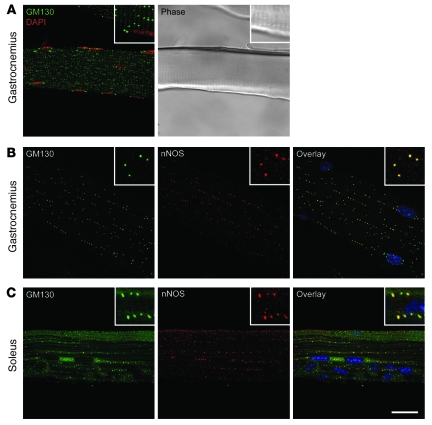
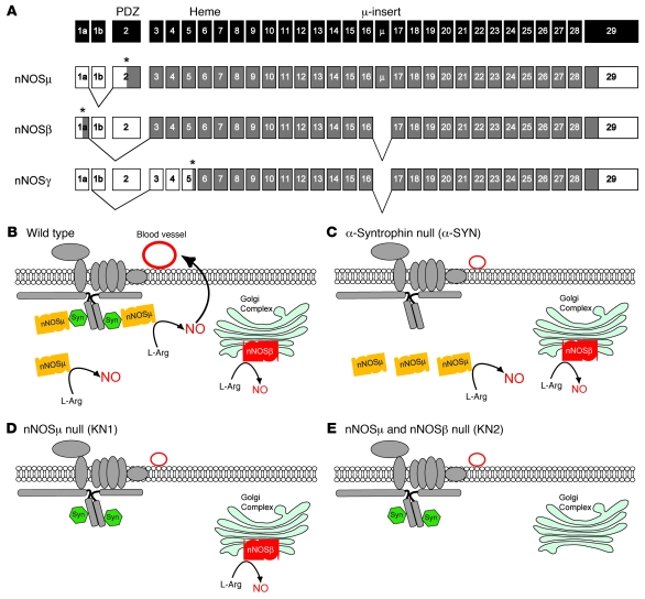
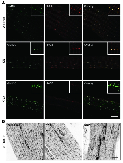
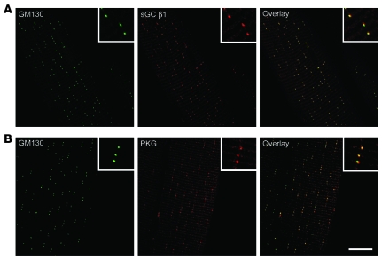
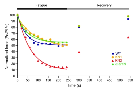
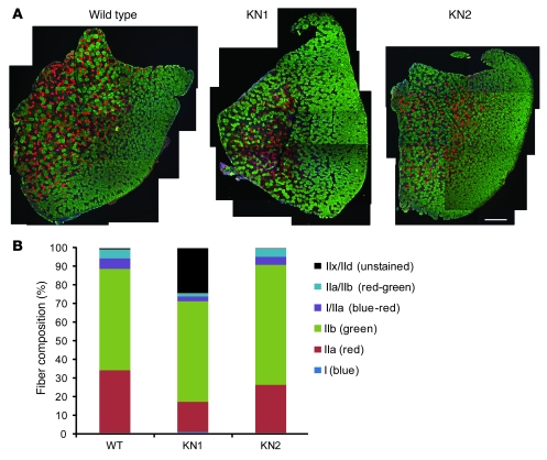
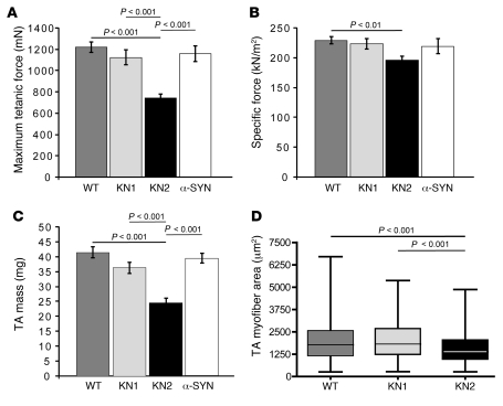
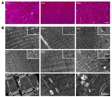
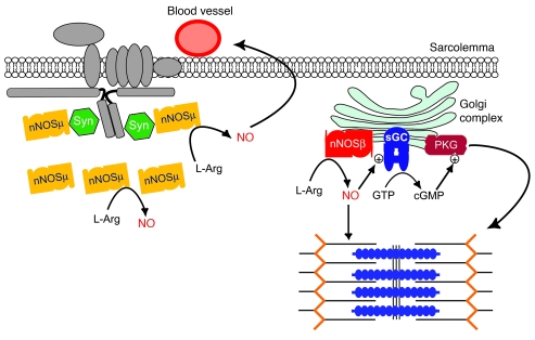
Signaling via the neuronal NOS (nNOS) splice variant nNOSmu is essential for skeletal muscle health and is commonly reduced in neuromuscular disease. nNOSmu is thought to be the predominant source of NO in skeletal muscle. Here we demonstrate the existence of what we believe to be a novel signaling pathway, mediated by the nNOS splice variant nNOSbeta, localized at the Golgi complex in mouse skeletal muscle cells. In contrast to muscles lacking nNOSmu alone, muscles missing both nNOSmu and nNOSbeta were severely myopathic, exhibiting structural defects in the microtubule cytoskeleton, Golgi complex, and mitochondria. Skeletal muscles lacking both nNOSmu and nNOSbeta were smaller in mass, intrinsically weak, highly susceptible to fatigue, and exhibited marked postexercise weakness. Our data indicate that nNOSbeta is a critical regulator of the structural and functional integrity of skeletal muscle and demonstrate the existence of 2 functionally distinct nNOS microdomains in skeletal muscle, created by the differential targeting of nNOSmu to the sarcolemma and nNOSbeta to the Golgi. We have previously shown that sarcolemmal nNOSmu matches the blood supply to the metabolic demands of active muscle. We now demonstrate that nNOSbeta simultaneously modulates the ability of skeletal muscle to maintain force production during and after exercise. We conclude therefore that nNOS splice variants are critical regulators of skeletal muscle exercise performance.


Figures

Figure 1
Figure 1. nNOS is localized to the Golgi complex in fast- and slow-twitch skeletal muscle fibers.
(A) Golgi complex distribution in fast-twitch gastrocnemius skeletal muscle cells (left panel). The Golgi complex (green) is immunolabeled with a FITC-conjugated GM130 antibody, a marker of cis-Golgi membranes. Myonuclei are counterstained with DAPI (red). A phase contrast image of the fiber is shown in the right panel. (B) nNOS (red) localizes to the Golgi (green) in gastrocnemius myofibers. Surface myonuclei (blue) are labeled with DAPI. The high degree of colocalization is evident in the inset image (yellow) of the overlay panel. (C) nNOS also localizes to the Golgi complex in slow-twitch myofibers of soleus muscle. Higher-magnification images are shown in the inset panels. Scale bar: 20 μm; 6 μm (insets). n ≥ 6.
Figure 2
Figure 2. nNOS splice variants and nNOS mutant mouse lines used to analyze NO signaling in skeletal muscle.
(A) Exon structure of the murine Nos1 gene, which encodes 31 exons (black boxes) (39). Relevant isozymes are shown here. The coding sequence is shown in gray, and asterisks mark translation initiation sites. The 5ι- and 3ι-untranslated sequences are shown as white boxes. In skeletal muscle, the predominant transcript is nNOSμ containing exon 2 (encoding the PSD-95, discs-large, ZO-1 [PDZ] domain) and the 34–amino acid μ-insert. nNOSβ is formed by the splicing of exon 1a to exon 3, creating a unique 6–amino acid N terminus. nNOSγ is produced by translation initiation at an internal ATG site in exon 5. (B) WT muscle can express all nNOS splice variants. nNOSμ (yellow) localizes to the sarcolemma by binding α-syntrophin (Syn; green), a member of the dystrophin-glycoprotein complex (gray). nNOSμ also localizes to the cytoplasm. In contracting muscles, sarcolemmal nNOSμ-derived NO overrides sympathetic vasoconstriction maintaining blood vessel dilation (large black arrow) (7). In the present study, we identify a splice variant of nNOS localized to the Golgi complex, presumed to be nNOSβ (red). The small black arrows represent the synthesis of NO from l-arginine by nNOS. (C) Muscles lacking α-syntrophin cannot localize nNOSμ to the sarcolemma and thus cannot attenuate vasoconstriction, despite expression of cytosolic nNOSμ and Golgi nNOSβ. (D) The muscles of KN1 mice do not express nNOSμ, due to deletion of exon 2, but nNOSβ and nNOSγ splice variant production is unaffected (24). (E) KN2 mice do not express any full-length active nNOS splice variants due to deletion of exon 6 encoding the heme-binding domain (25).
Figure 3
Figure 3. A nNOS splice variant localizes to the Golgi complex and regulates microtubule cytoskeleton integrity in skeletal muscle.
(A) In WT gastrocnemius muscles, nNOS (red) colocalizes with GM130 (green). The inset overlay image emphasizes the high degree of overlap between GM130 and Golgi nNOS. In KN1 muscles, Golgi nNOS labeling is unaffected, indicating that the Golgi nNOS is not nNOSμ but nNOSβ and/or nNOSγ. Golgi nNOS immunolabeling is absent in KN2 muscles, demonstrating nNOS antibody specificity. The loss of all nNOS variants perturbs the distribution and morphology of the Golgi complex. Higher-magnification images are shown in the inset panels. (B) nNOS splice variants differentially regulate the structural integrity of the microtubule cytoskeleton. Microtubules were labeled with FITC-conjugated α-tubulin antibody. The characteristic microtubule lattice is evident in WT myofibers (left panel). Loss of nNOSμ has a minor impact on microtubule organization (middle panel), which was insufficient to disrupt Golgi distribution. The loss of all nNOS splice variants severely compromises the integrity of the microtubule cytoskeleton (right panel). Scale bar: 20 μm; 6 μm (insets). n ≥ 4.
Figure 4
Figure 4. A NO-cGMP signaling microdomain that we believe to be novel at the Golgi complex in skeletal muscle.
(A) Gastrocnemius myofibers were coincubated with antibodies against the cis-Golgi marker GM130 and the NO-binding β1 subunit of sGC (sGC β1). GM130 and the NO-binding β1 subunit of sGC showed a high degree of overlap in gastrocnemius muscle cells (right panel). (B) Isolated myofibers were separately colabeled with antibodies against GM130 and PKG. GM130 and PKG showed a high degree of overlap in skeletal muscle cells (right panel). These data demonstrate that compartmentalization of nNOS and effector proteins occurs at the Golgi complex in skeletal muscle. Higher-magnification images are shown in the inset panels. Scale bar: 20 μm; 6 μm (insets). n ≥ 6.
Figure 5
Figure 5. nNOS splice variants differentially regulate contraction-induced fatigue and postexercise force recovery.
TA muscles were subjected to a series of maximal tetanic stimulations every 2 seconds for 4 minutes to simulate exercise. Muscles were rested, then maximally stimulated once at 1 and 5 minutes to measure postexercise strength recovery. Representative traces for each nNOS mutant fitted with exponential decay curves are shown. TA muscles of α-syntrophin (α-SYN) knockout mice (green diamonds) or KN1 muscles (yellow circles) exhibit normal muscle fatigue indistinguishable from WT controls (blue squares). In contrast, KN2 muscles (red triangles) exhibited significantly increased susceptibility to contraction-induced muscle fatigue, with force output levels leveling off at approximately 60% lower than those from WT, KN1, and α-syntrophin muscles. Force recovery at 1 and 5 minutes was significantly decreased by approximately 50% after simulated exercise, indicating substantial postexercise weakness. Po is the tetanic force in millinewtons generated at time t during the stimulation protocol. Pi is the initial force output in millinewtons at time 0, at the beginning of the experiment.
Figure 6
Figure 6. nNOS-deficient KN2 skeletal muscles exhibit a more fatigue-susceptible fiber composition.
(A) Antibodies specific for MyHC type 1, type IIa, and type IIb were used to identify slow oxidative type 1 (blue, most fatigue resistant), fast oxidative type IIa (red, intermediate fatigue resistance), and fast glycolytic type IIb (green, least fatigue resistant) fiber types in TA muscles from WT, KN1, and KN2 mice. Unlabeled fibers (black) were designated fast oxidative type IIx/IId fibers, which also exhibit a high degree of fatigue resistance. Images are composites. Scale bar: 400 μm. (B) Quantitation of fiber composition. In nNOSμ-deficient TA muscles from KN1 mice, type IIa fibers were significantly decreased 50% (P < 0.01), while there was a remarkable 3,000% increase (P < 0.001) in type IIx/IId fibers relative to controls. In contrast, the loss of all nNOS splice variants in KN2 TA muscles resulted in a significant 45% (P < 0.05) increase in type IIb fibers relative to WT. nNOS splice variants differentially regulate fiber composition in skeletal muscle. n ≥ 4.
Figure 7
Figure 7. Golgi nNOS splice variant is required to maintain normal skeletal muscle strength, muscle mass, and myofiber size.
(A) Maximum isometric force-producing capacity of TA muscles from KN1 and α-syntrophin mice did not differ significantly from WT controls. nNOS-deficient KN2 muscles exhibited significantly reduced maximum force-generating capacity (P < 0.001) compared with controls, KN1, and α-syntrophin mice. (B) Mean specific force did not differ statistically between WT controls, KN1, and α-syntrophin mice. The additional loss of nNOSβ in KN2 mice lead to a significant 14% decrease (P < 0.01) in specific force, indicating that nNOSβ is required for normal muscle strength. (C) TA muscle weight was unaffected in α-syntrophin mice and KN1 mice; however, loss of nNOSμ and nNOSβ lead to a significant 40% reduction (P < 0.001) in muscle mass. (D) The CSA of KN1 myofibers was similar to WT controls. KN2 muscle cells were significantly smaller in area (P < 0.001), suggesting that decreased myofiber size could at least partially account for decreased KN2 muscle mass. Values represent mean ± SEM (A, B, and C). The median is represented by the line within the box plot in D. Group sizes in A, B, C, and D are n ≥ 6, n ≥ 6, n ≥ 9, and n ≥ 4, respectively.
Figure 8
Figure 8. nNOS splice variant-deficiency leads to myopathic changes in intermyofibrillar mitochondria and skeletal muscle cytoarchitecture.
(A) Decreased myofiber CSA was evident in hematoxylin and eosin–stained KN2 TA muscles. (B) Electron microscopic analysis of KN1 muscles revealed dramatic changes in mitochondrion morphology (bottom row), including irregular swelling, disruption of internal cristae, and markedly decreased matrix density (increased electron lucency). Sarcomere integrity and registration were unaffected in KN1 muscle. KN2 muscles exhibited more severe mitochondrion morphological abnormalities, including substantial swelling and variability in size, disrupted cristae, and reduced matrix density. The characteristic intermyofibrillar localization of mitochondria at the Z-I band interface was also disrupted in KN2 muscle (bottom row). Furthermore, loss of all nNOS splice variants often impaired alignment of sarcomere contractile units. Higher-magnification images are shown in the inset panels. Scale bar: 40 μm (A); 2 μm (top and middle rows of B); 1 μm (bottom row of B); 1.3 μm (insets). n = 4.
Figure 9
Figure 9. Model for nNOS splice variant microdomain signaling in skeletal muscle.
The differential targeting of nNOSβ and nNOSμ creates 2 spatially and functionally distinct NO signaling compartments at the Golgi and sarcolemma, respectively. Spatial confinement of nNOSβ and its effectors (sGC and PKG) creates a NO-cGMP signaling microdomain at the Golgi complex. During exercise, nNOSμ-derived NO attenuates vasoconstriction, thus serving to match blood and oxygen delivery with the increased metabolic demands of skeletal muscle tissue. At the same time, nNOSβ signaling functions to maintain the ability of contracting muscles to generate force during and after exercise. nNOSβ-derived NO may regulate contractility through cGMP-dependent mechanisms (mediated by PKG) or by cGMP-independent mechanisms (such as nitrosylation).

Similar articles

Cited by

References

    1. Bredt DS, Snyder SH. Isolation of nitric oxide synthetase, a calmodulin-requiring enzyme. Proc Natl Acad Sci U S A. 1990;87(2):682–685. doi: 10.1073/pnas.87.2.682. - DOI - PMC - PubMed
    1. Silvagno F, Xia H, Bredt DS. Neuronal nitric–oxide synthase–mu, an alternatively spliced isoform expressed in differentiated skeletal muscle. J Biol Chem. 1996;271(19):11204–11208. doi: 10.1074/jbc.271.19.11204. - DOI - PubMed
    1. Brenman JE, Chao DS, Xia H, Aldape K, Bredt DS. Nitric oxide synthase complexed with dystrophin and absent from skeletal muscle sarcolemma in Duchenne muscular dystrophy. Cell. 1995;82(5):743–752. doi: 10.1016/0092-8674(95)90471-9. - DOI - PubMed
    1. Brenman JE, et al. Interaction of nitric oxide synthase with the postsynaptic density protein PSD–95 and alpha1–syntrophin mediated by PDZ domains. Cell. 1996;84(5):757–767. doi: 10.1016/S0092-8674(00)81053-3. - DOI - PubMed
    1. Stamler JS, Meissner G. Physiology of nitric oxide in skeletal muscle. Physiol Rev. 2001;81(1):209–237. - PubMed

Publication types

MeSH terms