. 2010 Mar;120(3):735-43.
doi: 10.1172/JCI41360.

High-mobility group box 1 is involved in the initial events of early loss of transplanted islets in mice

Affiliations

High-mobility group box 1 is involved in the initial events of early loss of transplanted islets in mice

Nobuhide Matsuoka et al. J Clin Invest. 2010 Mar.

Abstract

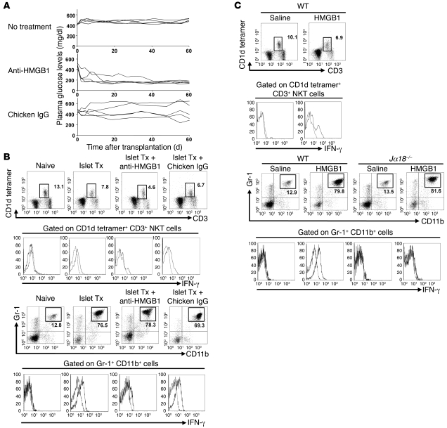
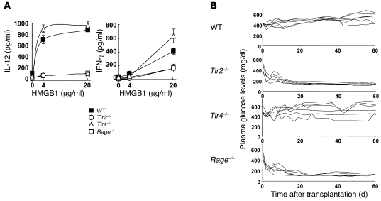
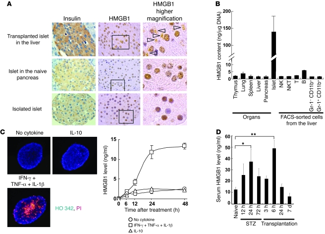
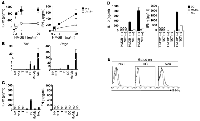
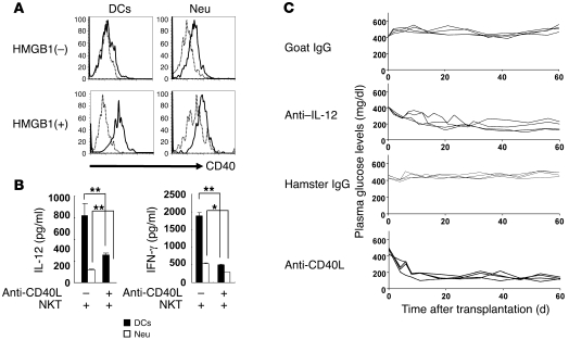
Islet transplantation for the treatment of type 1 diabetes mellitus is limited in its clinical application mainly due to early loss of the transplanted islets, resulting in low transplantation efficiency. NKT cell-dependent IFN-gamma production by Gr-1(+)CD11b(+) cells is essential for this loss, but the upstream events in the process remain undetermined. Here, we have demonstrated that high-mobility group box 1 (HMGB1) plays a crucial role in the initial events of early loss of transplanted islets in a mouse model of diabetes. Pancreatic islets contained abundant HMGB1, which was released into the circulation soon after islet transplantation into the liver. Treatment with an HMGB1-specific antibody prevented the early islet graft loss and inhibited IFN-gamma production by NKT cells and Gr-1(+)CD11b(+) cells. Moreover, mice lacking either of the known HMGB1 receptors TLR2 or receptor for advanced glycation end products (RAGE), but not the known HMGB1 receptor TLR4, failed to exhibit early islet graft loss. Mechanistically, HMGB1 stimulated hepatic mononuclear cells (MNCs) in vivo and in vitro; in particular, it upregulated CD40 expression and enhanced IL-12 production by DCs, leading to NKT cell activation and subsequent NKT cell-dependent augmented IFN-gamma production by Gr-1(+)CD11b(+) cells. Thus, treatment with either IL-12- or CD40L-specific antibody prevented the early islet graft loss. These findings indicate that the HMGB1-mediated pathway eliciting early islet loss is a potential target for intervention to improve the efficiency of islet transplantation.


Figures

Figure 1
Figure 1. Essential roles of HMGB1 in early loss of transplanted islets.
(A) Nonfasting plasma glucose levels in STZ-induced diabetic mice received 200 syngeneic islets (top panel) and those treated with chicken anti-HMGB1 antibody or control chicken IgG. Individual lines represent glucose levels of each animal. (B) FACS profiles of liver MNCs from naive mice, STZ-induced diabetic mice that received 200 syngenic islets (Islet Tx), and islet transplanted mice treated with anti-HMGB1 antibody or with chicken IgG. NKT cells (top 2 rows) and Gr-1+CD11b+ cells (bottom 2 rows) were analyzed for IFN-γ (second and fourth rows). The numbers in the figures represent the percentage of cells in the corresponding square areas. Representative data from 4 experiments are shown. (C) FACS profiles of NKT cells and Gr-1+CD11b+ cells after HMGB1 treatment. Liver MNCs from wild-type or Jα18–/– mice treated with i.v. injection of saline or HMGB1 (100 μg/mouse) were isolated 2 hours after the injection and examined by flow cytometry for IFN-γ production by NKT cells and Gr-1+CD11b+ cells. The numbers in the figures represent the percentage of cells in the corresponding square areas. Representative data from 4 experiments are shown.
Figure 2
Figure 2. HMGB1 receptors involved in early loss of transplanted islets.
(A) In vitro cytokine production by liver MNCs. Liver MNCs (2 × 10⁶/well) isolated from wild-type, Tlr2–/–, Tlr4–/–, or Rage–/– mice were cultured with indicated doses of HMGB1 in vitro for 48 hours, and IL-12 or IFN-γ levels in the culture supernatant were measured. Representative data from 2 experiments are shown. (B) Nonfasting plasma glucose levels of STZ-induced diabetic wild-type, Tlr2–/–, Tlr4–/–, or Rage–/– mice that received 200 syngeneic islets. Individual lines represent the glucose level of each animal.
Figure 3
Figure 3. HMGB1 production in tissues and cell types.
(A) Photomicrographs of islets. Islet cells 3 hours after transplantation in the liver (top row) and those of naive pancreas or isolated islets were examined. Sections stained with anti-insulin or anti-HMGB1 followed by staining with hematoxylin are shown. In general, HMGB1 was detected in the nucleus (brown), while some was detected in cytoplasm, as indicated by arrowheads. Original magnification: ×100 (first and second columns) and ×800 (third column). Boxed regions in the second column were enlarged. (B) HMGB1 contents (ng/μg DNA) of individual organs (n = 5), isolated islets, or FACS-sorted liver MNCs (n = 3). (C) Left panels: Fluorescence photomicrographs of isolated islets (original magnification, ×200) stained with HO 342 (blue) and PI (red) at 24 hours after in vitro culture with or without IFN-γ, TNF-α, and IL-1β (20 ng/ml each) or IL-10 (20 ng/ml). Right panels: HMGB1 levels in the culture (200 cells/dish) were also measured at the indicated time points in the absence of cytokine or the presence of cytokine mixtures or of control cytokine (IL-10). The values are expressed as the mean ± SD in each group (n = 5). (D) Serum HMGB1 levels were measured after STZ injection and also after transplantation of 400 syngeneic islets, which had been performed 72 hours after STZ injection (n = 5–6). The values are expressed as the mean ± SD. *P < 0.05; **P < 0.01.
Figure 4
Figure 4. NKT cell–dependent IL-12 and IFN-γ production by liver MNCs in response to HMGB1.
(A) Liver MNCs (2 × 10⁶/well) isolated from wild-type or Jα18–/– mice were cultured with the indicated doses of HMGB1 in vitro for 48 hours and measured for IL-12 and IFN-γ. Representative data from 2 experiments are shown. (B) PCR analysis on HMGB1 receptors. FACS-sorted liver MNCs (2 × 10³ for Tlr2, Rage, or Hprt) were analyzed for mRNA levels by quantitative real-time PCR. Data were analyzed by the ΔΔCt method using the expression level in Mo/Mϕ as normalized control. (C) Cytokine production in FACS-sorted liver MNCs upon stimulation with HMGB1. FACS-sorted cells were cultured in vitro (1 × 10⁵ cells/well) for 48 hours in the presence of HMGB1 (20 μg/ml). The amounts of IL-12 and IFN-γ were measured by CBA (n = 3). (D) Cytokine production by DCs, Mo/Mϕ, or Neu in the presence of NKT cells. FACS-sorted Gr-1CD11b+CD11c+ DCs, Gr-1CD11b+CD11c Mo/Mϕ, and Gr-1+CD11b+CD11c Neu (4 × 10⁴) were cocultured in vitro with NKT cells (2 × 10⁵) in the presence of HMGB1 (20 μg/ml) for 48 hours. The amounts of IL-12 and IFN-γ were measured by CBA (n = 3). (E) Intracellular cytokine staining of liver MNCs after HMGB1 treatment. Liver MNCs (2 × 10⁶) were cultured with HMGB1 (20 μg/ml) for 24 hours, and the indicated cells were gated and analyzed for their production of IFN-γ by intracellular cytokine staining.
Figure 5
Figure 5. Involvement of CD40-CD40L interaction in production of IL-12 and IFN-γ in early loss of transplanted islets.
(A) CD40 expression in DCs and Neu before or after treatment with HMGB1. Liver MNCs (2 × 10⁶) were treated without or with HMGB1 (20 μg/ml) for 24 hours and analyzed for CD40 expression (n = 3). (B) Requirement of CD40-CD40L interaction in the production of IL-12 and IFN-γ in the presence of NKT cells. DCs or Neu (4 × 10⁴) were cocultured in vitro with NKT cells (2 × 10⁵) in the presence of HMGB1 (20 μg/ml) for 48 hours with or without addition of anti-CD40L antibody. IL-12 and IFN-γ levels were measured by CBA (n = 3). The values are expressed as the mean ± SD. *P < 0.05; **P < 0.01. (C) Nonfasting plasma glucose levels of STZ-induced diabetic mice that had received 200 syngeneic islets and were treated with control goat IgG or goat anti-mouse IL-12 antibody and those with control hamster IgG or hamster anti-mouse CD40L antibody with 200 μg intraperitoneal injection per mouse at the time of transplantation. Individual lines represent the nonfasting plasma glucose levels of each diabetic mouse after islet transplantation.

Similar articles

Cited by

References

    1. Shapiro AM, et al. Islet transplantation in seven patients with type 1 diabetes mellitus using a glucocorticoid-free immunosuppressive regimen. N Engl J Med. 2000;343(4):230–238. doi: 10.1056/NEJM200007273430401. - DOI - PubMed
    1. Yasunami Y, et al. Vα14 NKT cell-triggered IFN-γ production by Gr-1+CD11b+ cells mediates early graft loss of syngeneic transplanted islets. . J Exp Med. 2005;202(7):913–918. doi: 10.1084/jem.20050448. - DOI - PMC - PubMed
    1. Muller S, Ronfani L, Bianchi ME. Regulated expression and subcellular localization of HMGB1, a chromatin protein with a cytokine function. J Internal Med. 2004;255(3):332–343. doi: 10.1111/j.1365-2796.2003.01296.x. - DOI - PubMed
    1. Seong SY, Matzinger P. Hydrophobicity: an ancient damage-associated molecular pattern that initiates innate immune responses. Nat Rev Immunol. 2004;4(6):469–478. doi: 10.1038/nri1372. - DOI - PubMed
    1. Lotze MT, Tracey KJ. High-mobility group box 1 protein (HMGB1): nuclear weapon in the immune arsenal. Nat Rev Immunol. 2005;5(4):331–342. doi: 10.1038/nri1594. - DOI - PubMed

Publication types

MeSH terms