. 2010 Mar 15;89(5):492-9.
doi: 10.1097/TP.0b013e3181c6ffd3.

Photochemotherapy induces the apoptosis of monocytes without impairing their function

Affiliations

Photochemotherapy induces the apoptosis of monocytes without impairing their function

Dalil Hannani et al. Transplantation. .

Abstract

Background: Extracorporeal photopheresis (ECP) is a powerful therapy currently used to treat various hematological disorders as in graft versus host disease. Clinical data clearly demonstrate its efficacy and immunomodulation toward the pathogenic T cells. However, ECP mechanism of action is still poorly understood. Monocytes represent up to 30% of the total amount of treated cells and are known to play an important role in adaptive immunity. However, data from previous reports analyzing the effect of psoralen and UV-A irradiation (PUVA) on their functions are heterogeneous. In this study, we focused on the effect of PUVA on human monocytes functions in adaptive immunity.

Design and methods: Purified human monocytes were treated in vitro by PUVA. We measured their kinetic of apoptosis after the treatment. We also determine whether their phenotype and functionalities were modified. Finally, we assessed the functionalities of PUVA-treated monocytes-derived dendritic cells (DC).

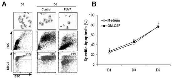
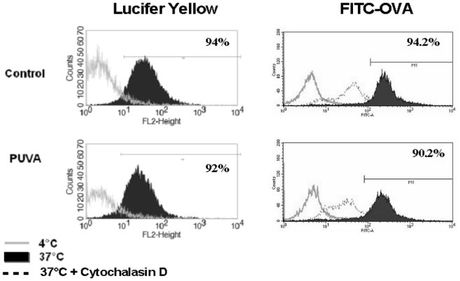
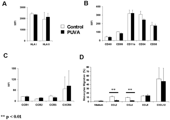
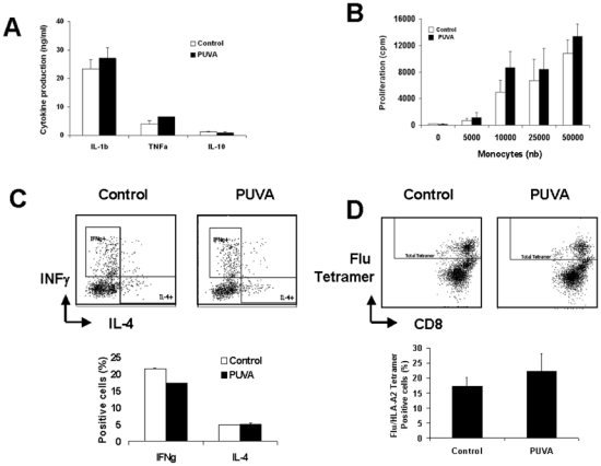
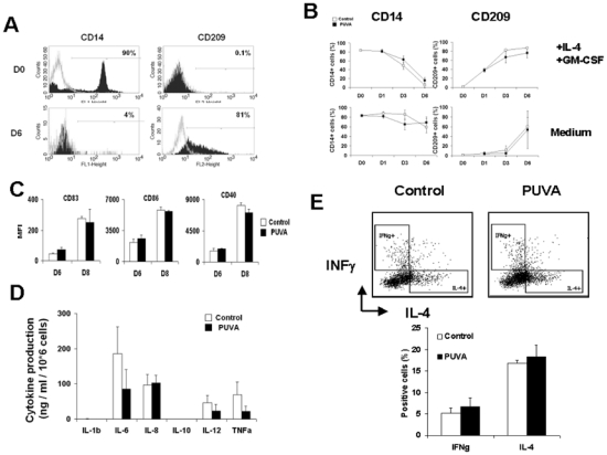
Results: PUVA treatment sentenced purified monocytes to die in 6 days and immediately altered their migratory capacities without impairing their ability of endocytosis. It also up-regulated co-stimulatory molecules and production of inflammatory cytokines on activation and consequently stimulated allogeneic or autologous T cells as efficiently as untreated monocytes. Moreover, PUVA-treated monocytes retained their ability to differentiate into fully functional DC that maturated and stimulated T cells as well as normal DC.

Conclusions: Our data demonstrate that monocytes undergo apoptosis and loose a part of their migratory capacity after ECP and the surviving cell functionalities are not impaired, suggesting that monocytes have a minor effect on ECP-mediated immunomodulation.


Figures

Figure 1
Figure 1. PUVA treatment induces apoptosis in monocytes
Treated (PUVA) or untreated (Control) monocytes cultured in the medium with or without GM-CSF for 6 days were labeled by DioC6. Dead cells are defined as DioC6 negative cells. (A) May-Grünwald-Giemsa staining and dot plots from GM-CSF condition are shown which are representative of 3 independent experiments. (B) The graphs represent the kinetic of monocytes apoptosis after PUVA treatment. FSC: Forward Scatter; SSC: Side Scatter
Figure 2
Figure 2. Endocytic capacity of monocytes is not impaired following PUVA treatment
Endocytosis assays were performed 24h after PUVA treatment in the presence of either Lucifer Yellow (left panel) or FITC-OVA (right panel) at 37°C for 3 hours (black histogram). Negative controls were performed at 4°C (empty histogram) or at 37°C in presence of 10μg/ml of Cytochalasin D. One experiment histogram is a representative of 2 performed with 2 donors.
Figure 3
Figure 3. Chemokine receptors, MHC molecules and activation and adhesion markers expression are not modified on monocytes by PUVA treatment but their migratory capacities are impaired
PUVA effects were determined on the expression of class I and II MHC molecules (A), on the activation and adhesion markers (B) and on the chemokine receptors (C) on purified monocytes. PUVA treated or untreated cells were cultured and then analyzed by flow cytometry. The migration capacities of treated monocytes were evaluated by transwell assay (D) Data represent the mean +/− s.d. of 2 independent experiments performed with monocytes from 2 donors. MFI: Mean Fluorescence Intensity.
Figure 4
Figure 4. PUVA treatment does not modify either cytokine production under activation or allostimulation or antigen specific stimulatory capacities of monocytes
(A) Cytokine production of PUVA-treated monocytes activated by LPS for 24h was measured by CBA inflammation kit. Data represent the mean +/− s.d. of 3 independent experiments performed with 3 donors. (B) Allogeneic mixed lymphocyte reaction was performed by mixing increasing amount of irradiated monocytes and purified allogeneic T lymphocytes. T lymphocytes proliferation was measured by the incorporation of [3H]thymidine after 6 days of culture. Data represent the cpm (mean +/− s.d.) of 2 independent experiments performed with 2 donors. (C) T cell polarization after 6-day culture with control or PUVA allogenic monocytes. Data represent the mean +/− s.d. of 3 independent experiments performed with 3 donors. (D) Amplification of Flu-specific T cells following 7-day culture with Influenza M1 peptide pulsed control or PUVA-treated monocytes.
Figure 5
Figure 5. PUVA treatment neither impairs the differentiation of monocytes into potent DC in the presence of cytokines nor induces their spontaneous differentiation
PUVA treated and untreated monocytes were cultured with or without GM-CSF and IL-4 for 6 days. CD14 and CD209 are specific markers for monocytes and dendritic cells respectively. (A) Representative histograms of three independent experiments performed with 3 donors represent CD14 and CD209 histogram at day 0 and 6, (B) CD14 and CD209 membrane expression on monocytes at day 0, 1, 3 and 6. Data represent the percentage of positive cells (mean +/− s.d.) of 2 independent experiments performed with 2 donors. (C) Immature DCs were obtained from PUVA treated and untreated monocytes cultured with GM-CSF and IL-4 for 6 days. DCs were matured by activation with LPS and IFNγ for 48h and the expression of activation markers were then analyzed by flow cytometry. The percentages of positive cells are represented. (D) Supernatants from mature DC were collected and the measurements cytokines were performed by CBA inflammation kit. Data represent the percentages of positive cells (mean +/− s.d.) of 3 independent experiments performed with 3 donors. (E) T cell polarization after 6-day culture with PUVA treated and control allogenic monocytes derived DC. Data represent the mean +/− s.d. of 3 independent experiments performed with 3 donors.

Similar articles

Cited by

References

    1. Miller JD, Kirkland EB, Domingo DS, et al. Review of extracorporeal photopheresis in early-stage (IA, IB, and IIA) cutaneous T-cell lymphoma. Photodermatol Photoimmunol Photomed. 2007;23 (5):163. - PubMed
    1. Marshall SR. Technology insight: ECP for the treatment of GvHD--can we offer selective immune control without generalized immunosuppression? Nat Clin Pract Oncol. 2006;3 (6):302. - PubMed
    1. Dall’Amico R, Murer L. Extracorporeal photochemotherapy: a new therapeutic approach for allograft rejection. Transfus Apheresis Sci. 2002;26 (3):197. - PubMed
    1. Plumas J, Manches O, Chaperot L. Mechanisms of action of extracorporeal photochemotherapy in the control of GVHD: involvement of dendritic cells. Leukemia. 2003;17 (11):2061. - PubMed
    1. Kanne D, Straub K, Rapoport H, Hearst JE. Psoralen-deoxyribonucleic acid photoreaction. Characterization of the monoaddition products from 8-methoxypsoralen and 4,5′8-trimethylpsoralen. Biochemistry. 1982;21 (5):861. - PubMed

Publication types

MeSH terms