. 2010 Mar 10;21(4):303-8.
doi: 10.1097/WNR.0b013e328337310a.

Dorsal horn neurons expressing NK-1 receptors mediate scratching in rats

Affiliations

Dorsal horn neurons expressing NK-1 receptors mediate scratching in rats

Earl E Carstens et al. Neuroreport. .

Abstract

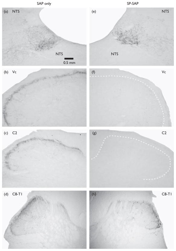
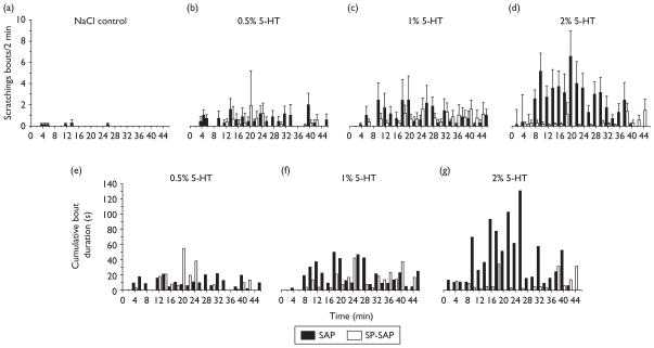
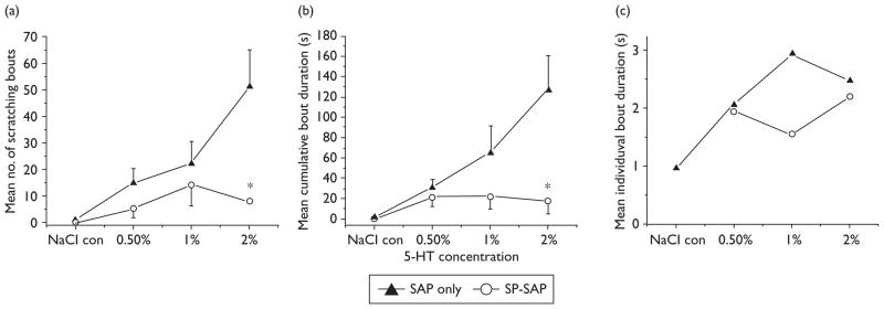
Itch is thought to be signaled by pruritogen-responsive neurons in the superficial spinal dorsal horn. Many neurons here express the substance P NK-1 receptor. We investigated whether neurotoxic destruction of spinal NK-1-expressing neurons affected itch-related scratching behavior. Rats received intracisternal substance P conjugated to saporin (SP-SAP), or saporin (SAP) only (controls), and were subsequently tested for scratching behavior elicited by intradermal 5-hydroxytryptamine. SAP controls exhibited dose-related hindlimb scratching, which was significantly attenuated in SP-SAP-treated rats. There was a virtual absence of NK-1 immunoreactive neurons in superficial laminae of the upper cervical and medullary dorsal horn in SP-SAP-treated rats. These results indicate that superficial dorsal horn neurons expressing NK-1 receptors play a key role in spinal itch transmission.


Figures

Fig. 1
Fig. 1
NK-1 receptor immunoreactivity in the superficial medullary and cervical spinal dorsal horn. Left column (a–d) shows sections from a rat pretreated with saporin (SAP), at the levels of (a) nucleus of solitary tract (NTS), (b) trigeminal subnucleus caudalis (Vc) in caudal medulla, (c) second cervical (C2) segment, and (d) lower cervical-upper thoracic (C8-T1) segment. Note extensive NK-1 immunoreactivity in superficial laminae of the medullary and spinal dorsal horns at all levels, as well as in NTS. This pattern of NK-1 staining was very similar in all 7 animals receiving intracisternal SAP. Calibration bar applies to all photomicrographs. Right column (e–h) similarly shows NK-1 immunoreactivity in an animal pretreated with substance P conjugated to saporin (SP-SAP) at the levels of (e) NTS, (f) Vc, (g) C2, and (h) C8-T1. All 11 animals receiving intracisternal SP-SAP exhibited a virtual absence of NK-1 immunoreactivity in the superficial dorsal horn down to mid-cervical levels, with heavy NK-1 staining in NTS comparable to that observed in SAP-treated animals. Three animals exhibited some degree of NK-1 immunoreactivity at lower cervical and upper thoracic levels (h).
Fig. 2
Fig. 2
Reduced 5-hydroxytryptamine (5-HT)-evoked scratching in rats receiving substance P conjugated to saporin (SP-SAP). (a–d) Bar graphs show, from left to right, the mean number of scratching bouts/2 min intervals evoked by intradermal injection of NaCl (a, vehicle control) and 5-HT at concentrations of (b) 0.5% (23.5 mM), (c) 1% (47 mM), and (d) 2% (94 mM). Filled bars: SAP; open bars, SP-SAP. Error bars: SEM. (e–g) Bar graphs plot total cumulative duration of 5-HT-evoked scratching bouts (recorded at 2-min intervals).
Fig. 3
Fig. 3
Summary of scratching data. (a) Graph plots mean number of scratching bouts versus 5-hydroxytryptamine (5-HT) concentration to show significant reduction in substance P conjugated to saporin (SP-SAP)-treated rats. Error bars: SEM. *Overall significant between-group difference (analysis of variance, P<0.01) with the 2% treatment group significantly different from vehicle and 0.5% (least significant difference; P<0.05). (b) Graph of mean cumulative duration of scratching bouts. *Significant (analysis of variance, P<0.01) between-group difference. (c) The mean individual bout duration did not significantly vary between groups across the range of 5-HT concentrations tested.

References

    1. Sun YG, Zhao ZQ, Meng XL, Yin J, Liu XY, Chen ZF. Cellular basis of itch sensation. Science. 2009;325:1531–1534. - PMC - PubMed
    1. Sun YG, Chen ZF. A gastrin-releasing peptide receptor mediates the itch sensation in the spinal cord. Nature. 2007;448:700–703. - PubMed
    1. Brown JL, Liu H, Maggio JE, Vigna SR, Mantyh PW, Basbaum AI. Morphological characterization of substance P receptor-immunoreactive neurons in the rat spinal cord and trigeminal nucleus caudalis. J Comp Neurol. 1995;356:327–344. - PubMed
    1. Mantyh PW, Rogers SD, Honore P, Allen BJ, Ghilardi JR, Li J, et al. Inhibition of hyperalgesia by ablation of lamina I spinal neurons expressing the substance P receptor. Science. 1997;278:275–279. - PubMed
    1. Hill R. NK1 (substance P) receptor antagonists – why are they not analgesic in humans? Trends Pharmacol Sci. 2000;21:244–246. - PubMed

Publication types

MeSH terms