Review
. 2010 Mar;8(3):231-41.
doi: 10.1038/nrmicro2296. Epub 2010 Feb 2.

Viral shape-shifting: norovirus evasion of the human immune system

Affiliations
Review

Viral shape-shifting: norovirus evasion of the human immune system

Eric F Donaldson et al. Nat Rev Microbiol. 2010 Mar.

Abstract

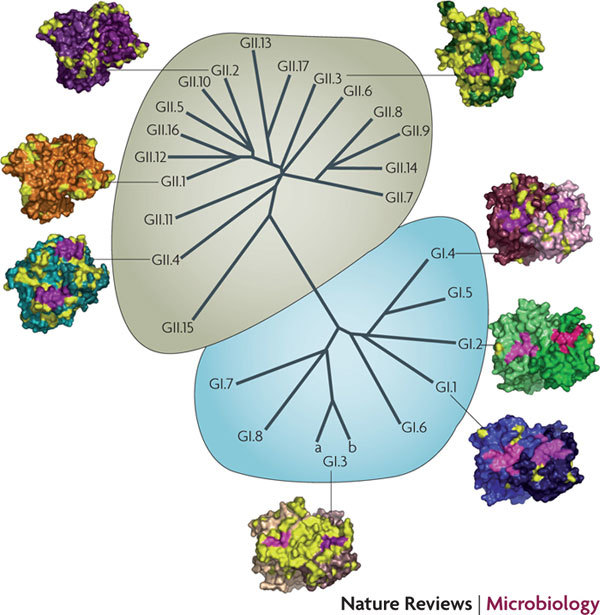
Noroviruses are the most common cause of food-borne gastroenteritis worldwide, and explosive outbreaks frequently occur in community settings, where the virus can immobilize large numbers of infected individuals for 24-48 hours, making the development of effective vaccines and antiviral therapies a priority. However, several challenges have hampered therapeutic design, including: the limitations of cell culture and small-animal model systems; the complex effects of host pre-exposure histories; differential host susceptibility, which is correlated with blood group and secretor status; and the evolution of novel immune escape variants. In this Review, we discuss the molecular and structural mechanisms that facilitate the persistence of noroviruses in human populations.


Conflict of interest statement

The authors declare no competing financial interests.

Figures

Figure 1
Figure 1. Genome organization and capsid structure.
a| The norovirus genome is composed of three open reading frames (ORFs). ORF1 (∼5 kb) is located in the first two-thirds of the genome and encodes a ∼200 kDa polyprotein that is auto-processed by a virally encoded 3C-like protease (3C) to yield the non-structural replicase proteins that are essential for viral replication. The resultant proteins are: p48, an amino-terminal protein of unknown function (∼48 kDa); nucleoside triphosphatase (NTP), a 2C-like protein; p22, a 22 kDa 3A-like protein; viral genome-linked protein (VPG), a protein that is covalently linked to the 5′ end of the genome; and RNA-directed RNA polymerase (RdRp), a 3D-like protein. ORF2 is 1.8kb in length and encodes the 57 kDa major structural capsid protein, viral protein 1 (VP1). VP1 is divided into two domains, the shell domain (yellow) and the protruding domain, which is further divided into two subdomains known as P1 (blue) and P2 (red). ORF3 is ∼0.6 kb in length and encodes a 22 kDa minor basic structural protein, VP2 (Ref. 89). b | The structure of the VP1 monomer is shown, with protein domains coloured as for part a. c | Two capsid protein monomers form the A–B dimer (indicated with the A monomer in lighter shades and the B monomer in darker shades), which allows the P2 domain to protrude from the viral particle. d |The virus-like particle is formed of 180 monomers of the capsid protein that assemble through different dimers. The A–B dimer, shown in colour, extends away from the capsid and provides the receptor-binding region and the sites of antigenic variation. In the virus particle, VP2 is incorporated in low copy number. Structural models were generated and pictures were rendered using MacPyMOL (Delano Scientific LLC, Palo Alto, California, USA).
Figure 2
Figure 2. Phylogenetics of the norovirus capsid protein.
A Bayesian phylogenetic tree of representative norovirus capsid protein sequences from all major genogroups and genotypes. Labels indicate the GI and GII genogroups that predominantly infect humans, and red brackets represent genotypes that seem to be evolving. The tree was generated with MRBAYES, using sapovirus Nongkhai 50 to root the tree.
Figure 3
Figure 3. Variation in noroviruses that infect humans.
The GI genogroup is highlighted in blue, and the GII genogroup is highlighted in green. The P domain dimer structures show the predominant genotypes that have infected humans over the past decade in the United States, recognizing that this may vary elsewhere, globally. Fuschia represents the histo-blood group antigen-binding sites and yellow indicates variation in the P2 subdomains. Structural models were generated using the program Modeller, and pictures were generated using MacPyMOL (Delano Scientific LLC, Palo Alto, California, USA).
Figure 4
Figure 4. Genogroup variation.
A | Variation in the GI genogroup. The crystal structure of the P domain of the genogroup I, genotype 1 Norwalk virus (GI.1-NV) was used to generate representative models of all eight GI genotypes. P domain chains are in blue, the histo-blood group antigen (HBGA)-binding sites are in magenta, a conserved structural domain is shown in red and all additional GI models are in black mesh. A a | The annotated GI.1-NV P domain structure. A b | The GI.1-NV P domain monomer with additional GI monomer models superimposed on the structure. A c | The GI.1-NV P domain dimer with additional GI dimer models superimposed on the structure. Note that the binding pocket and conserved domain are mostly preserved. B | Variation in the GII genogroup. The crystal structure of the P domain of GII.4 norovirus VA387 was used to generate representative models of all 17 GII P domains. GII.4 P domain chains are in cyan, the HBGA-binding sites are orange and all additional GII P domain models are in purple mesh. B a | The annotated GII.4 VA387 P domain structure. B b | The GII.4 VA387 P domain monomer with additional GII monomer models superimposed. B c | The GII.4 VA387 P domain dimer with additional GII P domain dimer models superimposed. Note that the binding pockets are structurally different in the GII models. C | Superimposing the GII and GI structural space. Ca | Superimposition of the GII.4 VA387 P domain dimer structure (shown in mesh) onto the GI. 1-NV P domain dimer structure indicates that GII.4 VA387 occupies more structural space than GI.1-NV (the colours of domains are as for parts A and B). Cb | Superimposing the GII monomer structure, indicated by the purple mesh, onto the GI monomer structure, shown in black, indicates that the GII genogroup occupies substantially more space. Cc | Superimposing the GII dimer structure, indicated by the purple mesh, onto the GI dimer structure, shown in black, indicates that the GII genogroup occupies more overall structural space. The additional structural space may allow the GII genogroup more structural flexibility, so that it could hypothetically tolerate more mutations while preserving its capacity to bind a differential receptor repertoire. Structural models were generated using the program Modeller and pictures were generated using MacPyMOL (Delano Scientific LLC, Palo Alto, California, USA).
Figure 5
Figure 5. Model of GI versus GII evolution in human populations.
a | Noroviruses of the GI genogroup seem to be limited in the amount of variation that occurs in the P2 subdomain of the capsid, which reduces the ability of the virus to generate antibody escape mutants. Therefore, viruses in the GI genogroup probably persist by evolving differential binding capacity or through original antigenic sin (OAS) of the host. b | By contrast, the GII genogroup contains more sequence information in the P2 subdomain, which can tolerate more substantial changes, probably allowing evolution to rearrange the surface and alter both binding capacity and antigenic properties. Therefore, as has been shown for GII.4, GII noroviruses are likely to persist by two mechanisms: receptor switching and antigenic drift.

References

    1. Atmar RL, Estes MK. The epidemiologic and clinical importance of norovirus infection. Gastroenterol. Clin. North Am. 2006;35:275–290. doi: 10.1016/j.gtc.2006.03.001. - DOI - PubMed
    1. CDC. Norovirus activity — United States, 2002. Morb. Mortal. Wkly Rep.52, 41–45 (2003). - PubMed
    1. Ike AC, et al. Molecular epidemiology of norovirus in outbreaks of gastroenteritis in southwest Germany from 2001 to 2004. J. Clin. Microbiol. 2006;44:1262–1267. doi: 10.1128/JCM.44.4.1262-1267.2006. - DOI - PMC - PubMed
    1. Jiang X, et al. Outbreaks of gastroenteritis in elderly nursing homes and retirement facilities associated with human caliciviruses. J. Med. Virol. 1996;50:335–341. doi: 10.1002/(SICI)1096-9071(199612)50:4<335::AID-JMV9>3.0.CO;2-9. - DOI - PubMed
    1. Mead PS, et al. Emerg. Infect. Dis. 1999;5:607–625. doi: 10.3201/eid0505.990502. - DOI - PMC - PubMed

Publication types