

doi: 10.1364/OL.35.000426.
High-speed line-scan confocal imaging of stimulus-evoked intrinsic optical signals in the retina
Affiliations
- PMID: 20125743
- PMCID: PMC2921995
- DOI: 10.1364/OL.35.000426
Item in Clipboard
High-speed line-scan confocal imaging of stimulus-evoked intrinsic optical signals in the retina
Opt Lett.
.
Abstract
A rapid line-scan confocal imager was developed for functional imaging of the retina. In this imager, an acousto-optic deflector was employed to produce mechanical vibration- and inertia-free light scanning, and a high-speed (68,000 Hz) linear CCD camera was used to achieve subcellular and submillisecond spatiotemporal resolution imaging. Two imaging modalities, i.e., frame-by-frame and line-by-line recording, were validated for the reflected light detection of intrinsic optical signals (IOSs) in visible light stimulus activated frog retinas. Experimental results indicated that fast IOSs were tightly correlated with retinal stimuli and could track visible light flicker stimulus frequency up to at least 2 Hz.
Figures
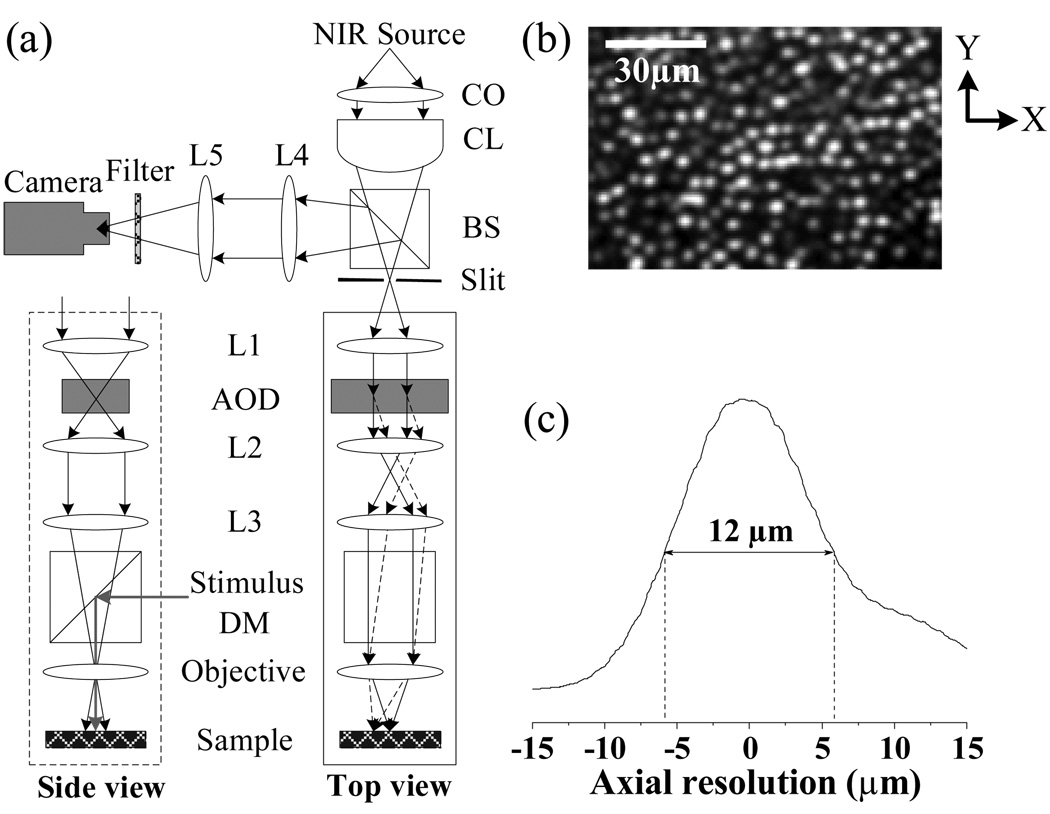
(a) Schematic diagram of line-scan confocal system. CO: collimator; CL: cylindrical lens BS: beam splitter; AOD: acousto-optic deflector; DM: dichroic mirror; Lx: spherical lenses. Focal length of the CL is 50 mm. Focal length of the lenses L1–L5 are 80mm, 60mm, 120mm, 60mm, and 100mm, respectively. (b) Confocal image showing clear cellular structure of individual photoreceptors. In the x direction (i.e., parallel to the focused scanning line), theoretical resolution of the imager is ~ 1.9 µm (0.61λ/NA). In the y direction (i.e., perpendicular to the focused scanning line), theoretical resolution of the imager is ~ 1.3 µm (0.4λ/NA). (c) Axial resolution of line-scan confocal imaging system was tested to be ~ 12 µm.
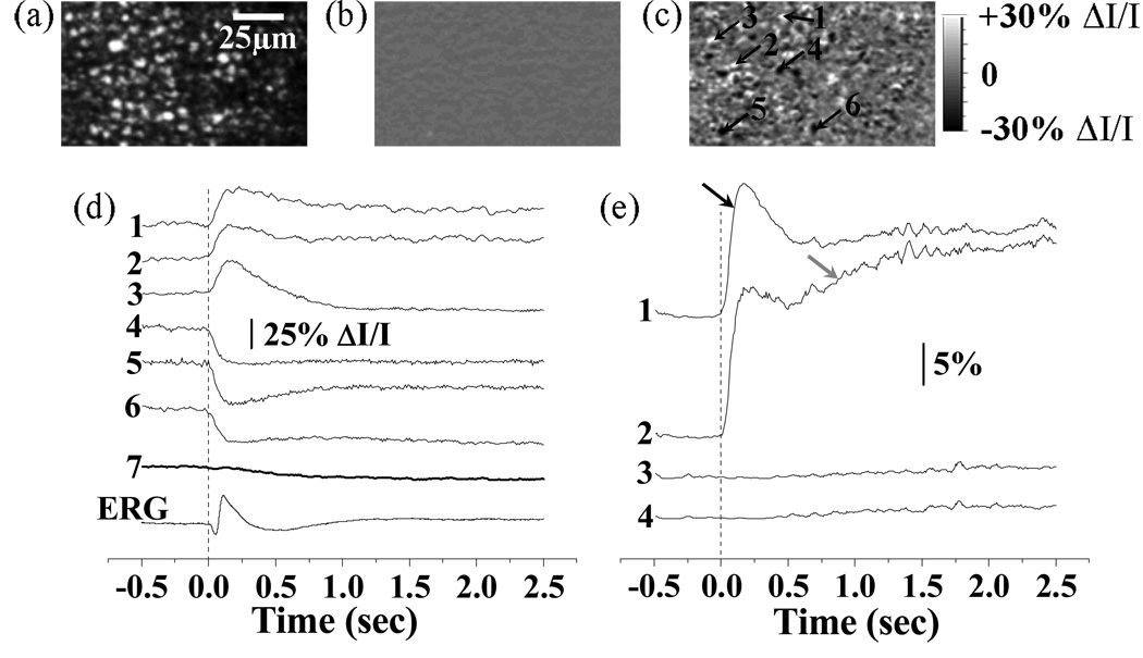
Frame-by-frame imaging of IOSs. (a) Raw image of photoreceptors. (b) Pre-stimulus IOS image. (c) Post-stimulus IOSs image. The raw confocal images were acquired at the speed of 100 frames/s, with NIR imaging light focused at photoreceptor layer. Both pre-stimulus and post-stimulus IOSs images were an average over 250 ms. (d) Tracings 1–6 revealed the IOSs variation property of local areas pointed by arrowheads 1–6 in (c). Tracing 7 represents integral IOSs by averaging all the pixels of each IOS image. Vertical line indicates the onset of the stimulus. (e) The percentage statistics of activated retinal areas with positive (> 5% ΔI/I) and negative (< −5% ΔI/I) IOSs. The threshold (5%ΔI/I) was used to reduce the effect of background noise on the statistics. Trace 1 (positive IOSs) and 2 (negative IOSs) are statistic results of experiment trial with stimulus delivered. Trace 3 (positive IOSs) and 4 (negative IOSs) are statistic results of control trial without stimulus.
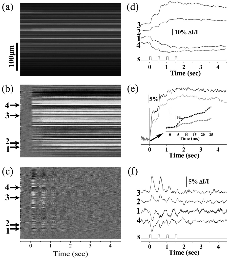
Line-by-line imaging of IOSs. (a) Line-by-line raw image; (b) Line-by-line IOS image; (c) Line-by-line DIOS image. (d) Representative localized IOSs (arrows 1–4). S: visible light stimulus. (e) The statistics of activated retinal area ratios with positive (black) and negative (grey) IOSs. Inset panel shows enlarged picture of early optical response marked by the gray bar. Rapid IOSs occurred within 5ms after stimulus onset (inset panel). (f) Representative localized DIOSs (arrows 1–4).
Similar articles
-
In vivo confocal imaging of fast intrinsic optical signals correlated with frog retinal activation.Opt Lett. 2011 Dec 1;36(23):4692-4. doi: 10.1364/OL.36.004692. Opt Lett. 2011. PMID: 22139286 Free PMC article.
-
In vivo confocal intrinsic optical signal identification of localized retinal dysfunction.Invest Ophthalmol Vis Sci. 2012 Dec 13;53(13):8139-45. doi: 10.1167/iovs.12-10732. Invest Ophthalmol Vis Sci. 2012. PMID: 23150616 Free PMC article.
-
Intrinsic optical signal imaging of retinal activation.Jpn J Ophthalmol. 2009 Jul;53(4):327-33. doi: 10.1007/s10384-009-0685-4. Epub 2009 Sep 8. Jpn J Ophthalmol. 2009. PMID: 19763749 Review.
-
High spatiotemporal resolution imaging of fast intrinsic optical signals activated by retinal flicker stimulation.Opt Express. 2010 Mar 29;18(7):7210-8. doi: 10.1364/OE.18.007210. Opt Express. 2010. PMID: 20389742 Free PMC article.
-
In Vivo Observations of Rapid Scattered Light Changes Associated with Neurophysiological Activity.In: Frostig RD, editor. In Vivo Optical Imaging of Brain Function. 2nd edition. Boca Raton (FL): CRC Press/Taylor & Francis; 2009. Chapter 5. In: Frostig RD, editor. In Vivo Optical Imaging of Brain Function. 2nd edition. Boca Raton (FL): CRC Press/Taylor & Francis; 2009. Chapter 5. PMID: 26844322 Free Books & Documents. Review.
Cited by
-
Investigation of the hyper-reflective inner/outer segment band in optical coherence tomography of living frog retina.J Biomed Opt. 2012 Jun;17(6):060504. doi: 10.1117/1.JBO.17.6.060504. J Biomed Opt. 2012. PMID: 22734727 Free PMC article.
-
Two-photon excited autofluorescence imaging of freshly isolated frog retinas.Biomed Opt Express. 2011 Jun 1;2(6):1494-503. doi: 10.1364/BOE.2.001494. Epub 2011 May 11. Biomed Opt Express. 2011. PMID: 21698013 Free PMC article.
-
In vivo optical coherence tomography of stimulus-evoked intrinsic optical signals in mouse retinas.J Biomed Opt. 2016 Sep 1;21(9):96010. doi: 10.1117/1.JBO.21.9.096010. J Biomed Opt. 2016. PMID: 27653936 Free PMC article.
-
Functional imaging of retinal photoreceptors and inner neurons using stimulus-evoked intrinsic optical signals.Methods Mol Biol. 2012;884:277-85. doi: 10.1007/978-1-61779-848-1_20. Methods Mol Biol. 2012. PMID: 22688714 Free PMC article.
-
In vivo intrinsic optical signal imaging of mouse retinas.Proc SPIE Int Soc Opt Eng. 2016 Feb 13;9693:96930H. doi: 10.1117/12.2212810. Epub 2016 Mar 4. Proc SPIE Int Soc Opt Eng. 2016. PMID: 28163346 Free PMC article.
References
-
- Cohen LB, Keynes RD, Hille B. Light scattering and birefringence changes during nerve activity. Nature. 1968;218(5140):438–441. - PubMed
-
- Nelson DA, Krupsky S, Pollack A, Aloni E, Belkin M, Vanzetta I, Rosner M, Grinvald A. Special report: Noninvasive multi-parameter functional optical imaging of the eye. Ophthalmic Surg Lasers Imaging. 2005;36(1):57–66. - PubMed
Publication types
MeSH terms
Grants and funding
LinkOut - more resources
Full Text Sources
