. 2010 Feb 2;5(2):e9020.
doi: 10.1371/journal.pone.0009020.

Role of the HCF-1 basic region in sustaining cell proliferation

Affiliations

Role of the HCF-1 basic region in sustaining cell proliferation

Marco Mangone et al. PLoS One. .

Abstract

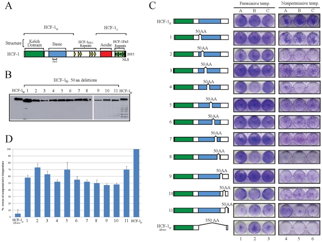
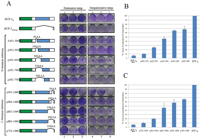
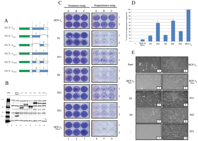
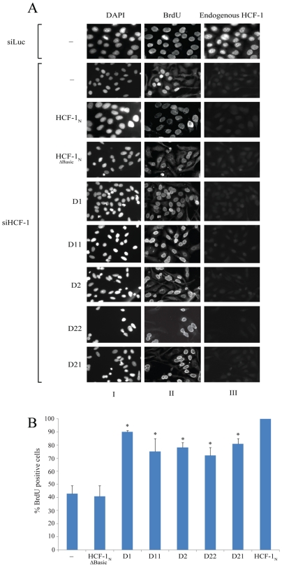
Background: The human herpes simplex virus-associated host cell factor 1 (HCF-1) is a conserved human transcriptional co-regulator that links positive and negative histone modifying activities with sequence-specific DNA-binding transcription factors. It is synthesized as a 2035 amino acid precursor that is cleaved to generate an amino- (HCF-1(N)) terminal subunit, which promotes G1-to-S phase progression, and a carboxy- (HCF-1(C)) terminal subunit, which controls multiple aspects of cell division during M phase. The HCF-1(N) subunit contains a Kelch domain that tethers HCF-1 to sequence-specific DNA-binding transcription factors, and a poorly characterized so called "Basic" region (owing to a high ratio of basic vs. acidic amino acids) that is required for cell proliferation and has been shown to associate with the Sin3 histone deacetylase (HDAC) component. Here we studied the role of the Basic region in cell proliferation and G1-to-S phase transition assays.

Methodology/principal findings: Surprisingly, much like the transcriptional activation domains of sequence-specific DNA-binding transcription factors, there is no unique sequence within the Basic region required for promoting cell proliferation or G1-to-S phase transition. Indeed, the ability to promote these activities is size dependent such that the shorter the Basic region segment the less activity observed. We find, however, that the Basic region requirements for promoting cell proliferation in a temperature-sensitive tsBN67 cell assay are more stringent than for G1-to-S phase progression in an HCF-1 siRNA-depletion HeLa-cell assay. Thus, either half of the Basic region alone can support G1-to-S phase progression but not cell proliferation effectively in these assays. Nevertheless, the Basic region displays considerable structural plasticity because each half is able to promote cell proliferation when duplicated in tandem. Consistent with a potential role in promoting cell-cycle progression, the Sin3a HDAC component can associate independently with either half of the Basic region fused to the HCF-1 Kelch domain.

Conclusions/significance: While conserved, the HCF-1 Basic region displays striking structural flexibility for controlling cell proliferation.


Conflict of interest statement

Competing Interests: The authors have declared that no competing interests exist.

Figures

Figure 1
Figure 1. The HCF-1 Basic region does not contain a unique essential region to promote cell proliferation.
A) Schematic structure of human HCF-1. Structural elements and subunit boundaries are shown above. The small bracket below the figure highlights the Sin3 binding site. B) Western blot analysis of the expression of the eleven 50 AA deletion-scanning mutants (labeled 1 to 11) in COS7 cells. C) Schematic of the eleven 50 AA scanning deletion mutants (labeled 1 to 11) and tsBN67 colony assay. Plates resulting from growth at the control permissive (left) and selective nonpermmissive (right) temperatures are shown. Each clone has been tested in triplicate as shown in columns A, B, and C. D) Spectrophotometric quantitation of the ability of deletion mutants to rescue cell proliferation at the nonpermissive temperature. The error bars are standard error of the mean.
Figure 2
Figure 2. HCF-1 Basic region deletion mutants rescue cell proliferation in a size-dependent manner.
A) Schematics of the nested set of N-terminal and C-terminal HCF-1 deletion mutants and tsBN67 colony assay. B and C) Spectrophotometric quantitation of the ability of N- and C-terminal deletion mutants to rescue cell proliferation at the nonpermissive temperature. The error bars are standard error of the mean.
Figure 3
Figure 3. HCF-1 Basic region duplications can rescue tsBN67 colony forming activity at the nonpermissive temperature.
A) Schematic of the HCF-1 Basic region duplication mutants. Each domain, labeled 1 and 2, has been selectively deleted, duplicated or placed in inverted position. B) Western blot analysis of the expression of HCF-1 Basic region mutants in 293 cells. Red asterisks indicate the expected proteins based on their predicted size. C) tsBN67 colony formation assays. Each clone has been tested at permissive and nonpermissive temperature in triplicate as shown in columns A, B, and C and plated in duplicate. D) Spectrophotometric quantitation of the ability of deletion mutants to rescue cell proliferation at the nonpermissive temperature. The error bars are standard error of the mean. E) Colony morphology of selected clones at nonpermissive temperature.
Figure 4
Figure 4. HCF-1 Basic region Domains 1 and 2 can individually promote HeLa cell G1-to-S phase progression.
HeLa cells that stably express different HCF-1 silencing resistance mutants were tested for their ability to bypass G1-to-S arrest induced by silencing of endogenous HCF-1 with RNAi. A) Immunofluorescence analysis of HeLa cells silenced with Luciferase RNAi oligos and with HCF-1 siRNA oligos. Three panels are showed for each clone tested. The left panel shows DNA staining with DAPI. The middle panel shows cells probed with antibody for BrdU incorporation. The right panel shows the level of endogenous HCF-1 remaining after siRNA treatment as detected by the HCF-1C antibody (αH12). B) Percentage of BrdU positive cells after 72 hours of siRNA silencing as determined by the immunofluorescence results normalized to HCF-1N1011. Sample sizes were as follows: HeLa, n = 1127; HCF-1N1011-ΔBasic, n = 881; HCF-1N1011-D1, n = 1920; HCF-1N1011-D11, n = 859; HCF-1N1011-D2, n = 705; HCF-1N1011-D22, n = 1798; HCF-1N1011-D21, n = 1478; and HCF-1N1011, n = 1475. The error bars are standard error of the mean. *p<0.05 (Mann-Whitney U test), comparing the HCF-1N mutants to HCF-1N1011-ΔBasic.
Figure 5
Figure 5. Multiple regions within the HCF-1 Basic region associate with Sin3a.
αFLAG immunoprecipitates of the stable HeLa cell lines with the single, duplicated, and swapped Basic region Domains 1 and 2 were analyzed by immunoblot with an αSin3a antisera. A) Recovery of the FLAG tagged HCF-1 recombinant proteins was verified by immunoblot analysis with the αFLAG antibody. B) Sin3a in the supernatant (bottom) and immunoprecipitates (top) was determined by αSin3a immunoblotting.

References

    1. Wilson AC, LaMarco K, Peterson MG, Herr W. The VP16 accessory protein HCF is a family of polypeptides processed from a large precursor protein. Cell. 1993;74:115–125. - PubMed
    1. Wilson AC, Peterson MG, Herr W. The HCF repeat is an unusual proteolytic cleavage signal. Genes Dev. 1995;9:2445–2458. - PubMed
    1. Kristie TM, Pomerantz JL, Twomey TC, Parent SA, Sharp PA. The cellular C1 factor of the herpes simplex virus enhancer complex is a family of polypeptides. J Biol Chem. 1995;270:4387–4394. - PubMed
    1. Goto H, Motomura S, Wilson AC, Freiman RN, Nakabeppu Y, et al. A single-point mutation in HCF causes temperature-sensitive cell-cycle arrest and disrupts VP16 function. Genes Dev. 1997;11:726–737. - PubMed
    1. Reilly PT, Herr W. Spontaneous reversion of tsBN67 cell proliferation and cytokinesis defects in the absence of HCF-1 function. Exp Cell Res. 2002;277:119–130. - PubMed

Publication types

MeSH terms

Substances