. 2010 Feb 2;5(2):e8999.
doi: 10.1371/journal.pone.0008999.

Planar cell polarity enables posterior localization of nodal cilia and left-right axis determination during mouse and Xenopus embryogenesis

Affiliations

Planar cell polarity enables posterior localization of nodal cilia and left-right axis determination during mouse and Xenopus embryogenesis

Dragana Antic et al. PLoS One. .

Abstract

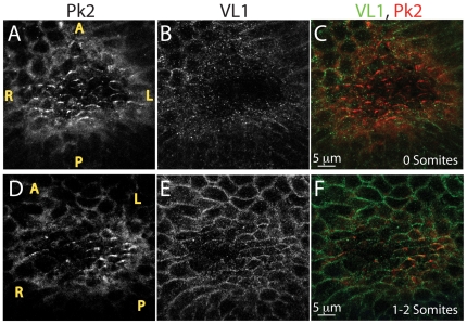
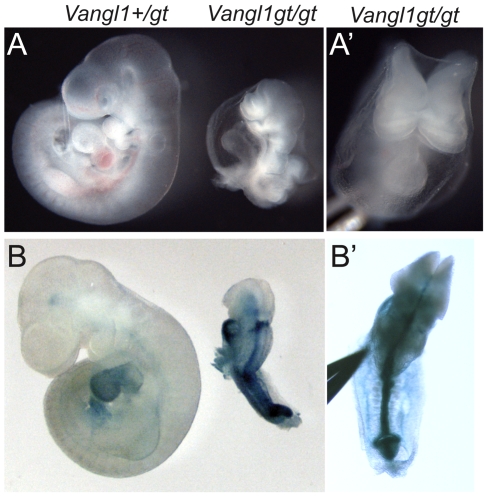
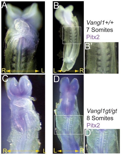
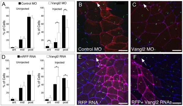
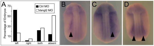
Left-right asymmetry in vertebrates is initiated in an early embryonic structure called the ventral node in human and mouse, and the gastrocoel roof plate (GRP) in the frog. Within these structures, each epithelial cell bears a single motile cilium, and the concerted beating of these cilia produces a leftward fluid flow that is required to initiate left-right asymmetric gene expression. The leftward fluid flow is thought to result from the posterior tilt of the cilia, which protrude from near the posterior portion of each cell's apical surface. The cells, therefore, display a morphological planar polarization. Planar cell polarity (PCP) is manifested as the coordinated, polarized orientation of cells within epithelial sheets, or as directional cell migration and intercalation during convergent extension. A set of evolutionarily conserved proteins regulates PCP. Here, we provide evidence that vertebrate PCP proteins regulate planar polarity in the mouse ventral node and in the Xenopus gastrocoel roof plate. Asymmetric anterior localization of VANGL1 and PRICKLE2 (PK2) in mouse ventral node cells indicates that these cells are planar polarized by a conserved molecular mechanism. A weakly penetrant Vangl1 mutant phenotype suggests that compromised Vangl1 function may be associated with left-right laterality defects. Stronger functional evidence comes from the Xenopus GRP, where we show that perturbation of VANGL2 protein function disrupts the posterior localization of motile cilia that is required for leftward fluid flow, and causes aberrant expression of the left side-specific gene Nodal. The observation of anterior-posterior PCP in the mouse and in Xenopus embryonic organizers reflects a strong evolutionary conservation of this mechanism that is important for body plan determination.


Conflict of interest statement

Competing Interests: The authors have declared that no competing interests exist.

Figures

Figure 1
Figure 1. VANGL1 and PRICKLE2 set up PCP in the mouse ventral node.
(A–F) Localization of VL1 and PK2 proteins in the ventral node: PRICKLE2 is expressed in the node of 0 somite embryos (A, C red) prior to VANGL1, which is detected in node cells (E, F, green) of 1–2 somite embryos. VL1 and PK2 co-localize in node cells and form crescents pointed toward the anterior (F, yellow). Motile cilia can be visualized above the plane of VL1 or PK2 localization (not shown). Yellow letters mark anterior (A), posterior (P), Left (L) and right (R). As nodes were imaged from the ventral side, left side of the embryo is on the right side of each panel and right side of the embryo is on the left side of each panel.
Figure 2
Figure 2. Vangl1gt/gt homozygous embryos fail to turn.
Vangl1+/gt heterozygous embryos are normal and they develop into viable, fertile adults. In contrast, ∼14% of Vangl1gt/gt mutants die at E9.5–10.5. (A' , B') Cranial (A') and dorsal (B') views of Vangl1gt/gt mutant embryos shown in A and B showing open neural tube defects. (B, B') One mutant and its heterozygous littermate were stained for VANGL1-βGEO fusion protein. Each of five phenotypically mutant Vangl1gt/gt homozygotes isolated at this stage failed to turn from lordotic to fetal position.
Figure 3
Figure 3. Aberrant Pitx2 expression in a Vangl1gt/gt homozygous embryo.
(A, B) Wild-type embryos with 7 somites (A, B, B') have Pitx2 expressed in the left lateral plate mesoderm (LPM). (C, D) A Vangl1gt/gt homozygous embryo with 8 somites has Pitx2 expressed bilaterally but predominant expression is in the right LPM. (D') Somites of the Vangl1gt/gt homozygous mutant are narrow and compressed in the anterior-posterior direction, in comparison to the wild-type embryo (B'). The mis-expression of Pitx2 and the turning defect suggest that Vangl1 might regulate L-R asymmetry establishment in the mouse ventral node. (B') and (D') are the boxed regions from (B) and (D) and are at the same magnification.
Figure 4
Figure 4. Vangl2 Morpholino or RNA alters positioning of cilia in the GRP.
(A, D) A clone of cells in the GRP was generated by injecting one dorsal blastomere of 8-cell stage embryos with Vangl2 MO or a Vangl2 RNA, along with RFP RNA as a tracer or as a control. At stages 17/18, the dorsal half of the embryo was removed, fixed, and stained with antibodies directed to ZO-1 and acetylated–tubulin to mark cell boundaries and cilia, respectively. GRP cilia both inside (injected) and outside (uninjected) the clone were scored based on the location in GRP cells that were divided into three equal zones (anterior, middle and posterior). Shown are data from a minimum of 3 GRPs where 100–200 injected and uninjected cells were scored. Asterisks (* = p<0.05, ** = p<0.01.) represent p-values obtained using a two-tailed t-test. (B–C, E–F) Shown are confocal images of the GRP injected with the Vangl2 MO and RFP RNA (C), with Control MO and RFP RNA (B), with Vangl2 and RFP RNAs (F) or with RFP RNA alone (E). RFP (red) marks the position of the injected clone, cilia (green) are stained with the acetylated-tubulin antibody, and cell boundaries (blue) are stained with an antibody to ZO-1. Anterior is indicated by direction of the arrow in B, C, E, F.
Figure 5
Figure 5. Vangl2 MO knockdown disrupts left-right patterning.
(A) Xenopus embryos were injected with Vangl2 MO, or Control MO alone, targeting one dorsal blastomere at the 8-cell stage to generate a clone of injected cells within the GRP. At stages 20/21, embryos were fixed and probed by in-situ hybridization with an antisense Xenopus nodal-related1 (Xnr1) probe. Sidedness of staining was scored for at least 60 embryos for each condition. Data from one experiment are shown. (B–D) Shown are embryos injected with either Vangl2 or Control MOs and then probed with Xnr1 in-situ probe. Xnr1 staining is evident on the left (B) right (C) or both (D) sides of the midline. Black arrowheads indicate staining.

Similar articles

Cited by

References

    1. Basu B, Brueckner M. Cilia multifunctional organelles at the center of vertebrate left-right asymmetry. Curr Top Dev Biol. 2008;85:151–174. - PubMed
    1. Shiratori H, Hamada H. The left-right axis in the mouse: from origin to morphology. Development. 2006;133:2095–2104. - PubMed
    1. Nonaka S, Yoshiba S, Watanabe D, Ikeuchi S, Goto T, et al. De novo formation of left-right asymmetry by posterior tilt of nodal cilia. PLoS Biol. 2005;3:e268. - PMC - PubMed
    1. Hirokawa N, Tanaka Y, Okada Y, Takeda S. Nodal flow and the generation of left-right asymmetry. Cell. 2006;125:33–45. - PubMed
    1. Tabin CJ. The key to left-right asymmetry. Cell. 2006;127:27–32. - PubMed

Publication types

MeSH terms

LinkOut - more resources