. 2010 Feb 2;5(2):e9023.
doi: 10.1371/journal.pone.0009023.

The high affinity IgE receptor Fc epsilonRI is expressed by human intestinal epithelial cells

Affiliations

The high affinity IgE receptor Fc epsilonRI is expressed by human intestinal epithelial cells

Eva Untersmayr et al. PLoS One. .

Abstract

Background: IgE antibodies play a paramount role in the pathogenesis of various intestinal disorders. To gain insights in IgE-mediated pathophysiology of the gut, we investigated the expression of the high affinity IgE receptor Fc epsilonRI in human intestinal epithelium.

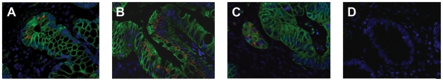
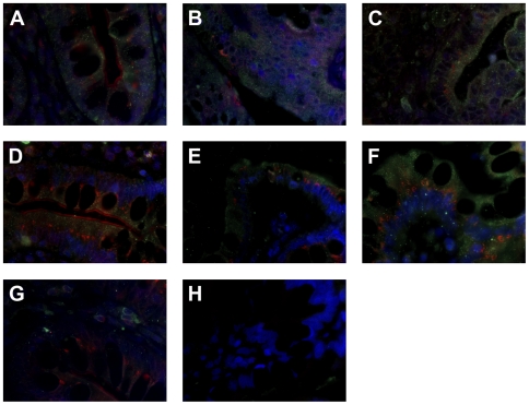
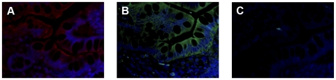
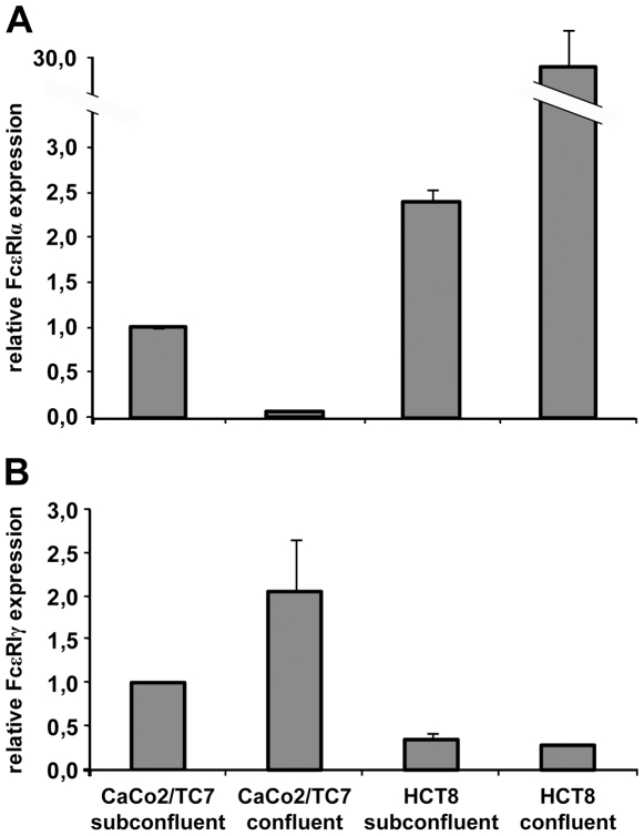
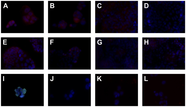
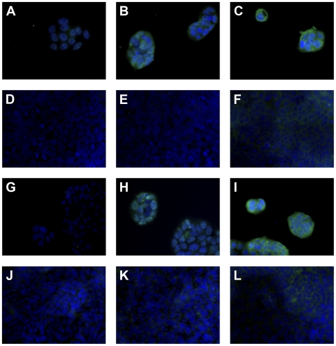
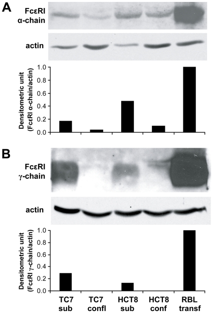
Methodology/principal findings: Fc epsilonRI alpha-chain, as detected by immunohistochemistry, was positive in epithelial cells for eight of eleven (8/11) specimens from colon cancer patients and 5/11 patients with inflammation of the enteric mucosa. The Fc epsilonRIalpha positive epithelial cells co-expressed Fc epsilonRIgamma, whereas with one exception, none of the samples was positive for the beta-chain in the epithelial layer. The functionality of Fc epsilonRI was confirmed in situ by human IgE binding. In experiments with human intestinal tumor cell lines, subconfluent Caco-2/TC7 and HCT-8 cells were found to express the alpha- and gamma-chains of Fc epsilonRI and to bind IgE, whereas confluent cells were negative for gamma-chains.

Conclusions/significance: Our data provide the first evidence that the components of a functional Fc epsilonRI are in vitro expressed by the human intestinal epithelial cells depending on differentiation and, more importantly, in situ in epithelia of patients with colon cancer or gastrointestinal inflammations. Thus, a contribution of Fc epsilonRI either to immunosurveillance or pathophysiology of the intestinal epithelium is suggested.


Conflict of interest statement

Competing Interests: The authors have declared that no competing interests exist.

Figures

Figure 1
Figure 1. Positive staining for FcεRI α-chain and defensin-5 is observed in serial sections from intestinal tissue.
(A) FcεRI α-chain is detected on the membrane, as well as in the cytoplasm of epithelial cells in small intestine of cancer patient No. 16. FcεRI α-chain positive cells are also found in (B) the colon and (C) a tumor sample from the same patient. (D) Staining with anti-defensin-5 antibodies confirmed that FcεRIα expressing cells at the basis of the small intestinal crypts are Paneth cells. Defensin-5 is expressed also in (E) colon and (F) tumor sample in same areas, but to a lesser extend when compared with FcεRI α-chain staining. Similar staining pattern are observed also for the CD patient No. 8, as FcεRI α-chain positive cells are detected in (G) the epithelium of small intestinal tissue, (H) colon and (I) lesional region. Defensin-5 positive cells are located at (J) the crypt basis of small intestine, (K) along the colon crypt, as well as in (L) the lesional region. Original magnification ×10, inset ×40.
Figure 2
Figure 2. FcεRI α-chain is expressed in epithelial cells.
Co-staining with anti- FcεRI α-chain (red) and anti-keratin 8 antibodies (green) verifies the epithelial expression of FcεRI in (A) the crypts of small intestinal section, where FcεRIα is found primarily in the supranuclear region, in (B) colon tissue and in (C) tumor sample of cancer patient No. 16. (D) Negative control with mouse IgG2b and rabbit IgG. The blue fluorescence DAPI staining indicates the nuclei. Original magnification ×40.
Figure 3
Figure 3. Co-expression of FcεRI α- and γ-chain, but not FcεRI α- and β-chain is observed in epithelial cells of intestinal tissue.
In sections from cancer patient No. 23 FcεRI γ-chain (green) is found to be co-expressed in FcεRIα (red) positive epithelial cells of (A) the small intestine, as well as in (B) colon tissue and (C) tumor sample. In addition, sections from CD patient No. 8 are double-positive for FcεRI α- and γ-chain in (D) the small intestinal, (E) colon and in (F) lesional tissue. (G) Only in the subepithelial tissue FcεRI β-chain (green) positive cells are detected as shown here in the crypts of small intestinal tissue. (H) Negative control with mouse IgG2b and goat IgG isotype control antibodies. The blue fluorescence DAPI staining indicates the nuclei. Original magnification ×64.
Figure 4
Figure 4. FcεRI positive tissue reveals IgE binding activity.
Immunofluorescence staining of serial sections from patient No. 8 revealed IgE binding in (A) FcεRI α-chain (red) positive epithelial cells being incubated with (B) monoclonal NiP-specific humanized IgE antibodies (green). (C) Negative control with PBS. The blue fluorescence DAPI staining indicates the nuclei. Original magnification ×40.
Figure 5
Figure 5. Expression of FcεRI α- and γ-chain but not β-chain mRNA in human intestinal epithelial cells.
The expression pattern of the FcεRI complex was analyzed by real-time PCR analysis using specific primers for detection of (A) FcεRI α-, β- (not shown), and (B) γ-chain. Target gene expression levels were normalized to the average of housekeeping genes and are depicted relative to the value of subconfluent Caco-2/TC7 cells. The values are presented as means +/− SD (n = 3) from one experiment. The results are representative of two independent experiments.
Figure 6
Figure 6. Abundant surface and cytoplasmatic FcεRI α-chain expression only in subconfluent human intestinal tumor cell lines.
Immunofluoresence staining for (A–H) FcεRIα is performed in (A, B) subconfluent and (C, D) confluent Caco2/TC7 as well as in (E, F) subconfluent and (G, H) confluent HCT-8. Triton-X-100 permeabilized (A, C, E, G) and untreated cells (B, D, F, H) were compared. (I–L) Representative control staining in subconfluent Caco2/TC7 with the (I, J) anti-lamin A/C or (K, L) unspecific murine IgG2b, (I, K) permeabilized or (J, L) untreated. The blue fluorescence DAPI staining indicates the nuclei. Original magnification ×40.
Figure 7
Figure 7. Abundant FcεRI γ-chain expression and IgE binding in subconfluent human intestinal tumor cell lines.
Immunofluorescence staining of (A, D, G, J) FcεRI β- and (B, E, H, K) FcεRI γ-chain are performed in (A–C) subconfluent and (D–F) confluent Caco2/TC7 and in (G–I) subconfluent and (J–L) confluent HCT-8. (C, F, I, L) According to the expression pattern of FcεRI in the subconfluent cells, IgE binding is observed exclusively in subconfluent, non-mature intestinal cells. The blue fluorescence DAPI staining indicates the nuclei. Original magnification ×40.
Figure 8
Figure 8. Western blot analysis reveals FcεRI in human intestinal tumor cell lines.
(A) FcεRI α-chain and (B) FcεRI γ-chain expression is investigated in Caco2/TC7 and HCT8 subconfluent and confluent cell lines. In all experiments RBL cells transfected with the human FcεRI receptor served as positive controls and the protein expression signal was normalized to the expression of house-keeping protein actin.

Similar articles

Cited by

References

    1. Brandtzaeg P. History of oral tolerance and mucosal immunity. Ann N Y Acad Sci. 1996;778:1–27. - PubMed
    1. Gounni AS, Lamkhioued B, Ochiai K, Tanaka Y, Delaporte E, et al. High-affinity IgE receptor on eosinophils is involved in defence against parasites. Nature. 1994;367:183–186. - PubMed
    1. Gurish MF, Bryce PJ, Tao H, Kisselgof AB, Thornton EM, et al. IgE enhances parasite clearance and regulates mast cell responses in mice infected with Trichinella spiralis. J Immunol. 2004;172:1139–1145. - PubMed
    1. Huber A, Genser D, Spitzauer S, Scheiner O, Jensen-Jarolim E. IgE/anti-IgE immune complexes in sera from patients with Crohn's disease do not contain food-specific IgE. Int Arch Allergy Immunol. 1998;115:67–72. - PubMed
    1. van Spreeuwel JP, Lindeman J, van Maanen J, Meyer CJ. Increased numbers of IgE containing cells in gastric and duodenal biopsies. An expression of food allergy secondary to chronic inflammation? J Clin Pathol. 1984;37:601–606. - PMC - PubMed

Publication types

MeSH terms