. 2010 Jan 29;6(1):e1000745.
doi: 10.1371/journal.ppat.1000745.

Protection of mice against lethal challenge with 2009 H1N1 influenza A virus by 1918-like and classical swine H1N1 based vaccines

Affiliations

Protection of mice against lethal challenge with 2009 H1N1 influenza A virus by 1918-like and classical swine H1N1 based vaccines

Balaji Manicassamy et al. PLoS Pathog. .

Abstract

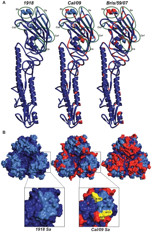
The recent 2009 pandemic H1N1 virus infection in humans has resulted in nearly 5,000 deaths worldwide. Early epidemiological findings indicated a low level of infection in the older population (>65 years) with the pandemic virus, and a greater susceptibility in people younger than 35 years of age, a phenomenon correlated with the presence of cross-reactive immunity in the older population. It is unclear what virus(es) might be responsible for this apparent cross-protection against the 2009 pandemic H1N1 virus. We describe a mouse lethal challenge model for the 2009 pandemic H1N1 strain, used together with a panel of inactivated H1N1 virus vaccines and hemagglutinin (HA) monoclonal antibodies to dissect the possible humoral antigenic determinants of pre-existing immunity against this virus in the human population. By hemagglutinination inhibition (HI) assays and vaccination/challenge studies, we demonstrate that the 2009 pandemic H1N1 virus is antigenically similar to human H1N1 viruses that circulated from 1918-1943 and to classical swine H1N1 viruses. Antibodies elicited against 1918-like or classical swine H1N1 vaccines completely protect C57B/6 mice from lethal challenge with the influenza A/Netherlands/602/2009 virus isolate. In contrast, contemporary H1N1 vaccines afforded only partial protection. Passive immunization with cross-reactive monoclonal antibodies (mAbs) raised against either 1918 or A/California/04/2009 HA proteins offered full protection from death. Analysis of mAb antibody escape mutants, generated by selection of 2009 H1N1 virus with these mAbs, indicate that antigenic site Sa is one of the conserved cross-protective epitopes. Our findings in mice agree with serological data showing high prevalence of 2009 H1N1 cross-reactive antibodies only in the older population, indicating that prior infection with 1918-like viruses or vaccination against the 1976 swine H1N1 virus in the USA are likely to provide protection against the 2009 pandemic H1N1 virus. This data provides a mechanistic basis for the protection seen in the older population, and emphasizes a rationale for including vaccination of the younger, naïve population. Our results also support the notion that pigs can act as an animal reservoir where influenza virus HAs become antigenically frozen for long periods of time, facilitating the generation of human pandemic viruses.


Conflict of interest statement

The authors have declared that no competing interests exist.

Figures

Figure 1
Figure 1. Differences in pathogenicity of 2009 pandemic H1N1 virus isolates in mice.
(A) Body weight of BALB/c mice infected with the Cal/09 isolate. Six-week old BALB/c mice were infected with indicated dose of virus and body weights were monitored everyday as measure of virus induced disease. (B) Neth/09 isolate is more pathogenic than Cal/09 isolate in mice. C57B/6 mice were infected with either Cal/09 or Neth/602 isolate of 2009 pandemic H1N1 virus at indicated doses. (C) Comparison of survival rates of BALB/c and C57B/6 mice infected with Cal/04 and Neth/602 viruses. (D) Viral titers in the lungs of BALB/c mice infected with either 1×10³ or 5×10⁴ pfu of the Cal/09 isolate. (E) Viral titers in the lungs of C57B/6 mice infected with either 1×10³ or 5×10⁴ pfu of Cal/09 or 5×10⁴ pfu of Neth/09. ND indicates that data was not determined. The body weights were measured everyday and are represented as a percentage of body weight prior to infection (n = 4 or 5 mice/group). Mice showing more than 25% of body weight loss were considered to have reached the experimental end point and were humanely euthanized. The lungs of 2–3 infected mice were excised and viral loads in the homogenates were measured by performing plaque assay in MDCK cells.
Figure 2
Figure 2. Prior infection with Cal/09 virus cross-protects mice against the lethal challenge with Neth/09 isolate.
(A, B) Determination of LD50 for the Neth/09 isolate in C57B/6 mice (n = 5/group). Mice were infected with the indicated doses of virus and their body weight (A) and survival (B) were monitored for 14 days. (C, D) Re-challenge of Cal/09 infected mice with Neth/09 (n = 4/group). Six weeks old C57B/6 mice were inoculated with PBS (Mock) or Cal/09 isolate (103 or 5×10⁴ pfu). After 21 days the mice were challenged with 1×10⁶ pfu of Neth/09 isolate. The body weight (C) and survival (D) were monitored for 14 days as in Fig. 1.
Figure 3
Figure 3. Vaccination of mice with 1918, 1943 and classical swine H1N1 virus-based inactive vaccines protects against Neth/09 lethal infection.
Five weeks old C57B/6 were immunized with 15 µg of the indicated inactivated viruses or with 1918 VLP vaccine followed by a boost (15 µg) after two weeks. Four weeks after the first immunization, vaccinated mice were challenged with Neth/09 isolate at a dose of 50 LD50 (7.9×10⁵ pfu). Mice were monitored for body weight loss and survival for 14 days as in Fig. 1. (A) Body weight of classical swine H1N1 (Sw/30, NJ/76), 1918 VLP and human H1N1 Wei/43 vaccinated mice after challenge with Neth/09 (n = 4 or 5 mice/group) (D) Body weight of mice vaccinated with contemporary (from 1977–2007) H1N1 or H3N2 inactivated virus after challenge with Neth/09 (n = 4 or 5 mice/group). (B, E) Survival of mice vaccinated with H1N1 and H3N2 viruses post-challenge. (C, F) Viral titers in H1N1 and H3N2 vaccinated mice on days 3 and 6 p.c. Each data point represents the average viral titers from 2 or 3 mice. A single group of control (no vaccine) mice were used and are included in all the panels for direct comparison. The Cal/09 vaccinated group is also included on (A) and (B) for comparison.
Figure 4
Figure 4. Passive immunization of mice with 1918 and 2009 H1N1 HA specific cross-reactive antibodies protects against Neth/09.
Nine week old, C57B/6 mice were passively immunized with a total 150 µg of indicated monoclonal antibody (intraperitoneal route) 24 hr prior to viral challenge. After antibody administration, the mice were challenged with Neth/09 isolate at a dose 50 LD50. (A) Body weight of passively immunized mice challenged with Neth/09 (n = 5/group). (B) Survival of passively immunized mice post-challenge with Neth/09. (C) Viral Titers in the lungs at days 3 and 6 p.c. Viral load in the lungs homogenates were quantified as in Fig. 1. Control (No Ab) mice are the same as controls in Fig. 3 and are included for direct comparison.
Figure 5
Figure 5. Comparison of the conservation of antigenic sites in HA of H1N1 viruses.
Alignment of HA sequences from USSR/77, Tx/91, Bris/59/07 and 2009 human H1N1 viruses (top panel). Alignment of HA sequences from 1918, Sw/30, Wei/43, NJ/76 and 2009 H1N1 viruses (bottom panel). The antigenic sites are indicated in colored shaded boxes and were previously described by Brownlee and Fodor , using H3 numbering, as Cb (78–83), Sa (128–129, 156–160, 162–167), Sb (187–198), Ca1 (169–173, 206–208, 238–240) and Ca2 (140–145, 224–226).
Figure 6
Figure 6. Comparison of antigenic differences in the HA structures of 1918, Cal/09 and Bris/59/07 viruses.
(A) Ribbon representation of an HA monomer (H1 and H2) of the three viruses. Individual antigenic sites are highlighted by green ellipses in the HA monomers. (B) Top panel: Zenithal surface top view of HA trimeric complexes depicting antigenic sites and their differences among 1918, Cal/09 and Bris/59/07 viruses. Blue color corresponds to conserved amino acids and red color represents amino acids that differ from 1918 HA. The antigenic sites are colored in light blue. Bottom panel: Close-up view of antigenic site Sa in monomeric 1918 and Cal/04 HAs. Amino acids K171, G172 and K180 (K157, G158 and K166 by H3 numbering) corresponding to mAb escape mutations are shown in yellow.

References

    1. Fiore AE, Shay DK, Broder K, Iskander JK, Uyeki TM, et al. Prevention and control of influenza: recommendations of the Advisory Committee on Immunization Practices (ACIP), 2008. MMWR Recomm Rep. 2008;57:1–60. - PubMed
    1. Palese P, Shaw ML. Orthomyxoviridae: the viruses and their replication. In: Howley DMKPM, editor. Fields virology. 5th edition. Philadelphia, PA: Lippincott Williams & Wilkins; 2007. pp. 1647–1689.
    1. Cox NJ, Neumann G, Donis RO, Kawaoka Y. Orthomyxoviruses: influenza. In: Mahy BWJ, ter Meulen V, editors. Topley and Wilson's microbiology and microbial infections. London: Hodder Arnold Press; 2005. pp. 634–698.
    1. Gillim-Ross L, Subbarao K. Emerging respiratory viruses: challenges and vaccine strategies. Clin Microbiol Rev. 2006;19:614–636. - PMC - PubMed
    1. Skehel JJ, Wiley DC. Receptor binding and membrane fusion in virus entry: the influenza hemagglutinin. Annu Rev Biochem. 2000;69:531–569. - PubMed

Publication types