

Evaluation of skeletal and cardiac muscle function after chronic administration of thymosin beta-4 in the dystrophin deficient mouse
- PMID: 20126456
- PMCID: PMC2813286
- DOI: 10.1371/journal.pone.0008976
Evaluation of skeletal and cardiac muscle function after chronic administration of thymosin beta-4 in the dystrophin deficient mouse
Abstract
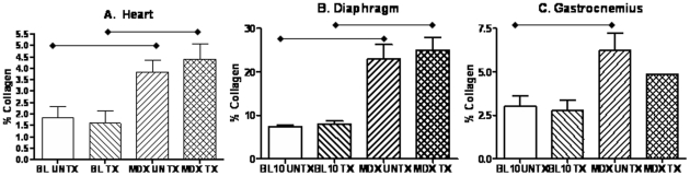
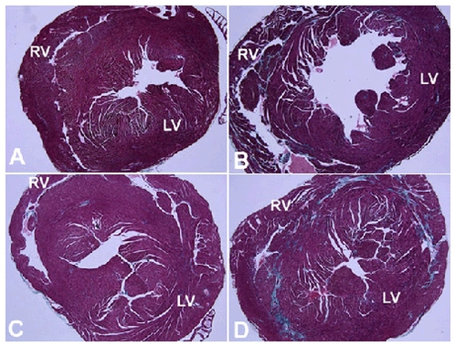
Thymosin beta-4 (Tbeta4) is a ubiquitous protein with many properties relating to cell proliferation and differentiation that promotes wound healing and modulates inflammatory mediators. We studied the effects of chronic administration of Tbeta4 on the skeletal and cardiac muscle of dystrophin deficient mdx mice, the mouse model of Duchenne muscular dystrophy. Female wild type (C57BL10/ScSnJ) and mdx mice, 8-10 weeks old, were treated with 150 microg of Tbeta4 twice a week for 6 months. To promote muscle pathology, mice were exercised for 30 minutes twice a week. Skeletal and cardiac muscle function were assessed via grip strength and high frequency echocardiography. Localization of Tbeta4 and amount of fibrosis were quantified using immunohistochemistry and Gomori's tri-chrome staining, respectively. Mdx mice treated with Tbeta4 showed a significant increase in skeletal muscle regenerating fibers compared to untreated mdx mice. Tbeta4 stained exclusively in the regenerating fibers of mdx mice. Although untreated mdx mice had significantly decreased skeletal muscle strength compared to untreated wild type, there were no significant improvements in mdx mice after treatment. Systolic cardiac function, measured as percent shortening fraction, was decreased in untreated mdx mice compared to untreated wild type and there was no significant difference after treatment in mdx mice. Skeletal and cardiac muscle fibrosis were also significantly increased in untreated mdx mice compared to wild type, but there was no significant improvement in treated mdx mice. In exercised dystrophin deficient mice, chronic administration of Tbeta4 increased the number of regenerating fibers in skeletal muscle and could have a potential role in treatment of skeletal muscle disease in Duchenne muscular dystrophy.
Conflict of interest statement
Figures


References
-
- Hoffman EP, Brown RH, Jr, Kunkel LM. Dystrophin: the protein product of the Duchenne muscular dystrophy locus. Cell. 1987;51:919–928. - PubMed
-
- Sicinski P, Geng Y, Ryder-Cook AS, Barnard EA, Darlison MG, et al. The molecular basis of muscular dystrophy in the mdx mouse: a point mutation. Science. 1989;244:1578–1580. - PubMed
-
- Anderson JE, Bressler BH, Ovalle WK. Functional regeneration in the hindlimb skeletal muscle of the mdx mouse. J Muscle Res Cell Motil. 1988;9:499–515. - PubMed
-
- De Luca A, Pierno S, Liantonio A, Cetrone M, Camerino C, et al. Enhanced dystrophic progression in mdx mice by exercise and beneficial effects of taurine and insulin-like growth factor-1. J Pharmacol Exp Ther. 2003;304:453–463. - PubMed
Publication types
MeSH terms
Substances
Grants and funding
LinkOut - more resources
Full Text Sources
