. 2010 Jan 29;5(1):e8966.
doi: 10.1371/journal.pone.0008966.

Identification of trkH, encoding a potassium uptake protein required for Francisella tularensis systemic dissemination in mice

Affiliations

Identification of trkH, encoding a potassium uptake protein required for Francisella tularensis systemic dissemination in mice

Khaled Alkhuder et al. PLoS One. .

Abstract

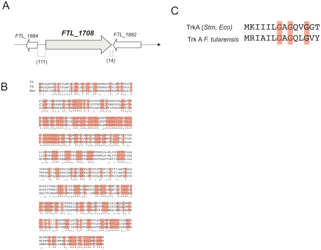
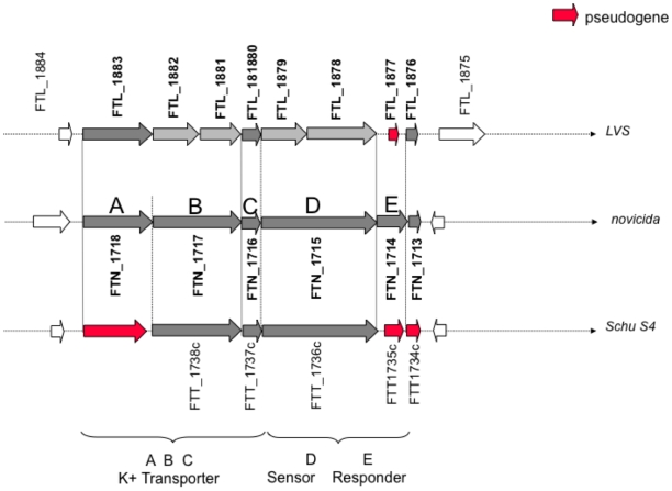
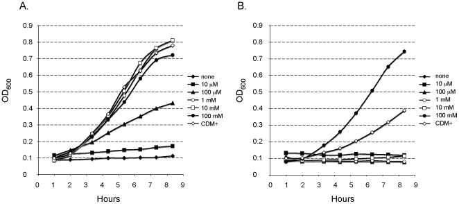
Francisella tularensis is a highly infectious bacterium causing the zoonotic disease tularaemia. During its infectious cycle, F. tularensis is not only exposed to the intracellular environment of macrophages but also resides transiently in extracellular compartments, in particular during its systemic dissemination. The screening of a bank of F. tularensis LVS transposon insertion mutants on chemically defined medium (CDM) led us to identify a gene, designated trkH, encoding a homolog of the potassium uptake permease TrkH. Inactivation of trkH impaired bacterial growth in CDM. Normal growth of the mutant was only restored when CDM was supplemented with potassium at high concentration. Strikingly, although not required for intracellular survival in cell culture models, TrkH appeared to be essential for bacterial virulence in the mouse. In vivo kinetics of bacterial dissemination revealed a severe defect of multiplication of the trkH mutant in the blood of infected animals. The trkH mutant also showed impaired growth in blood ex vivo. Genome sequence analyses suggest that the Trk system constitutes the unique functional active potassium transporter in both tularensis and holarctica subspecies. Hence, the impaired survival of the trkH mutant in vivo is likely to be due to its inability to survive in the low potassium environment (1-5 mM range) of the blood. This work unravels thus the importance of potassium acquisition in the extracellular phase of the F. tularensis infectious cycle. More generally, potassium could constitute an important mineral nutrient involved in other diseases linked to systemic dissemination of bacterial pathogens.


Conflict of interest statement

Competing Interests: The authors have declared that no competing interests exist.

Figures

Figure 1
Figure 1. Genetic organzation of the trkH region and multiple alignments of the TrkH, TrkA proteins.
(A) The gene FTL_1708 (trkH) is flanked by two genes in opposite orientation. Parenthetic numbers give the sizes (in base pairs) of the intergenic regions flanking FTL_1708. (B) Alignment of the TrkH proteins of F. tularensis strains LVS (designated Ftl), Schu S4 (designated ftu) and TrkH of S. typhimurium LT2 (designated Stm) were performed using the Clustal W program. Residues that are identical in all strains (boxed in pink) are marked by “*”, conserved substitutions by a “:”, and semi-conserved substitutions by a “.” (C) Alignment of the N-terminal end of TrkA (from E. coli K12 and S. typhimurium LT2, upper line) and F. tularensis (from Schu S4 and LVS, lower line) encompassing the characteristic nucleotide binding motif GXGXXG at the N-terminus of TrkA. Pink residues are the conserved glycines involved in nucleotide binding.
Figure 2
Figure 2. The kdp locus in F. tularensis subsp. holartica (LVS), novicida, and tularensis.
The prototypical Kdp potassium transport system comprises: i) the transporter, composed of KdpA, KdpB and KdpC; and ii) the TCS, regulating its expression, and composed of KdpD, the sensor kinase; and KdpE, the response regulator. Upper line, the LVS locus. Whenever an FTL number has been attributed, it is indicated above the orf. Middle line, F. tularensis subsp. novicida which comprises an intact kdp locus. A, B, D and E, above each gene refer to kdpA, kdpB, kdpC, kdpD and kdpE, respectively. Lower line, F. tularensis subsp. tularensis. In dark grey, the intact genes; in light grey, the interrupted genes (but annotated as genes in the KEGG database); in red, the interrupted genes annotated as pseudogenes (in KEGG). KdpB. In F. tularensis subsp. novicida, the kdpB gene (FTN_1717; 2,040 bp) encodes a 679 aas protein. In LVS, the proximal portion of the gene carries: i) an in frame 63 bp deletion (deletion of nucleotides 509 to 571); and ii) a single nucleotide insertion (between nucleotides 1,096 and 1,097; FTN_1717 numbering), leading to a premature termination of the coding sequence (creation of a TGA stop codon 9 bp downstream of the insertion). The resulting 374 aas truncated protein is designated FTL_1882. An ATG codon (in frame with the rest of the kdpB sequence of FTN_1717) is found immediately downstream of the stop codon (29 bp) of FTL_1882, leading to the prediction of a second open reading frame of 301 aas designated FTL_1881. Blastn analysis of the nucleotide sequence of LVS corresponding to gene FTN_1717 (i.e. 1,978 bp from the start codon of FTL_1882 to the stop codon of FTL_1881) reveals that the entire LVS sequence is 100% identical to F. tularensis subsp. holarctica FTNF002-00 genome region 1,811,308 to 1,809,331 and >99% identical to F. tularensis subsp. holarctica OSU18 genome region 1,815,047 to 1,813,070 (with a unique C to A substitution at position 71 of FTL_1882). Thus, in the three subsp. holarctica genomes available, the proximal part of the kdpB gene carries the same in frame deletion and the gene is interrupted by the same single nucleotide insertion. KdpD. In F. tularensis subsp. novicida, the kdpD gene (FTN_1715; 2,682 bp) encodes a 893 aas protein. In LVS, the corresponding gene is interrupted by a single nucleotide insertion (between nucleotides 981 and 982; FTN_1715 numbering), leading to a premature termination of the coding sequence (creation of a TGA stop codon 32 bp downstream of the insertion). The resulting truncated predicted orf is designated FTL_1879 (337 aa). An ATG codon (in frame with the rest of the kdpD sequence of FTN_1715) is found downstream of the stop codon (92 bp) of FTL_1879, leading to the prediction of a second orf of 525 aas, designated FTL_1878. Blastn analysis of the nucleotide sequence of LVS (2,683 bp) corresponding to gene FTN_1715 reveals that the entire LVS sequence is 100% identical to F. tularensis subsp. holarctica OSU18 genome region 1,812,395 to 1,809,713100 and 99% identical to F. tularensis subsp. holarctica FTNF002-00 genome region 1,808,656 to 1,805,974 (with 3 single nucleotide substitutions). Thus, in the three subsp. holarctica genomes available, the kdpD gene is interrupted by the same single nucleotide insertion. KdpE. In F. tularensis subsp. novicida, the kdpE gene (FTN_1714; 687 bp) encodes a 228 aas protein. In LVS, the corresponding region is not predicted to encode a protein (FTL_1877 is designated pseudogene). Comparison of the F. tularensis subsp. novicida and LVS nucleotide sequences reveal the presence of a 13 bp deletion in the proximal portion of the LVS sequence (deletion of nucleotides 45 to 57, FTN_1714 numbering), resulting in a frame shift and the premature termination of the protein sequence (a TGA stop codon is found 11 bp downstream of the deletion). Blastn analysis of the nucleotide sequence of LVS (671 bp) shows a 100% identity with the F. tularensis subsp. holarctica OSU18 genome region 1,809,671 to 1,809,001; and >99% identity with F. tularensis subsp. holarctica FTNF002-00 genome region 1,805,932 to 1,805,262 (1 single nucleotide substitution) as well as with F. tularensis subsp. tularensis Schu S4 genome region 1,823,954 to 1,823, 284 (2 single nucleotide substitution). Thus, in the three subsp. holarctica genomes available and in Schu S4, the kdpE gene is a pseudogene resulting from the same 13 bp deletion. The regions containing the mutations in genes kdpB (FTL_1881-1882), kdpD (FTL_1878-1879) and kdpE (FTL_1877), have been resequenced by sequencing of cloned PCR products. This analysis fully confirmed the published sequence of LVS.
Figure 3
Figure 3. Growth properties of the trkH mutant in chemically definded medium.
LVS (A) and trkH (B) were grown in chemically defined medium (CDM+) or in CDMx buffered with sodium phosphate and supplemented with KCl as potassium source (at concentrations indicated in legend).
Figure 4
Figure 4. Functional complementation.
Bacteria were grown in Schaedler K3 until an OD600 of 0.3. Ten µl of each culture (nd: non diluted, corresponding to ca. 107 b) and of serial ten-fold dilutions, were spotted onto: CDM agar plate (A) and Chocolate agar plate (B). LVS: wild-type strain; TrkH: the trkH mutant derivative; TrkH/pC: the trkH mutant transformed with plasmid pC-trkH (a pFNLTP6 derivative carrying a wild-type trkH allele under control of the gro promoter).
Figure 5
Figure 5. Resistance to serum and high osmolarity.
Survival of LVS (closed symbols) and trkH mutant (open symbols) after 1 hour incubation in human non-decomplemented serum (0–20%) (A). Growth of LVS (closed symbols) and trkH (open symbols) in CDM supplemented with NaCl as indicated in legend (B).
Figure 6
Figure 6. Intracellular replication of LVS and trkH strains.
Intracellular bacterial replication was monitored over a 48 h-period in J774 macrophage like cells (A), in THP1 human macrophages (B), and in mouse bone marrow-derived macrophages (BMM) from BALB/c mice (C). Results are shown as the average of log10 (CFU well−1) ± standard deviation. Closed symbols designate the LVS strain and open symbols the trkH strain.
Figure 7
Figure 7. The trkH mutant is attenuated for virulence in mice.
Groups of five BALB/c mice were infected with LVS (closed symbols) or trkH (open symbols) bacteria at different doses by the i.p. route. The log10 of the numbers of bacteria used for infection are shown in legend in parenthesis. The survival of mice (in %) was followed for 10 days after infection.
Figure 8
Figure 8. In vivo dissemination of LVS and trkH.
BALB/c mice were infected with approximately 104 LVS or trkH bacteria by the i.p. route. After 2, 3, 4, and 7 days of infection, groups of five mice were sacrificed and the bacterial burden in the spleen, liver, and blood was determined by plating tissue homogenates on chocolate agar. *: all mice infected by LVS died before day 7.
Figure 9
Figure 9. Multiplication of trkH mutant is impaired in murine blood ex vivo.
Heparinized blood samples from eight mice were inoculated with ∼5×10⁴ bacteria ml−1 and incubated at 37°C with shaking for 24 and 48 h, at which point the number of viable bacteria was determined. Closed symbols correspond to the LVS strain and open symbols the trkH mutant.

Similar articles

Cited by

References

    1. Titball RW, Petrosino JF. Francisella tularensis genomics and proteomics. Ann N Y Acad Sci. 2007;1105:98–121. - PubMed
    1. Ellis J, Oyston PC, Green M, Titball RW. Tularemia. Clin Microbiol Rev. 2002;15:631–646. - PMC - PubMed
    1. Sjostedt A. Tularemia: history, epidemiology, pathogen physiology, and clinical manifestations. Ann N Y Acad Sci. 2007;1105:1–29. - PubMed
    1. Barker JR, Klose KE. Molecular and genetic basis of pathogenesis in Francisella tularensis. Ann N Y Acad Sci. 2007;1105:138–159. - PubMed
    1. Brotcke A, Weiss DS, Kim CC, Chain P, Malfatti S, et al. Identification of MglA-regulated genes reveals novel virulence factors in Francisella tularensis. Infect Immun. 2006;74:6642–6655. - PMC - PubMed

Publication types