. 2010 Jan 29;5(1):e8959.
doi: 10.1371/journal.pone.0008959.

CD28 down-regulation on circulating CD4 T-cells is associated with poor prognoses of patients with idiopathic pulmonary fibrosis

Affiliations

CD28 down-regulation on circulating CD4 T-cells is associated with poor prognoses of patients with idiopathic pulmonary fibrosis

Syed R Gilani et al. PLoS One. .

Abstract

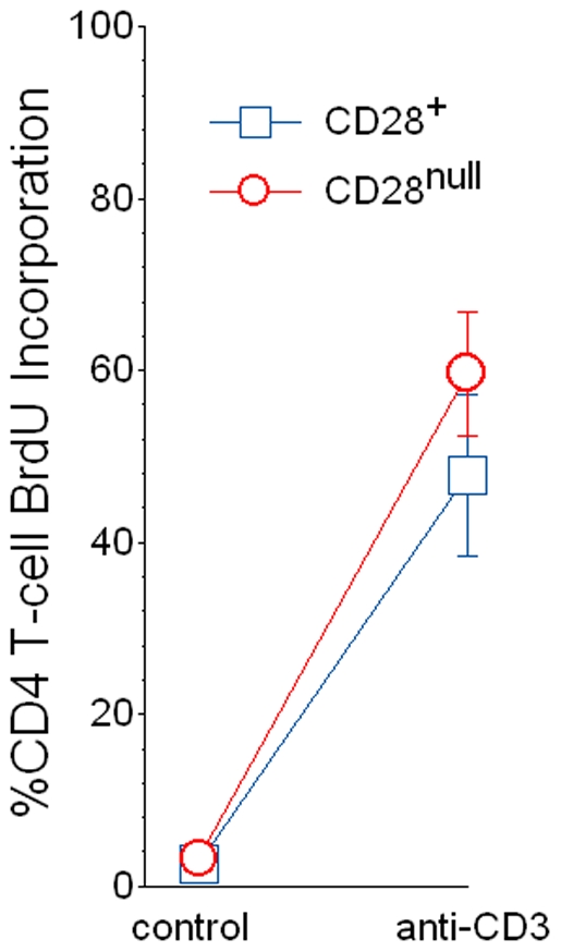
Background: Although the etiology of idiopathic pulmonary fibrosis (IPF) remains perplexing, adaptive immune activation is evident among many afflicted patients. Repeated cycles of antigen-induced proliferation cause T-cells to lose surface expression of CD28, and we hypothesized this process might also occur in IPF.

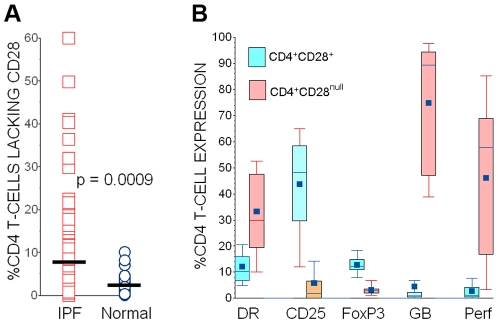
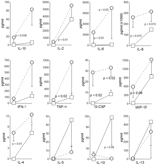
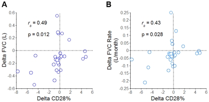
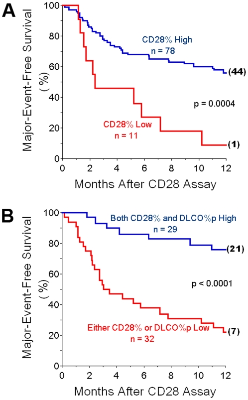
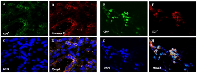
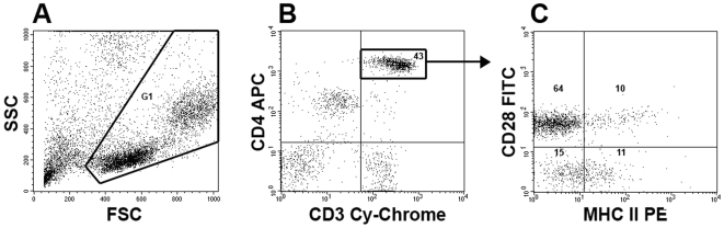
Methodology/principal findings: Peripheral blood CD4 T-cells from 89 IPF patients were analyzed by flow cytometry and cytokine multiplex assays, and correlated with clinical events. In comparison to autologous CD4(+)CD28(+)cells, the unusual CD4(+)CD28(null) lymphocytes seen in many IPF patients had discordant expressions of activation markers, more frequently produced cytotoxic mediators perforin (2.4+/-0.8% vs. 60.0+/-7.4%, p<0.0001) and granzyme B (4.5+/-2.8% vs.74.9+/-6.5%, p<0.0001), produced greater amounts of many pro-inflammatory cytokines, and less frequently expressed the regulatory T-cell marker FoxP3 (12.9+/-1.1% vs. 3.3+/-0.6% p<0.0001). Infiltration of CD4(+)CD28(null) T-cells in IPF lungs was confirmed by confocal microscopy. Interval changes of CD28 expression among subjects who had replicate studies were correlated with conterminous changes of their forced vital capacities (r(s) = 0.49, p = 0.012). Most importantly, one-year freedom from major adverse clinical events (either death or lung transplantation) was 56+/-6% among 78 IPF patients with CD4(+)CD28(+)/CD4(total)>or=82%, compared to 9+/-9% among those with more extensive CD28 down-regulation (CD4(+)CD28(+)/CD4(total)<82%) (p = 0.0004). The odds ratio for major adverse events among those with the most extensive CD28 down-regulation was 13.0, with 95% confidence intervals 1.6-111.1.

Conclusions/significance: Marked down-regulation of CD28 on circulating CD4 T-cells, a result of repeated antigen-driven proliferations, is associated with poor outcomes in IPF patients. The CD4(+)CD28(null) cells of these patients have potentially enhanced pathogenic characteristics, including increased productions of cytotoxic mediators and pro-inflammatory cytokines. These findings show proliferative T-cell responses to antigen(s) resulting in CD28 down-regulation are associated with progression and manifestations of IPF, and suggest assays of circulating CD4 T-cells may identify patients at greatest risk for clinical deterioration.


Conflict of interest statement

Competing Interests: The contributing author (SRD) has received a one-time ∼$50,000 grant from APT Pharmaceuticals to help in development of a humanized mouse model, in which immunodeficient animals are reconstituted with human T-cells. However, this work has no direct bearing on the data or conclusions presented in this paper. In addition, SRD, in conjunction with the University of Pittsburgh, now have patents pending that include the use of T-cell characteristics as surrogate biomarkers for lung diseases, including IPF.

Figures

Figure 1
Figure 1. Characteristics of CD4 T-cell subpopulations in IPF patients.
A: The proportions of circulating CD4 T-cells that also expressed CD28 (CD28%) were reduced in many IPF patients. The horizontal line denotes the population means. B: In contrast to autologous CD4+CD28+ cells, the CD4+CD28null T-cells of IPF patients more often express major histocompatibility antigen (MHC) Class II (DR), but less frequently express CD25. CD4+CD28null T-cells of IPF patients less frequently produce transcription factor FoxP3 (a putative marker of regulatory T-cells), but much more frequently produce cytotoxic mediators granzyme B (GB) and perforin (Perf). For each measure n = 24, and p values for all intergroup comparisons (CD4+CD28+ vrs. CD4+CD28null cells) are <0.0001.
Figure 2
Figure 2. Cytokine elaborations by autologous CD4 subpopulations of IPF patients.
Initial (left) data point in each series represents control unstimulated (basal) condition, while second (right) data point delineates productions of cells after stimulation with plate bound anti-CD3 antibody. These paired specimens (control and stimulated) are also connected by lines. CD4+CD28null cells from IPF patients (open circles with paired specimens connected by dashed lines) tend to elaborate greater amounts of pro-inflammatory and TH1 cytokines (top two rows), whereas CD4+CD28+ cells (open squares with paired specimens connected by solid lines) have an apparent TH2 bias, with the exception of IL-4 production (bottom row) (n = 6 randomly-selected specimens in each measure).
Figure 3
Figure 3. Segregated autologous CD4+CD28+ and CD4+CD28null cells from peripheral blood of IPF patients (n = 9) had similar proliferations (determined by BrdU incorporation) in 5-day control (unstimulated) cultures, as well as after stimulation with plate-bound anti-CD3 monoclonal antibody.
Figure 4
Figure 4. Associations of CD28 expression with pulmonary function.
Changes of CD28% with replicate testing (Delta CD28%) were significantly correlated with concomittant interval changes of A.) absolute FVC (Delta FVC) and B.) FVC as a time-dependent rate (Delta FVC/month).
Figure 5
Figure 5. Associations of CD28% espression with clinical outcome.
A.) Survival curves show cumulative freedom from major adverse events (lung transplantation or death) of IPF patients. Those subjects with the most extreme CD28 down-regulation, with CD28 expressed on <82% of their circulating CD4 T-cells (CD28% Low), had much worse outcomes than the cohort with greater proportions of CD4-Tcells that expressed CD28 (CD28% High). Numbers in parenthesis at the ends of survival curves denote remaining, unafflicted subjects that were censored at 12 months of observation. B.) Survival curves showing that cummulative freedom from major adverse events of IPF patients who have either significant CD28 downregulation (CD28% Low) or diffusing capacities for carbon monoxide, as percentages of predicted normal values (DLCO%p) <38, had worse outcomes than the IPF cohort who were both CD28% High and had more normal DLCO%p.
Figure 6
Figure 6. Localization of CD4, CD3, and granzyme B expressing cells in IPF lungs.
A.) and B.) Cells expressing CD4 (Green) and granzyme B (red), respectively, are widely distributed in IPF lung sections. C.) DAPI (blue) stains DNA within cell nuclei. D.) Co-localizations of CD4 and granzyme B (yellow) among nucleated cells are seen in the merged image. Nearly all the intrapulmonary CD4+ cells co-expressed granzyme B. Images E.), F.) and G.) similarly depict CD4 (green), CD3 (red) and DAPI (blue). H.) Merged image shows most of the CD4+ cells also co-expressed CD3. Similar results were present in both IPF lungs. The majority of CD4+ cells in these IPF lung sections co-expressed both granzyme B and CD3 and are, thus, CD4+CD28null T-cells (see also Figure 1B). All images are 60x.
Figure 7
Figure 7. Flow cytometry methodology (see also references 20, 52).
Aliquots of fresh, live peripheral blood mononuclear cells (PBMNC) were stained with anti-CD4-allophycocyanin (APC), anti-CD28-fluorescein isothiocyanate (FITC), and phycoerythrin (PE)-conjugated antibodies against other cell epitopes. A.) Ten thousand (10,000) or more live cells were selected for further study, based their side scatter (SSC) and forward scatter (FSC) characteristics (G1). B.) The brightly staining CD4 cells among these also expressed CD3 (Cy-Chrome) and, thus, are T-cells . C.) These CD4 T-cells were further characterized based on their expression of CD28. The proportions of CD4+CD28+ T-cells among the total CD4+ T-cell population (upper left and upper right quadrants) defines the CD28%. The respective proportions of CD4+CD28+ and CD4+CD28null cells that co-expressed other cell determinants of interest (in this case MHC Class II [DR]) were quantitated. Numbers within the delineated region/quadrants denote the proportions of cells with these respective characteristics.

Similar articles

Cited by

References

    1. American Thoracic Society. Idiopathic pulmonary fibrosis: diagnosis and treatment. International consensus statement. American Thoracic Society (ATS), and the European Respiratory Society (ERS). Am J Respir Crit Care Med. 2000;161:646–664. - PubMed
    1. Selman M, Thannickal VJ, Pardo A, Zisman DA, Martinez FJ, et al. Idiopathic pulmonary fibrosis: pathogenesis and therapeutic approaches. Drugs. 2004;64:406–430. - PubMed
    1. Kurosu K, Takiguchi Y, Okada O, Yumoto N, Sakao S, et al. Identification of annexin 1 as a novel autoantigen in acute exacerbation of idiopathic pulmonary fibrosis. J Immunol. 2008;181:756–767. - PubMed
    1. Takahashi T, Wada I, Ohtsuka Y, Munakata M, Homm Y, et al. Autoantibody to alanyl-tRNA synthetase in patients with idiopathic pulmonary fibrosis. Respirology. 2007;12:642–653. - PubMed
    1. Magro CM, Waldman WJ, Knight DA, Allen JN, Nadasdy T, et al. Idiopathic pulmonary fibrosis related to endothelial injury and antiendothelial cell antibodies. Hum Immunol. 2006;67:284–297. - PubMed

Publication types