. 2010 Feb;12(2):103-15.
doi: 10.1593/neo.91298.

A mutant of hepatitis B virus X protein (HBxDelta127) promotes cell growth through a positive feedback loop involving 5-lipoxygenase and fatty acid synthase

Affiliations

A mutant of hepatitis B virus X protein (HBxDelta127) promotes cell growth through a positive feedback loop involving 5-lipoxygenase and fatty acid synthase

Qi Wang et al. Neoplasia. 2010 Feb.

Abstract

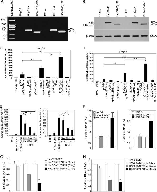
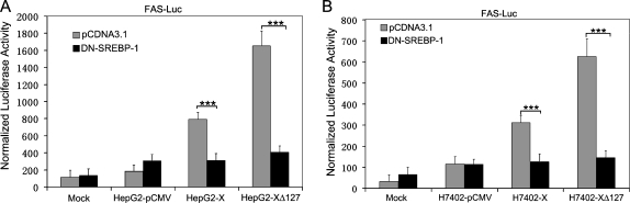
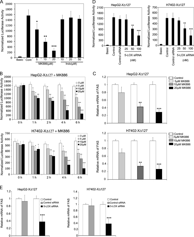
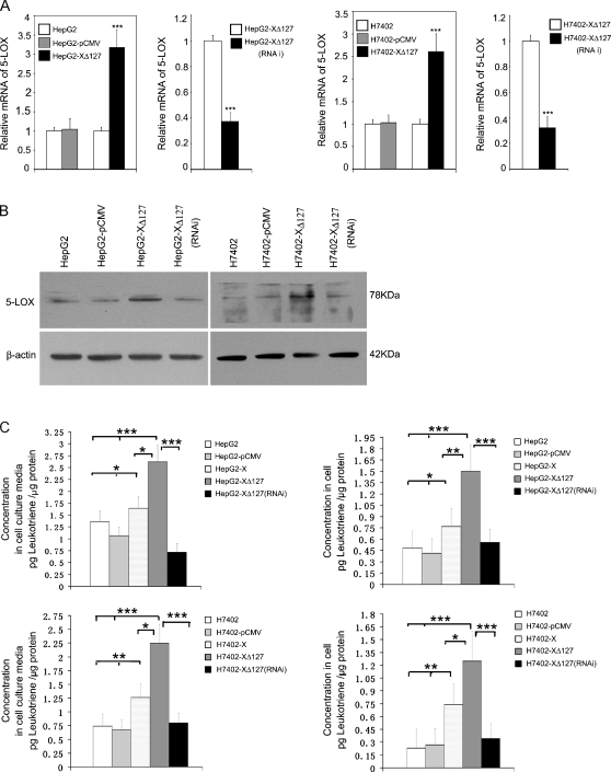
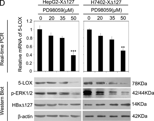
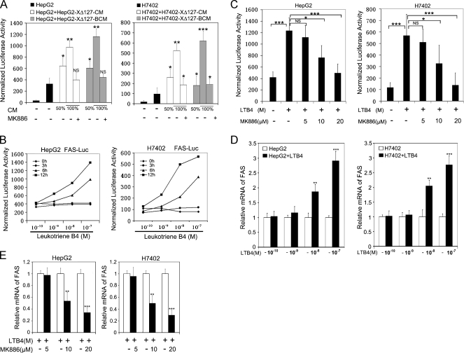
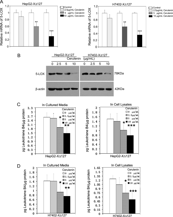
Hepatocellular carcinoma (HCC) is one of the most common malignant tumors worldwide. Hepatitis B virus X protein (HBx) contributes to the development of HCC, whereas HBx with COOH-terminal deletion is a frequent event in the HCC tissues. Previously, we identified a natural mutant of HBx-truncated 27 amino acids at the COOH-terminal (termed HBxDelta127), which strongly enhanced cell growth. In the present study, we focused on investigating the mechanism. Accordingly, fatty acid synthase (FAS) plays a crucial role in cancer cell survival and proliferation; thus, we examined the signaling pathways involving FAS. Our data showed that HBxDelta127 strongly increased the transcriptional activities of FAS in human hepatoma HepG2 and H7402 cells. Moreover, we found that 5-lipoxygenase (5-LOX) was responsible for the up-regulation of FAS by using MK886 (an inhibitor of 5-LOX) and 5-LOX small interfering RNA. We observed that HBxDelta127 could upregulate 5-LOX through phosphorylated extracellular signal-regulated protein kinases 1/2 and thus resulted in the increase of released leukotriene B4 (LTB4, a metabolite of 5-LOX) by ELISA. The additional LTB4 could upregulate the expression of FAS in the cells as well. Interestingly, we found that FAS was able to upregulate the expression of 5-LOX in a feedback manner by using cerulenin (an inhibitor of FAS). Collectively, HBxDelta127 promotes cell growth through a positive feedback loop involving 5-LOX and FAS, in which released LTB4 is involved in the up-regulation of FAS. Thus, our finding provides a new insight into the mechanism involving the promotion of cell growth mediated by HBxDelta127.


Figures

Figure 1
Figure 1
HBxΔ127 upregulates the expression of FAS in hepatoma HepG2 (or H7402) cells. (A) HBx and HBxΔ127 genes were identified in HepG2-X and HepG2-XΔ127 (or H7402-X and H7402-XΔ127) cells by RT-PCR. (B) The expression of HBx and HBxΔ127 was detected by immunoblot analysis. (C) Luciferase reporter gene assay showed that the transcriptional activities of FAS were higher in HepG2 cells when cotransfecting the plasmids of pFAS-WT-Luc and pCMV-X or pCMV-XΔ127 than controls by transient transfection, suggesting that HBxΔ127 has stronger ability to upregulate FAS than HBx. However, pCMV-X or pCMV-XΔ127 failed to stimulate pFAS-ΔSRE-Luc (a mutant of FAS promoter) as control. The data are representative of three independent experiments. Values represent mean ± SD (n = 3). *P < .05, **P < .01, ***P < .001 (Student's t test). (D) The previously mentioned experiments were repeatable in another hepatoma H7402 cells. (E) In stable transfection system, pSilencer-X plasmid (RNAi targeting mRNA of HBxΔ127) could abolish the enhancement of transcriptional activities of FAS in HepG2-XΔ127 (or H7402-XΔ127) cells by luciferase reporter gene assay in a dose-dependent manner. The data are representative of three independent experiments. Values represent mean ± SD (n = 3). *P < .05, **P < .01, ***P < .001 (Student's t test). (F) The expression levels of FAS mRNA in HepG2-XΔ127 (or H7402-XΔ127) cells were examined by real-time PCR. Plotted are the means ± SD of three samples normalized to β-actin. Statistically significant differences relative to FAS mRNA levels in HepG2 (or H7402) cells, arbitrarily set to 1.0, are indicated: *P < .05, **P < .01, ***P < .001 (Student's t test). (G, H) The expression levels of FAS mRNA in HepG2-XΔ127 (or H7402-XΔ127) cells transfected with a pSilencer-X plasmid were examined by real-time PCR. Plotted are the means ± SD of three samples normalized to β-actin. Statistically significant differences relative to FAS mRNA levels in HepG2-XΔ127 (or H7402-XΔ127) cells, arbitrarily set to 1.0, are indicated: *P < .05, **P < .01, ***P < .001 (Student's t test).
Figure 2
Figure 2
HBxΔ127 upregulates FAS promoter activity through SREBP-1. (A) The transcriptional activity of FAS was examined by luciferase reporter gene assay in HepG2-XΔ127 cells when cotransfection was performed using plasmids of pCDNA3.1 (empty vector) or DN-SREBP-1 (a mutant of SREBP-1). The data showed that DN-SREBP-1 was able to inhibit the transcriptional activity of FAS by competition with wild type SREBP-1 in HepG2-XΔ127 cells, suggesting that HBxΔ127 upregulates FAS promoter activity through SREBP-1. Data are representative of three independent experiments. Values represent mean ± SD (n = 3). ***P < .001 (Student's t test). (B) The previously mentioned experiments were repeatable in H7402-XΔ127 cells. Data are representative of three independent experiments. Values represent mean ± SD (n = 3). ***P < .001 (Student's t test).
Figure 3
Figure 3
5-LOX is responsible for the up-regulation of FAS mediated by HBxΔ127. (A) The transcriptional activities of FAS were examined by luciferase reporter gene assay in HepG2-XΔ127 cells when the MK886 (an inhibitor of 5-LOX) and Indo (an inhibitor of COX-2) were used, respectively. Data are representative of three independent experiments. Values represent mean ±S D (n = 3). *P < .05 versus control, **P < .01 versus control, ***P < .001 versus control (Student's t test). (B) The experiments were repeated in HepG2-XΔ127 (or H7402-XΔ127) cells in a dose-dependent and time course manner. Data are representative of three independent experiments. Values represent mean ± SD (n = 3). *P < .05 versus control, **P < .01 versus control, ***P < .001 versus control (Student's t test). (C) HepG2-XΔ127 (or H7402-XΔ127) cells (2 x 10⁵) were incubated with or without MK886 (5, 10, and 20 µM) for 6 hours. The mRNA levels of FAS were examined by real-time PCR. Plotted are the means ± SD of three samples normalized to β-actin. Statistically significant differences relative to FAS mRNA levels in HepG2-XΔ127 (or H7402-XΔ127) cells, arbitrarily set to 1.0, are indicated: *P < .05, **P < .01, ***P < .001 (Student's t test). (D) HepG2-XΔ127 cells were transfected with 100 nM 5-LOX siRNA or control siRNA for 48 hours. Then, the transcriptional activities of FAS were examined by luciferase reporter gene assay. Data are representative of three independent experiments. Values represent mean ± SD (n = 3). **P < .01 versus control (Student's t test). (E) Real-time PCR showed the expression levels of FAS mRNA in control cells and 5-LOX siRNA-transfected HepG2-XΔ127 (or H7402-XΔ127) cells. Plotted are the means ± SD of three samples normalized to β-actin. Statistically significant differences relative to FAS mRNA levels in HepG2-XΔ127 (or H7402-XΔ127) cells, arbitrarily set to 1.0, are indicated: *P < .05, **P < .01, ***P < .001 (Student's t test).
Figure 4
Figure 4
HBxΔ127 upregulates the expression levels of 5-LOX and increases the release of LTB4 (a metabolite of 5-LOX). (A) The expression level of 5-LOX was increased in HepG2-XΔ127 (or H7402-XΔ127) cells by real-time PCR, which was abolished by RNAi targeting mRNA of HBx gene using pSilencer3.0-X plasmid. Plotted are the means ± SD of three samples normalized to β-actin. Statistically significant differences relative to control, arbitrarily set to 1.0, are indicated: *P < .05, **P < .01, ***P < .001 (Student's t test). (B) The expression level of 5-LOX was examined by Western blot analysis as well. (C) The level of LTB4 was determined by ELISA in the conditioned medium or in the cell lysates from HepG2-X/HepG2-XΔ127 (or H7402-X/H7402-XΔ127) cells. The amount of LTB4 in HepG2/HepG2-pCMV (or H7402/H7402-pCMV) cells was used as control. Data are representative of three independent experiments. Values represent mean ± SD (n = 3). * P < .05, ** P < .01, ***P < .001 (Student's t test). (D) HepG2-XΔ127 (or H7402-XΔ127) cells were treated with or without PD98059 (20, 35, and 50 µM) for 4 hours. Then, we examined the mRNA levels of 5-LOX by real-time PCR. Plotted are the means ± SD of three samples normalized to β-actin. Statistically significant differences relative to FAS mRNA levels in HepG2-XΔ127 (or H7402-XΔ127) cells, arbitrarily set to 1.0, are indicated: *P < .05, **P < .01, ***P < .001 (Student's t test). The protein expression levels of 5-LOX were examined by Western blot analysis. The protein levels of HBxΔ127 and p-ERK1/2 were also determined as controls.
Figure 4
Figure 4
HBxΔ127 upregulates the expression levels of 5-LOX and increases the release of LTB4 (a metabolite of 5-LOX). (A) The expression level of 5-LOX was increased in HepG2-XΔ127 (or H7402-XΔ127) cells by real-time PCR, which was abolished by RNAi targeting mRNA of HBx gene using pSilencer3.0-X plasmid. Plotted are the means ± SD of three samples normalized to β-actin. Statistically significant differences relative to control, arbitrarily set to 1.0, are indicated: *P < .05, **P < .01, ***P < .001 (Student's t test). (B) The expression level of 5-LOX was examined by Western blot analysis as well. (C) The level of LTB4 was determined by ELISA in the conditioned medium or in the cell lysates from HepG2-X/HepG2-XΔ127 (or H7402-X/H7402-XΔ127) cells. The amount of LTB4 in HepG2/HepG2-pCMV (or H7402/H7402-pCMV) cells was used as control. Data are representative of three independent experiments. Values represent mean ± SD (n = 3). * P < .05, ** P < .01, ***P < .001 (Student's t test). (D) HepG2-XΔ127 (or H7402-XΔ127) cells were treated with or without PD98059 (20, 35, and 50 µM) for 4 hours. Then, we examined the mRNA levels of 5-LOX by real-time PCR. Plotted are the means ± SD of three samples normalized to β-actin. Statistically significant differences relative to FAS mRNA levels in HepG2-XΔ127 (or H7402-XΔ127) cells, arbitrarily set to 1.0, are indicated: *P < .05, **P < .01, ***P < .001 (Student's t test). The protein expression levels of 5-LOX were examined by Western blot analysis. The protein levels of HBxΔ127 and p-ERK1/2 were also determined as controls.
Figure 5
Figure 5
LTB4 is involved in the up-regulation of FAS. (A) HepG2 (or H7402) cells were transfected with 0.3 µg of plasmid of FAS promoter-luciferase reporter gene, respectively. We treated the transfected cells with conditioned medium (CM) from HepG2-XΔ127 (or H7402-XΔ127) cells or boiled conditioned medium (BCM) from HepG2-XΔ127 (or H7402-XΔ127) cells at indicated the different concentration for 12 hours before the cell lysates were obtained and luciferase activity was measured. In addition, HepG2 (or H7402) cells were also treated with 100% HepG2-XΔ127 CM (or H7402-XΔ127 CM), which were pretreated with 20 µM MK886 for 6 hours. Data are representative of three independent experiments. Values represent mean ± SD (n = 3). *P < .05 versus control, **P < .01 versus control, ***P < .001 versus control (Student's t test). (B) HepG2 (or H7402) cells were transfected with 0.3 µg of plasmid of FAS promoter-luciferase reporter gene, respectively. We treated the transfected cells with the addition of 0.1, 1, 10, or 100 nM LTB4 or without LTB4 at different time points (3, 6, and 12 hours) before cell lysates were obtained and luciferase activity was measured. Data are representative of three independent experiments. P < .01 (Student's t test) (C) HepG2 (or H7402) cells were incubated with the addition of 100 nM LTB4 together with 5, 10, or 20 µM MK886 for 6 hours before cell lysates were obtained and luciferase activity was measured. Data are representative of three independent experiments. Values represent mean ± SD (n = 3). *P < .05 versus control, **P < .01 versus control, ***P < .001 versus control (Student's t test). (D) HepG2 (or H7402) cells were treated with the addition of LTB4 (0.1, 1, 10, or 100 nM) for 12 hours. Then, the FAS mRNA was examined by real-time PCR, respectively. Plotted are the means ± SD of three samples normalized to β-actin. Statistically significant differences relative to FAS mRNA levels in HepG2 (or H7402) cells, arbitrarily set to 1.0, are indicated: *P < .05, **P < .01, ***P < .001 (Student's t test). (E) HepG2 (or H7402) cells were incubated with the addition of 100 nM LTB4 together with 5, 10, or 20 µM MK886 for 6 hours. The mRNA levels of FAS were examined by real-time PCR, respectively. Plotted are the means ± SD of three samples normalized to β-actin. Statistically significant differences relative to FAS mRNA levels in HepG2 (or H7402) cells without MK886, arbitrarily set to 1.0, are indicated: *P < .05, **P < .01, ***P < .001 (Student's t test).
Figure 6
Figure 6
FAS contributes to the up-regulation of 5-LOX in a positive feedback loop manner. HepG2-XΔ127 (or H7402-XΔ127) cells (2 x 10⁵) were incubated with or without cerulenin (2.5, 5, and 10 µg/ml) for 12 hours. (A) The mRNA level of 5-LOX was detected by real-time PCR. Plotted are the means ± SD of three samples normalized to β-actin. Statistically significant differences relative to 5-LOX mRNA levels in HepG2-XΔ127 (or H7402-XΔ127) cells without the action of cerulenin, arbitrarily set to 1.0, are indicated: *P < .05, **P < .01, ***P < .001 (Student's t test). (B) The protein expression level of 5-LOX was examined by Western blot analysis. (C, D) The amount of LTB4 was examined by ELISA in the conditioned medium or in the cell lysates from the previously mentioned treated HepG2-XΔ127 (or H7402-XΔ127) cells. Data are representative of three independent experiments. Values represent mean ±S D (n = 3). *P < .05 versus control, **P < .01 versus control, ***P < .001 versus control (Student's t test).

Similar articles

Cited by

References

    1. Lok AS. Chronic hepatitis B. N Engl J Med. 2002;346:1682–1683. - PubMed
    1. Murakami S. Hepatitis B virus X protein: a multifunctional viral regulator. J Gastroenterol. 2001;36:651–660. - PubMed
    1. Tang H, Oishi N, Kaneko S, Murakami S. Molecular functions and biological roles of hepatitis B virus X protein. Cancer Sci. 2006;97:977–983. - PMC - PubMed
    1. Liu XH, Wang L, Zhang SH, Lin J, Zhang SM, Feitelson MA, Gao HJ, Zhu MH. Mutations in the carboxyl terminus of the X protein of Hepatitis B Virus regulate Wnt-5a expression in hepatoma Huh7 cells: cDNA microarray and proteomic analyses. Carcinogenesis. 2008;29:1207–1214. - PMC - PubMed
    1. Liu XH, Lin J, Zhang SH, Zhang SM, Feitelson MA, Gao HJ, Zhu MH. COOH-terminal deletion of HBx gene is a frequent event in HBV-associated hepatocellular carcinoma. World J Gastroenterol. 2008;14:1346–1352. - PMC - PubMed

Publication types

MeSH terms

Substances