. 2010 Feb;12(2):116-27.
doi: 10.1593/neo.91384.

GAS6/AXL axis regulates prostate cancer invasion, proliferation, and survival in the bone marrow niche

Affiliations

GAS6/AXL axis regulates prostate cancer invasion, proliferation, and survival in the bone marrow niche

Yusuke Shiozawa et al. Neoplasia. 2010 Feb.

Abstract

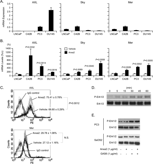
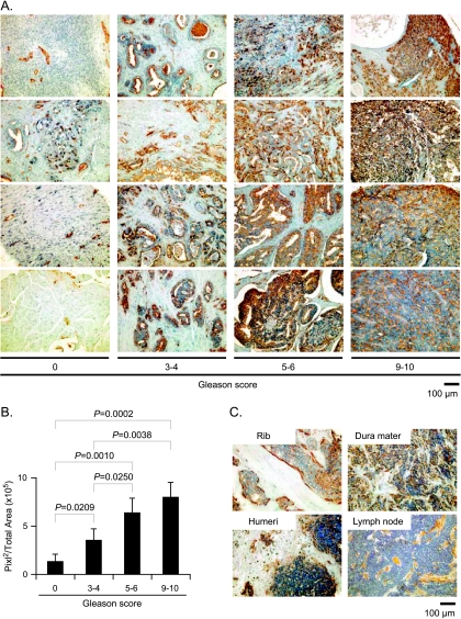
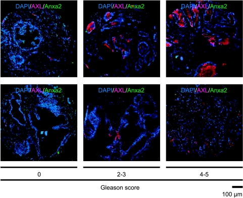
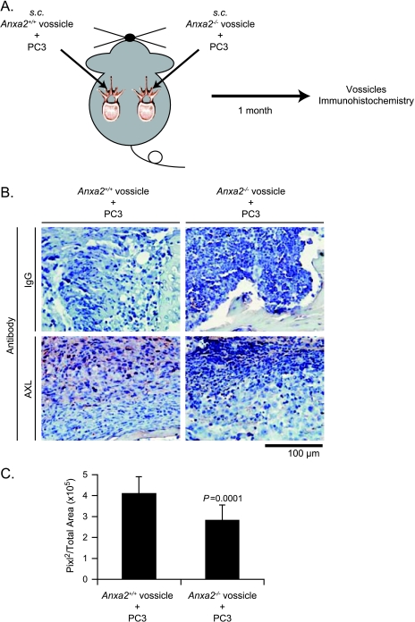
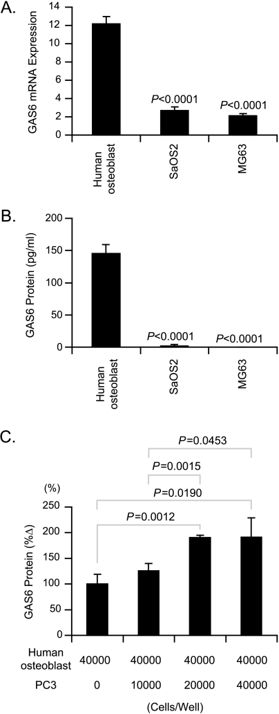
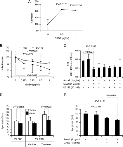
Our recent studies have shown that annexin II, expressed on the cell surface of osteoblasts, plays an important role in the adhesion of hematopoietic stem cells (HSCs) to the endosteal niche. Similarly, prostate cancer (PCa) cells express the annexin II receptor and seem to use the stem cell niche for homing to the bone marrow. The role of the niche is thought to be the induction and sustenance of HSC dormancy. If metastatic PCa cells occupy a similar or the same ecological niche as HSCs, then it is likely that the initial role of the HSC niche will be to induce dormancy in metastatic cells. In this study, we demonstrate that the binding of PCa to annexin II induces the expression of the growth arrest-specific 6 (GAS6) receptors AXL, Sky, and Mer, which, in the hematopoietic system, induce dormancy. In addition, GAS6 produced by osteoblasts prevents PCa proliferation and protects PCa from chemotherapy-induced apoptosis. Our results suggest that the activation of GAS6 receptors on PCa in the bone marrow environment may play a critical role as a molecular switch, establishing metastatic tumor cell dormancy.


Figures

Figure 1
Figure 1
Anxa2-induced AXL family in PCa. (A) AXL, Sky, and Mer mRNA levels of PCa cell lines (LNCaP, C4-2B, PC3, and DU145) were determined by real time RT-PCR. Data were normalized to β-actin and are presented as mean ± SD from three independent PCRs. (B) PCa cell lines (LNCaP, C4-2B, PC3, and DU145) were treated with 1 µg/ml of N-terminal Anxa2 peptide for 24 hours, and RNA was isolated. The levels of mRNA were determined by real-time RT-PCR. Data were normalized to β-actin and presented as the mean ± SD from three independent PCRs. *Significant difference from vehicle treatment. (C) Representative flow cytometric analyses of AXL and Mer expression on PC3. PC3 cells were treated with 1 µg/ml of an N-terminal Anxa2 peptide for 24 hours. The expression levels of AXL and Mer were determined by flow cytometry. Data are presented as mean ± SD from triplicate determinations. Data are presented as the mean ± SD from three independent experiments. *Significant difference from vehicle treatment. NS indicates not significant. (D) PC3 cells were cultured in medium without FBS for 5 hours. After serum starvation, the cells were treated with 1 µg/ml of GAS6 for 5, 15, 30, 45, and 60 minutes. Total protein was extracted and analyzed by Western blot for phosphorylated Erk1/2. Total Erk1/2 was used as an internal control for loading. (E) PC3 cells and SaOS2 cells were cultured in medium without FBS for 5 hours. After serum starvation, the cells were treated with 1 µg/ml of Anxa2 and/or 1 µg/ml of GAS6 for 60 minutes. Total protein was extracted and analyzed by Western blot for phosphorylated Erk1/2. Total Erk1/2 was used as an internal control for loading.
Figure 2
Figure 2
PCa expresses AXL. (A) Representative elements of a tissue microarray in PCa stained with anti-AXL antibodies. IgG staining control not shown. (B) Quantitative analysis of data presented in (A). (C) Representative elements of a tissue microarray in metastatic PCa stained with anti-AXL antibody. Original magnification, x20. Scale bar, 100 µm.
Figure 3
Figure 3
Interaction between the expression of AXL and Anxa2 in PCa. Representative elements of a tissue microarray in PCa costained with anti-AXL antibodies and anti-Anxa2 antibodies. Nuclei were identified by 4′,6-diamidino-2-phenylindole. Original magnification, x20. Scale bar, 100 µm.
Figure 4
Figure 4
Anxa2-induced AXL expression of prostate cancer in vivo. (A) Experimental design. (B) PC3 cells (1 x 10⁴ cells per 10 µl) were injected into vertebral bodies (vossicles) derived from Anxa2+/+ and Anxa2-/- mice and transplanted into immunodeficient mice (n = 12). At 1 month after transplantation, the vossicles were dissected, and the AXL expression in the vossicles was evaluated by immunohistochemistry. Original magnification, x60. Scale bar, 100 µm. (C) Quantitative analysis of AXL staining in (B). Data are presented as mean ± SD. *Significant difference from AXL staining of PCa grown in Anxa2+/+ vossicles.
Figure 5
Figure 5
Osteoblasts express GAS6. (A) GAS6 mRNA levels in human osteoblasts and the osteosarcoma cell lines SaOS2 and MG63 were determined by real-time RT-PCR. Data were normalized to β-actin and presented as mean ± SD from three independent PCRs. *Significant differences from mRNA expressed by human osteoblasts. (B) Human osteoblasts, SaOS2, and MG63 (4 x 10⁴ cells per well) were plated in 24-well plates in Dulbecco's modified Eagle medium (1 ml) without serum. (C) PC3 cells (0–4 x 10⁴ cells) were seeded directly onto primary human osteoblast monolayers (4 x 10⁴ cells) in 24-well tissue culture plates in serum-free conditions. At 48 hours, culture medium was collected. The levels of GAS6 were determined by ELISA, and supernatants were normalized by total protein concentration. Data are presented as mean ± SD from triplicate determinations. *Significant difference from human osteoblasts alone.
Figure 6
Figure 6
GAS6 regulates the invasion, proliferation, and apoptosis of PCa cell lines. (A) Matrigel invasion assays were performed to measure the treatment effect of GAS6 (0–1 µg/ml) on PC3 cell invasion. Data are presented as mean ± SD from triplicate determinations. *Significant different from spontaneous invasion. GAS6 inhibits cell proliferation in PCa cell lines (DU145 and PC3) in a dose-dependent manner. (B) DU145 and PC3 were seeded at 5000 cells per well in 96-well plates and were cultured with 0.1% FBS in the presence or absence of GAS6 (0–1 µg/ml). After 48 hours, cell proliferation was assessed using the XTT assay. Data are presented as mean ± SD from triplicate determinations. *Significant difference from vehicle treatment. (C) PC3 were seeded at 5000 cells per well in 96-well plates and were cultured with 0.1% FBS in the presence or absence of GAS6 (1 µg/ml) and/or Anxa2 (1 µg/ml). In some cases, the cells were pretreated with U0126 (10 µM) for 1 hour. After 48 hours, cell proliferation was assessed using XTT assay. Data are presented as mean ± SD from triplicate determinations. The effects of GAS6 on apoptosis and drug resistance of PCa were measured by annexin V staining. (D) Left: PC3 were treated with GAS6 (1 µg/ml) in serum-free medium for 24 hours. The percentage of apoptotic cells was assessed by flow cytometry. Right: PC3 were incubated with Taxotere after 24 hours with GAS6 (1 µg/ml) treatment in medium containing 5% FBS. Data are presented as mean ± SD from triplicate determinations. *Significant difference from vehicle treatment. (E) PC3 were incubated with Taxotere after 24 hours with GAS6 (1 µg/ml) and/or Anxa2 (1 µg/ml) treatment in medium containing 5% FBS. After 48 hours of incubation, the percentage of apoptotic cells was assessed by flow cytometry. Data are presented as mean ± SD from triplicate determinations.
Figure 7
Figure 7
Dormancy of PCas is regulated by Anxa2 and GAS6 in the marrow. PCa cells display a remarkable ability to invade and survive in bone. The metastatic process is functionally similar to the migrational or “homing” behavior of hematopoietic cells to the bone marrow. Numerous molecules have been implicated in regulating HSC homing, participating as both chemoattractants and regulators of cell growth. Our previous work has demonstrated that PCa cells, like HSCs, use the CXC chemokine stromal-derived factor 1 (SDF-1 or CXCL12) and its receptor (CXCR4 or CXCR7/RDC1 [not shown]) to gain access to the bone marrow for metastasis formation and growth in bone. Engagement of SDF-1 receptors on PCa cells leads to increased expression of αvβ3 integrins, CD164, and Anxa2 that bind PCa to osteoblasts. Binding of PCa cells to Anxa2 induces transcription of the GAS6 family of receptors including AXL. AXL binding to GAS6 produced by osteoblasts induces quiescence of PCa cells and protects them from chemotherapy.

Similar articles

Cited by

References

    1. Coleman RE. Clinical features of metastatic bone disease and risk of skeletal morbidity. Clin Cancer Res. 2006;12:6243s–6249s. - PubMed
    1. Taichman RS. Blood and bone: two tissues whose fates are intertwined to create the hematopoietic stem-cell niche. Blood. 2005;105:2631–2639. - PubMed
    1. Taichman RS, Emerson SG. Human osteoblasts support hematopoiesis through the production of granulocyte colony-stimulating factor. J Exp Med. 1994;179:1677–1682. - PMC - PubMed
    1. Arai F, Hirao A, Ohmura M, Sato H, Matsuoka S, Takubo K, Ito K, Koh GY, Suda T. Tie2/angiopoietin-1 signaling regulates hematopoietic stem cell quiescence in the bone marrow niche. Cell. 2004;118:149–161. - PubMed
    1. Calvi LM, Adams GB, Weibrecht KW, Weber JM, Olson DP, Knight MC, Martin RP, Schipani E, Divieti P, Bringhurst FR, et al. Osteoblastic cells regulate the haematopoietic stem cell niche. Nature. 2003;425:841–846. - PubMed

Publication types

MeSH terms

Substances