. 2010 Feb;12(2):139-49.
doi: 10.1593/neo.91446.

In vivo imaging of xenograft tumors using an epidermal growth factor receptor-specific affibody molecule labeled with a near-infrared fluorophore

Affiliations

In vivo imaging of xenograft tumors using an epidermal growth factor receptor-specific affibody molecule labeled with a near-infrared fluorophore

Haibiao Gong et al. Neoplasia. 2010 Feb.

Abstract

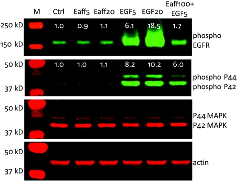
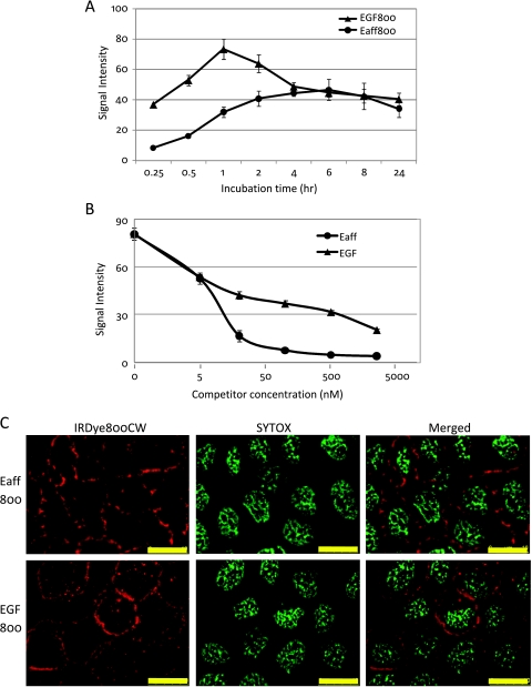
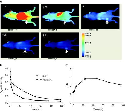
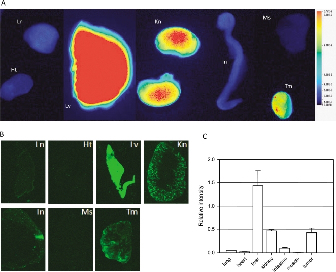
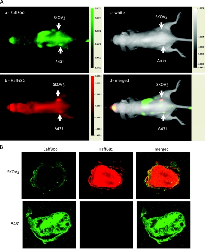
Overexpression of epidermal growth factor receptor (EGFR) is associated with many types of cancers. It is of great interest to noninvasively image the EGFR expression in vivo. In this study, we labeled an EGFR-specific Affibody molecule (Eaff) with a near-infrared (NIR) dye IRDye800CW maleimide and tested the binding of this labeled molecule (Eaff800) in cell culture and xenograft mouse tumor models. Unlike EGF, Eaff did not activate the EGFR signaling pathway. Results showed that Eaff800 was bound and taken up specifically by EGFR-overexpressing A431 cells. When Eaff800 was intravenously injected into nude mice bearing A431 xenograft tumors, the tumor could be identified 1 hour after injection and it became most prominent after 1 day. Images of dissected tissue sections demonstrated that the accumulation of Eaff800 was highest in the liver, followed by the tumor and kidney. Moreover, in combination with a human EGFR type 2 (HER2)-specific probe Haff682, Eaff800 could be used to distinguish between EGFR- and HER2-overexpressing tumors. Interestingly, the organ distribution pattern and the clearance rate of Eaff800 were different from those of Haff682. In conclusion, Eaff molecule labeled with a NIR fluorophore is a promising molecular imaging agent for EGFR-overexpressing tumors.


Figures

Figure 1
Figure 1
The effect of EGF and EGFR-specific Affibody (Eaff) on EGFR-mediated phosphorylation of EGFR and ERK1/2 (P44/42 MAPK) proteins. A431 cells were treated with either Eaff or EGF. Two concentrations (5 and 20 nM) for both Eaff (Eaff5 and Eaff20) and EGF (EGF5 and EGF20) were used. A combination of high concentration Eaff (100 nM, Eaff100) and 5 nM EGF was also used to treat cells. P44/42 MAPK and actin were used as internal controls. The relative expression levels were calculated by dividing the signal intensities of phospho-EGFR or phospho-P44/P42 by actin signal intensities. Note that the molecular weight markers in the second panel (phospho-P44/P42) and the third panel (P44/P42 MAPK) were the same. Ctrl indicates control without drug treatment.
Figure 2
Figure 2
Specific binding and uptake of IRDye800CW-labeled Affibody molecules. (A) The protein expression levels of EGFR and HER2 in MDA-MB-231 (MDA231), A431, SKOV3, and SKBR3 cells. Actin served as an internal control. The relative expression levels were calculated by dividing the signal intensities of EGFR or HER2 by actin signal intensities. (B) The binding and uptake of EGFR-specific Eaff800 and HER2-specific Haff800 by MDA231, A431, SKOV3, and SKBR3 cells. (C) Concentration-dependent binding and uptake of Eaff800-, Haff800-, or IRDye800CW-free dye by A431 cells.
Figure 3
Figure 3
The comparison of cellular binding and uptake between Eaff800 and EGF800. (A) Binding and uptake time course of Eaff800 (5 nM) and EGF800 (5 nM) by A431 cells. (B) The blocking of Eaff800 (5 nM) binding by increasing concentrations of unlabeled Eaff or EGF. (C) Microscopic examination of Eaff800 (20 nM) and EGF800 (20 nM) binding and uptake by A431 cells. Sytox green was used to stain the nuclei. Scale bar, 10 µm.
Figure 4
Figure 4
In vivo optical imaging of nude mice bearing A431 tumors using Eaff800. (A) A representative series of whole body images (dorsal view) acquired at different time points after injection of 0.5 nmol of Eaff800. The tumors were indicated with arrows. (B) Clearance of Eaff800 from the tumor and normal tissue. Average signal intensities were quantified using ROIs of equivalent-sized areas from the tumor sites and contralateral sites at indicated time points. Data were presented as mean ± SD of three individual mice. (C) TBR at different time points after probe injection. TBR was calculated by dividing the mean tumor signal by the mean background signal of the contralateral site.
Figure 5
Figure 5
Tissue distribution of Eaff800. (A) Nude mice bearing A431 tumors were killed 1 day after Eaff800 injection. The organs were collected and rinsed in PBS before imaging. Ht indicates heart; In, intestine; Kn, kidney; Ln, lung; Lv, liver; Ms, muscle; Tm, tumor. Note that the liver was imaged separately and merged to the picture because the liver signal was so strong that it illuminated the surrounding tissues if imaged together. (B) Fluorescence images of cryosections of dissected organs. The organs were snap-frozen in OCT compound and sectioned at 8-µm thickness. (C) Quantification of signal intensities of tissue sections. Average signal intensities were calculated using ROIs from different tissue sections.
Figure 6
Figure 6
Two-color in vivo optical imaging with Eaff800 and Haff682. (A) Nude mice bearing A431 and SKOV3 tumors on the left and right sides, respectively, were injected with 100 µl of PBS containing 0.5 nmol of Eaff800 and 0.5 nmol of Haff682. Whole body images (dorsal view) were acquired 1 day after agent injection. Green and red represent IRDye800CW and DY-682 fluorescence signals, respectively. The tumors were indicated with arrows. (B) Fluorescence images of cryosections of A431 and SKOV3 tumors. Mice bearing A431 and SKOV3 tumors were killed 1 day after agent injection. The tumors were snap-frozen in OCT compound and sectioned at 8-µm thickness.

References

    1. Ferguson KM, Darling PJ, Mohan MJ, Macatee TL, Lemmon MA. Extracellular domains drive homo- but not hetero-dimerization of erbB receptors. EMBO J. 2000;19:4632–4643. - PMC - PubMed
    1. Kari C, Chan TO, Rocha de Quadros M, Rodeck U. Targeting the epidermal growth factor receptor in cancer: apoptosis takes center stage. Cancer Res. 2003;63:1–5. - PubMed
    1. Yarden Y, Sliwkowski MX. Untangling the ErbB signalling network. Nat Rev Mol Cell Biol. 2001;2:127–137. - PubMed
    1. Baselga J. Targeting tyrosine kinases in cancer: the second wave. Science. 2006;312:1175–1178. - PubMed
    1. Speake G, Holloway B, Costello G. Recent developments related to the EGFR as a target for cancer chemotherapy. Curr Opin Pharmacol. 2005;5:343–349. - PubMed

LinkOut - more resources