. 2010 Feb;12(2):161-72.
doi: 10.1593/neo.91542.

Overexpression of elafin in ovarian carcinoma is driven by genomic gains and activation of the nuclear factor kappaB pathway and is associated with poor overall survival

Affiliations

Overexpression of elafin in ovarian carcinoma is driven by genomic gains and activation of the nuclear factor kappaB pathway and is associated with poor overall survival

Adam Clauss et al. Neoplasia. 2010 Feb.

Abstract

Ovarian cancer is a leading cause of cancer mortality in women. The aim of this study was to elucidate whether whey acidic protein (WAP) genes on chromosome 20q13.12, a region frequently amplified in this cancer, are expressed in serous carcinoma, the most common form of the disease. Herein, we report that a trio of WAP genes (HE4, SLPI, and Elafin) is overexpressed and secreted by serous ovarian carcinomas. To our knowledge, this is the first report linking Elafin to ovarian cancer. Fluorescence in situ hybridization analysis of primary tumors demonstrates genomic gains of the Elafin locus in a majority of cases. In addition, a combination of peptidomimetics, RNA interference, and chromatin immunoprecipitation experiments shows that Elafin expression can be transcriptionally upregulated by inflammatory cytokines through activation of the nuclear factor kappaB pathway. Importantly, using a clinically annotated tissue microarray composed of late-stage, high-grade serous ovarian carcinomas, we show that Elafin expression correlates with poor overall survival. These results, combined with our observation that Elafin is secreted by ovarian tumors and is minimally expressed in normal tissues, suggest that Elafin may serve as a determinant of poor survival in this disease.


Figures

Figure 1
Figure 1
A trio of WAP genes is overexpressed in ovarian carcinomas. (A) Expression of the WAP genes was determined by semiquantitative RT-PCR. Epididymis served as a positive control for WAP expression, and telomerase IOSE served as a cell-of-origin control. Elafin, SLPI, and HE4 are overexpressed in a panel of ovarian carcinomas. (B) Gene expression profiling of Elafin expression in different types of ovarian carcinomas and normal OSE. The tumors are divided into five groups: late-stage high-grade (LSHG) serous, early-stage high-grade (ESHG) serous, low-grade (LG) serous, and LSHG endometrioid. Elafin expression is significantly higher in all tumors compared with OSE. (C) Quantitative RT-PCR of Elafin expression in microdissected LSHG serous carcinomas (n = 20) compared with microdissected OSE (n = 10). LSHG serous cancers show a 70-fold increase of Elafin expression compared with OSE. (D) Expression of Elafin in OSE and serous carcinoma. Representative images from a panel of 8 normal ovaries and 20 late-stage high-grade serous carcinomas immunostained for Elafin. Original magnification, x20.
Figure 2
Figure 2
Expression of Elafin in human epididymis. IHC was performed on epididymis tissue using antibodies against Elafin, HE4, p63, and cytokeratin 7 (CK7). Elafin expression is limited to the p63- and CK7-positive basal cells, whereas HE4 is expressed in the luminal cell compartment.
Figure 3
Figure 3
Elafin is uniquely expressed by ovarian and squamous cancers. (A) RT-PCR analysis shows that Elafin is specifically expressed in ovarian cancer cell lines (OVCAR3, OVCAR5, CaoV3), but not in other common epithelial cancer cell lines, including breast (T47D, MCF7), kidney (293), colon (HCT-116, HCT-115), and osteosarcoma (U2OS) or in normal diploid fibroblasts (IMR90). (B) Northern dot blot analysis for Elafin expression in matched tumor (T) and normal (N) tissue RNA. Higher Elafin expression is detected in ovarian tumor compared with normal tissue; the converse is found in breast. Other tumors that show an increase of Elafin expression were found to be squamous cell carcinomas (marked by black arrowheads). (C) Elafin is a secreted protein. Conditioned medium was collected from cell lines expressing Elafin RNA as well as nonexpressors. Recombinant Elafin served as a positive control. The two bands that are observed on the Western blot of the recombinant protein are due to a mixture of protein with or without a signal peptide. Elafin is detected in the conditioned medium collected from Elafin-RNA-expressing cells but not from nonexpressors. (D) Primary tumors express and secrete Elafin. Elafin expression was determined by semiquantitative RT-PCR (27 cycles) in a collection of enriched primary tumors (DF lines). CK7 was used to validate the epithelial nature of the tumors. Actin served as a loading control. IMR90, HOSE, OVCAR3, and epididymis served as either negative or positive controls. (E) Primary tumors that express Elafin RNA also secreted it, as detected by Western blot analysis of conditioned medium collected from primary cell lines. The first three lanes are media from nonexpressing primary tumors and the last three are from high Elafin-expressing primary tumors. Recombinant Elafin is used as a positive control.
Figure 4
Figure 4
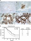
Elafin overexpression correlates with poor overall survival. IHC on 134 serous ovarian tumors (late-stage high-grade) with an Elafin antibody. (A) Examples of Elafin expression in ovarian tumors (10x). A few scattered positive cells (+1). Localized positive staining (+2). Large area of cells expressing Elafin (+3) and an example at a higher magnification (40x). (B) A Kaplan-Meier curve plotting overall survival of 77 patients with no or low Elafin expression (0 to +1, black) versus 57 high Elafin expression cases (+2 to +3, gray). Higher Elafin expression correlates with decreased survival (P = .023, N = 134) with a median survival of 47.3 months for patients with low Elafin expression versus 32.4 months for the patients that had tumors with higher Elafin expression. Scale used for scoring slides.
Figure 5
Figure 5
Genomic gains at the PI3 locus underlie Elafin overexpression in ovarian carcinomas. FISH for the PI3 locus. The G248P8772D4/G248P82920B4 fosmid probe (green) for PI3 was cohybridized with the control chromosome 20 centromeric probe D20Z1 (red) on tissue sections of ten ovarian carcinomas that express high levels of Elafin protein by IHC. (A, B) Representative examples of gains of the PI3 locus compared with the control probe. (C) Example of chromosome 20 trisomy. There are three foci of both the PI3 probe and the centromere probe. (D) Example of disomy at both the PI3 and centromeric loci.
Figure 6
Figure 6
NF-κB drives the expression of Elafin in response to inflammatory mediators. (A) Schematic representation of predicted transcription factor binding sites in the Elafin promoter. The location of these sites relative to the first base of the start codon for Elafin (+1) is indicated by the number above the square. (B) A semiquantitative RT-PCR before and after IL-1β stimulation of no- or low-Elafin expressors. Cells were treated with 5 ng/ml IL-1β for 48 hours before RNA was harvested. An increase of Elafin expression was detected in HeyA8, OVCAR8, and TOV21G cells but not in HOSE. Actin serves as a loading control. (C) Time course of Elafin expression after cytokine treatment. OVCAR8 cells were treated with IL-1β, and Elafin expression was monitored by quantitative RT-PCR for 48 hours. (D) Immunofluorescent localization of p65. The NF-κB subunit p65 is normally localized in the cytoplasm of OVCAR8 cells (left panel) but is translocated into the nucleus upon cytokine stimulation using IL-1β (middle panel). The translocation of p65 is inhibited by blocking the NF-κB pathway with a peptide that specifically binds to the NBD, thus sequestering p65 to the cytoplasm (right panel). (E) Elafin induction by IL-1β can be abrogated by NBD. Scramble peptide does not inhibit the induction by IL-1β. (F) OVCAR8 cells transfected with siRNA against NF-κB subunit p65 72 hours before IL-1β treatment. Transfected cells show almost no increase in Elafin expression compared with nontransfected or cells transfected with siRNA against GFP. Two unique siRNA give rise to similar results. Inlayed panel shows efficiency of siRNA knockdown for p65 by Western blot; GAPDH serves as a loading control (right graph). (G) Occupancy of NF-κB sites on Elafin promoter by ChIP. In OVCAR8 cells, NF-κB (p65) binds to the predicted binding sites 2 hours after IL-1β treatment. Anti-Golgi antibodies were used as negative controls for the IP. Primers located 5′ and 3′ of the NF-κB sites (-2 and +2 kb, respectively) served as controls and yielded no product. Input DNA shows that all primers give rise to products on genomic DNA. (H) IL-1β can act through NF-κB to induce further Elafin expression in ovarian cancer lines that constitutively express Elafin. OVCAR-3 cells constitutively express Elafin compared with OVCAR-8 cell (Figure W3). Stimulation of OVCAR-3 cells with IL-1β increases Elafin expression even further. Knockdown of NF-κB activity with siRNA against the p65 subunit (Western blot, inset) abrogates the stimulus-induced increase but not the baseline levels of Elafin expression.

Similar articles

Cited by

References

    1. Jemal A, Siegel R, Ward E, Hao Y, Xu J, Murray T, Thun MJ. Cancer statistics, 2008. CA Cancer J Clin. 2008;58:71–96. - PubMed
    1. Stewart B, Kleihues P. WHO World Cancer Report. Lyon, France: IARC Press; 2003.
    1. Cannistra SA. Cancer of the ovary. N Engl J Med. 2004;351:2519–2529. - PubMed
    1. Bast RC., Jr Status of tumor markers in ovarian cancer screening. J Clin Oncol. 2003;21:200–205. - PubMed
    1. Drapkin R, Clauss A, Skates S. Urokinase-type plasminogen activator receptor: a beacon of malignancy? Clin Cancer Res. 2008;14:5643–5645. - PMC - PubMed

Publication types

MeSH terms