. 2010 Feb;12(2):173-82.
doi: 10.1593/neo.91604.

Constitutive expression of the alpha4 integrin correlates with tumorigenicity and lymph node metastasis of the B16 murine melanoma

Affiliations

Constitutive expression of the alpha4 integrin correlates with tumorigenicity and lymph node metastasis of the B16 murine melanoma

Robert B Rebhun et al. Neoplasia. 2010 Feb.

Abstract

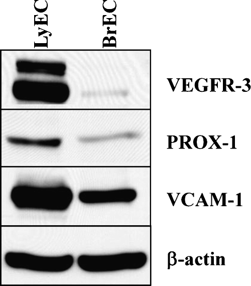
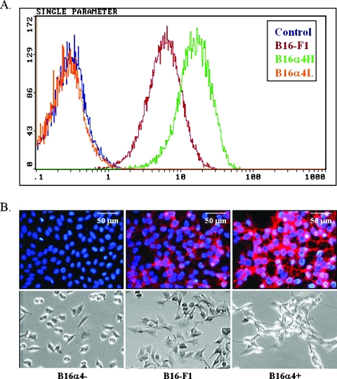
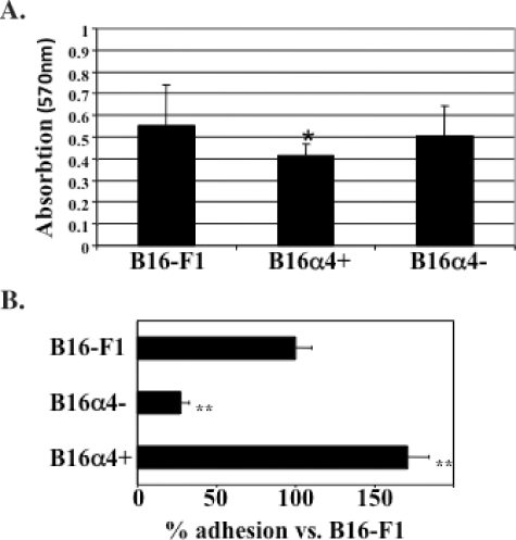
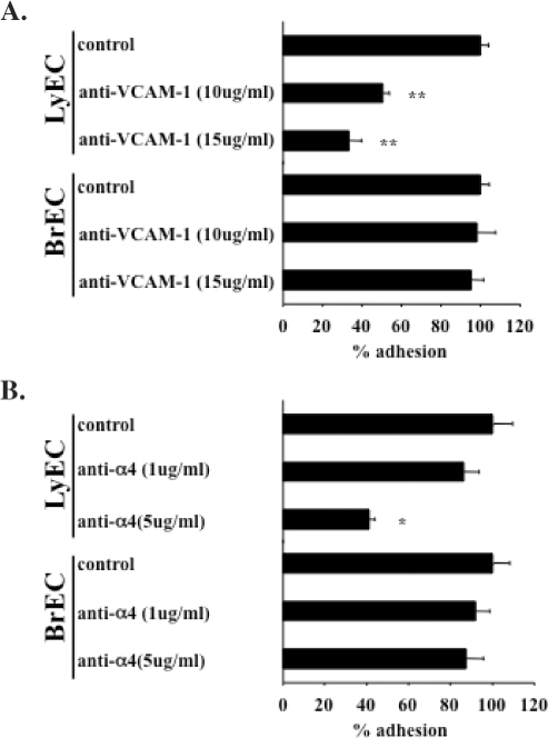
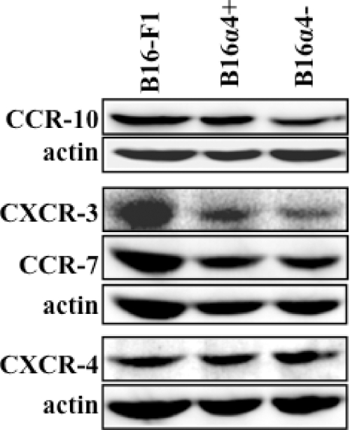
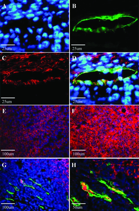
The lymphatic system plays a critical role in melanoma metastasis, and yet, virtually no information exists regarding the cellular and molecular mechanisms that take place between melanoma cells and the lymphatic vasculature. Here, we generated B16-F1 melanoma cells that expressed high (B16alpha(4)+) and negligible (B16alpha(4)-) levels of alpha(4) integrin to determine how the expression of alpha(4) integrins affects tumor cell interactions with lymphatic endothelial cells in vitro and how it impacts lymphatic metastasis in vivo. We found a direct correlation between alpha(4) integrin expression on B16-F1 melanoma cells and their ability to form adhesive interactions with monolayers of lymphatic endothelial cells. Adhesion of B16-F1 melanoma cells to lymphatic endothelial cells was mediated by the melanoma cell alpha(4) integrin binding to its counterreceptor, vascular cell adhesion molecule 1 (VCAM-1), that was constitutively expressed on the lymphatic endothelial cells. VCAM-1 was also expressed on the tumor-associated lymphatic vessels of B16-F1 and B16alpha(4)+ tumors growing in the subcutaneous space of C57BL/6J mice. B16-F1 tumors metastasized to lymph nodes in 30% of mice, whereas B16alpha(4)+ tumors generated lymph node metastases in 80% of mice. B16-F1 melanoma cells that were deficient in alpha(4) integrins (B16alpha(4)-) were nontumorigenic. Collectively, these data show that the alpha(4) integrin expressed by melanoma cells contributes to tumorigenesis and may also facilitate metastasis to regional lymph nodes by promoting stable adhesion of melanoma cells to the lymphatic vasculature.


Figures

Figure 1
Figure 1
Expression of VEGFR-3, Prox1, and VCAM-1 by lymphatic endothelial cells and brain-derived microvascular endothelial cells. Immunoblot analysis results show that lymphatic endothelial cells retain their characteristic features despite transfer to cell culture. Lymphatic endothelial cells (LyEC) express high levels of VEGFR-3, Prox1, and VCAM-1 compared with brain-derived vascular endothelial cells (BrEC). β-Actin is shown as an internal loading control.
Figure 2
Figure 2
Expression of α4 by B16 melanoma cell lines. (A) Selection of variant sublines from the parental B16-F1 melanoma cells was performed as described within the text. Successive selection from the B16-F1 cells yielded a subline that expressed high levels of α4 (B16α4+) and a subline that was deficient in α4 integrin (B16α4-). (B) Immunohistochemical staining of α4 and representative examples of morphologic differences as observed with phase-contrast microscopy. When compared with B16-F1 melanoma cells, B16α4- cells display decreased homotypic adhesion in vitro, whereas B16α4+ cells are more inclined to form cell-cell aggregates.
Figure 3
Figure 3
Analysis of B16 melanoma cell proliferation and melanoma cell adhesion to lymphatic endothelial cells. (A) Cell proliferation of B16-F1 melanoma cell lines in vitro was assessed by MTT. Cell division of both B16-F1 and B16α4- tumor cells was significantly greater than the growth of B16α4+ melanoma cells. (B) The adhesion of B16α4+ to lymphatic endothelial cells is seven times greater than the adhesion of B16α4- melanoma cells. Data are expressed as mean ± SEM and repeated at least four times with comparable results.
Figure 4
Figure 4
B16-F1 melanoma cell adhesion to lymphatic endothelial cells and brain microvascular endothelial cells. (A) Blockade of VCAM-1 expressed on lymphatic endothelial cells results in a significant reduction in number of adherent tumors cells to lymphatic endothelial cells, whereas the same treatment has no effect on tumor cell adhesion to brain endothelial cells. (B) Blockade of the melanoma cell α4 integrin also results in dose-dependent reduction in tumor cell adhesion to lymphatic endothelial cells. Identical treatment has no effect on ability of melanoma cells to adhere to brain endothelial cells. Data are expressed as mean ± SEM. *P < .05, **P < .001.
Figure 5
Figure 5
Chemokine receptor expression byB16 melanoma cell lines. Immunoblot analysis results demonstrate that B16-F1, B16α4+, and B16α4- express similar amounts of the chemokine receptors CCR-10, CCR-7, and CXCR-4. CXCR-3 expression was slightly greater in B16-F1 cells in comparison to B16α4+ and B16α4- melanoma cells. β-Actin is shown as an internal loading control.
Figure 6
Figure 6
VCAM-1 is expressed on B16 tumor-associated lymphatic endothelial cells. (A–D) Double immunostaining for the lymphatic specific marker LYVE-1 (green) and VCAM-1 (red) reveals colocalization (yellow) of VCAM-1 and LYVE-1 on B16 tumor-associated lymphatic vessels. Nuclei are stained with Hoescht (blue). (E) The intensity of α4 integrin expression (red) in parental B16-F1 tumors is significantly reduced in comparison to expression found in (F) B16α4+ tumors. (G, H) LYVE-1 (green) and α4 (red) expressions at the peritumoral region of B16α4+ tumors growing subcutaneously. B16α4+ tumor cells (confirmed with hematoxylin and eosin staining) can readily be identified around and within peritumoral lymphatic vessels.

Similar articles

Cited by

References

    1. Balch CM. The role of elective lymph node dissection in melanoma: rationale, results, and controversies. J Clin Oncol. 1988;6:163–172. - PubMed
    1. Balch CM, Buzaid AC, Soong SJ, Atkins MB, Cascinelli N, Coit DG, Fleming ID, Gershenwald JE, Houghton A Jr, Kirkwood JM, et al. New TNM melanoma staging system: linking biology and natural history to clinical outcomes. Semin Surg Oncol. 2003;21:43–52. - PubMed
    1. Balch CM, Buzaid AC, Soong SJ, Atkins MB, Cascinelli N, Coit DG, Fleming ID, Gershenwald JE, Houghton A Jr, Kirkwood JM, et al. Final version of the American Joint Committee on Cancer staging system for cutaneous melanoma. J Clin Oncol. 2001;19:3635–3648. - PubMed
    1. Balch CM, Soong SJ, Gershenwald JE, Thompson JF, Reintgen DS, Cascinelli N, Urist M, McMasters KM, Ross MI, Kirkwood JM, et al. Prognostic factors analysis of 17,600 melanoma patients: validation of the American Joint Committee on Cancer melanoma staging system. J Clin Oncol. 2001;19:3622–3634. - PubMed
    1. Callery C, Cochran AJ, Roe DJ, Rees W, Nathanson SD, Benedetti JK, Elashoff RM, Morton DL. Factors prognostic for survival in patients with malignant melanoma spread to the regional lymph nodes. Ann Surg. 1982;196:69–75. - PMC - PubMed

Publication types

Substances

LinkOut - more resources