Case Reports
. 2009 Dec:107:72-90.

Inferior oblique surgery for restrictive strabismus in patients with thyroid orbitopathy

Affiliations
Case Reports

Inferior oblique surgery for restrictive strabismus in patients with thyroid orbitopathy

Steven A Newman. Trans Am Ophthalmol Soc. 2009 Dec.

Abstract

Introduction: Thyroid orbitopathy is the most common cause of restrictive strabismus. Patients often present with vertical or horizontal double vision, or both, due to restriction involving most commonly the inferior and medial rectus muscles. Traditional muscle surgery involves release of the tight muscles. Previous literature has described a frequent need for secondary operations and an overcorrection incidence of up to 50%. Recognizing that the tight muscles are also limited in their excursion, it was proposed that operating on the better-moving eye, particularly the inferior oblique, might produce an improvement in binocularity and decrease the incidence of overcorrection.

Methods: A total of 37 patients with restrictive strabismus due to thyroid orbitopathy treated at the University of Virginia over 12 years with inferior oblique surgery were retrospectively reviewed.

Results: Eight patients were treated with a combination of inferior oblique surgery and horizontal muscle surgery at the same time. One patient was treated with simultaneous inferior oblique and superior rectus surgery. Seven patients had vertical correction with inferior oblique surgery alone. Twenty-three patients required secondary procedures. Eight patients were overcorrected but only one following primary surgery. At the time of last follow-up, ranging from 6 months to 8 years, 33 patients had no diplopia, 2 had minimal diplopia, and 2 had persistent diplopia. All but two were completely functional.

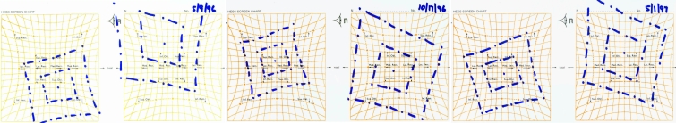
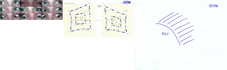
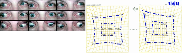
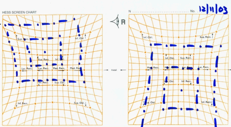
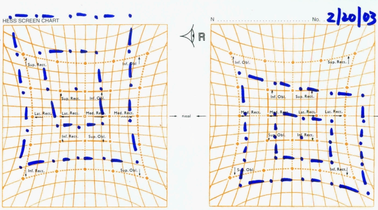
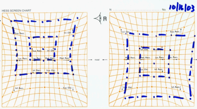
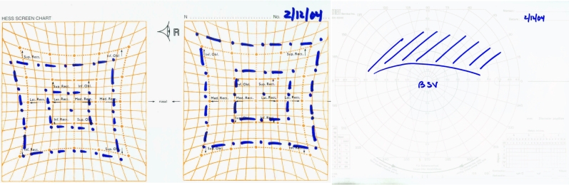
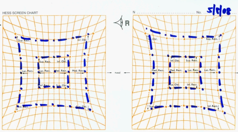
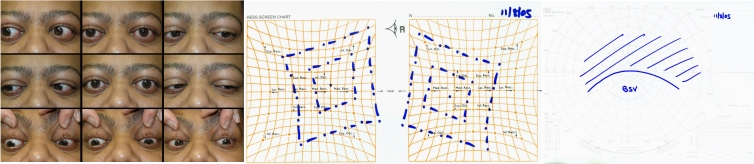
Conclusion: Inferior oblique surgery by balancing the overall excursion of extraocular muscles in thyroid patients may produce binocularity in primary position and down reading gaze. The amount of vertical correction from inferior oblique surgery alone is limited, often requiring ipsilateral superior or contralateral inferior rectus surgery. Inferior oblique surgery likely increases the area of binocular single vision and decreases the incidence of overcorrection. The use of Hess screen and binocular single vision fields is helpful in assessment and planning of surgery in these patients.


Figures

FIGURE 1
FIGURE 1
Case 1. Hess screens of 77-year-old woman with double vision (left), following left inferior rectus recession (middle), and following right inferior rectus recession (right).
FIGURE 2
FIGURE 2
Case 1. Left, Hess screen following right inferior oblique extirpation. Right, Binocular single vision field following right inferior oblique extirpation demonstrating double vision on down right gaze, but single vision up left.
FIGURE 3
FIGURE 3
Case 1. Left, Nine cardinal positions following bilateral inferior oblique extirpation. Right, Hess screen demonstrating minimal residual right hypertropia. At that time the patient had no double vision.
FIGURE 4
FIGURE 4
Case 2. Left, A 47-year-old patient with limitation in elevation bilaterally. Middle, Hess screen demonstrating secondary overaction of the inferior obliques due to restriction of the inferior rectus. Right, Binocular single vision fields done at the same time as the Hess screen illustrating single vision down and to the left and double vision up and to the right.
FIGURE 5
FIGURE 5
Case 2. Left, Nine cardinal positions following left inferior oblique extirpation. Right, Hess screen indicating residual right hypertropia on left gaze, but marked collapse of the previously noted left hypertropia on right gaze.
FIGURE 6
FIGURE 6
Case 2. Left, Nine cardinal positions following bilateral inferior oblique extirpation. At that time the patient was noting double vision only “when tired.” Right, Hess screen illustrating persistent problems with elevation of the right eye in right gaze.
FIGURE 7
FIGURE 7
Case 3. Left, Nine cardinal positions in a 69-year-old man with limitation in elevation particularly of the right eye in right gaze. Middle, Hess screen illustrating marked limitation in elevation on the right side. Right, Binocular single vision fields done at the same time as the previous Hess screen demonstrate single vision but only 30° down.
FIGURE 8
FIGURE 8
Case 3. Left, Nine cardinal positions following left inferior oblique extirpation showing similar limitation in elevation of the left eye in up right gaze. Middle, Hess screen illustrating persistent problems with elevation on the right, but decreased secondary overaction of the left inferior oblique following surgery. Right, Binocular single vision now reveals double vision only 10° above primary position.
FIGURE 9
FIGURE 9
Case 4. Hess screen illustrating limitation in elevation of the left eye with secondary overaction of the right inferior oblique.
FIGURE 10
FIGURE 10
Case 4. Left, Nine cardinal positions following right inferior oblique extirpation resulting in similar limitation in elevation of the right eye to the left eye in attempt at up left gaze. Middle, Hess screen following right inferior oblique surgery reveals residual limitation in down gaze in the right eye. Right, Binocular single vision fields following right inferior oblique reveals single vision above fixation, but double vision below primary gaze.
FIGURE 11
FIGURE 11
Case 4. Hess screen illustrating limitation in elevation of the left eye with secondary overaction of the right inferior oblique.
FIGURE 12
FIGURE 12
Case 5. Left, Nine cardinal positions of 63-year-old patient illustrating significant limitation in elevation particularly of the right eye in right gaze. Right, Hess screen done at the same time as the nine cardinal positions illustrates left hypertropia increasing on up gaze.
FIGURE 13
FIGURE 13
Case 5. Hess screen following left inferior oblique extirpation shows some limitation in overelevation of the left eye, but marked residual left hypertropia increasing on down gaze.
FIGURE 14
FIGURE 14
Case 5. Left, Nine cardinal positions following left superior rectus recession resulting in complete resolution of double vision. Right, Hess screen following left superior rectus recession shows minimal residual vertical limitation on the left.
FIGURE 15
FIGURE 15
Case 6. Hess screen of a 65-year-old woman with a 5-month history of double vision demonstrating a left hypertropia increasing on up right gaze secondary to restriction of the right inferior rectus.
FIGURE 16
FIGURE 16
Case 6. Hess screen following left inferior oblique extirpation shows reduction of the overacting inferior oblique on the left side with persistent left hyperdeviation.
FIGURE 17
FIGURE 17
Case 6. Left, Hess screen following left superior rectus recession shows slight overcorrection now with a right hypertropia on up gaze. Right, Binocular single vision fields done at the same time show binocular single vision in primary position and down reading gaze but double vision 18° above primary gaze.
FIGURE 18
FIGURE 18
Case 6. Recurrent left hyperdeviation following presumed slippage of right superior rectus suture resulting in increasing double vision on down gaze.
FIGURE 19
FIGURE 19
Case 6. Left, Nine cardinal positions immediately following right inferior rectus recession with slippage of the muscle. Right, Hess screen demonstrates a slight right hypertropia increasing on down gaze.
FIGURE 20
FIGURE 20
Case 6. Hess screen following reoperation with advancement of the slipped right inferior rectus. At that time the patient had no recurrent or residual problems with double vision.
FIGURE 21
FIGURE 21
Case 7. Left, Nine cardinal positions of a 34-year-old patient with severe proptosis and limitation in elevation on the left. Right, Hess screen done at the time of nine cardinal positions demonstrates a marked left hypotropia.
FIGURE 22
FIGURE 22
Case 7. Hess screen following bilateral orbital decompressive surgery with an increase in the relative left hypotropia.
FIGURE 23
FIGURE 23
Case 7. Left, Nine cardinal positions following right inferior oblique extirpation. Right, Hess screen following right inferior oblique extirpation demonstrates persistent limitation in elevation and abduction.
FIGURE 24
FIGURE 24
Case 7. Left, Nine cardinal positions following bilateral inferior rectus recessions with some minimal residual double vision when looking up and to either side. Middle, Hess screen done following bilateral inferior rectus recessions still shows effective overaction of the inferior obliques in spite of previous right inferior oblique extirpation. Right, Binocular single vision fields demonstrate single vision in primary position and in down reading gaze with persistent double vision up and to either side.

Similar articles

References

    1. Bok C, Hidalgo C, Morax S. Surgical management of diplopia in dysthyroid orbitopathy. J Fr Ophtamol. 2007;30:390–396. - PubMed
    1. Chu YK, Kim SJ, Lee SY. Surgical treatment modalities of thyroid ophthalmopathy. Korean J Ophthalmol. 2001;15:128–132. - PubMed
    1. Looi ALG, Luu CD, Wong TY, Seah LL, Rootman J. Factors associated with decompression and strabismus surgery in thyroid eye disease. Ann Acad Med Singapore. 2005;34:154–157. - PubMed
    1. Garrity JA, Fatourechi V, Bergstralh V, et al. Results of transantral orbital decompression in 428 patients with severe Graves’ orbitopathy. Am J Ophthalmol. 1993;116:533–547. - PubMed
    1. Werner SC, Coleman DJ, Franzen LA. Ultrasonographic evidence of a consistent orbital involvement in Graves’ disease. N Engl J Med. 1974;290:1447–1450. - PubMed

Publication types